Understanding the Anatomy of the Tongue
Explore the detailed anatomy of the tongue, a vital muscular organ in the mouth responsible for food manipulation, swallowing, taste perception, and speech production. Learn about its structure, measurements, weight, and functions, providing valuable insights into this essential component of the human body.

Download Presentation

Please find below an Image/Link to download the presentation.
The content on the website is provided AS IS for your information and personal use only. It may not be sold, licensed, or shared on other websites without obtaining consent from the author. If you encounter any issues during the download, it is possible that the publisher has removed the file from their server.
You are allowed to download the files provided on this website for personal or commercial use, subject to the condition that they are used lawfully. All files are the property of their respective owners.
The content on the website is provided AS IS for your information and personal use only. It may not be sold, licensed, or shared on other websites without obtaining consent from the author.
E N D
Presentation Transcript
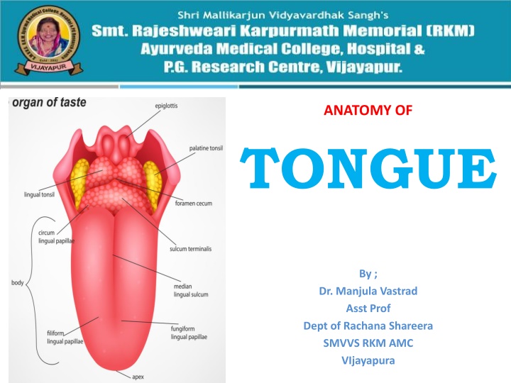
ANATOMY OF TONGUE By ; Dr. Manjula Vastrad Asst Prof Dept of Rachana Shareera SMVVS RKM AMC VIjayapura
INTRODUCTION :The TONGUE is a muscular organ in the mouth of that manipulates food for mastication and is used in the act of swallowing. It has importance in the digestive system and is the primary organ of taste. A major function of the tongue is the enabling of speech .
STRUCTURE The tongue is a muscular hydrostat that forms part of the floor of the oral cavity. The left and right sides of the tongue are separated by a vertical section of fibrous tissue known as the lingual septum. This division is along the length of the tongue save for the very back of the pharyngeal part and is visible as a groove called the median sulcus. The human tongue is divided into anterior and posterior parts by the terminal sulcus which is a V-shaped groove. The apex of the terminal sulcus is marked by a blind foramen, the foramen cecum, which is a remnant of the median thyroid diverticulum in early embryonic development. The anterior oral part is the visible part situated at the front and makes up roughly two-thirds the length of the tongue. The posterior pharyngeal part is the part closest to the throat, roughly one-third of its length.
The anterior tongue is, at its apex, thin and narrow. It is directed forward against the lingual surfaces of the lower incisor teeth. The posterior part is, at its root, directed backward, and connected with the hyoid bone by the hyoglossiand genioglossi muscles and the hyoglossal membrane, with the epiglottis three glossoepiglottic folds of mucous membrane, with the soft palate by the glossopalatine arches, and with the pharynx by the superior pharyngeal constrictor muscle and the mucous membrane. It also forms the anterior wall of the oropharynx. by
Measurement and Weight: The average length of the human tongue from the oropharynx to the tip is 10 cm.The average weight of the human tongue from adult males is 70g and for adult females 60g.
The upper surface of the tongue is called the dorsum, and is divided by a groove into symmetrical halves by the median sulcus. The foramen cecum marks the end of this division (at about 2.5 cm from the root of the tongue) and the beginning of the terminal sulcus. The foramen cecum is also the point of attachment of the thyroglossal duct and is formed during the descent of the thyroid diverticulum in embryonic development.
The terminal sulcus is a shallow groove that runs forward as a shallow groove in a V shape from the foramen cecum, forwards and outwards to the margins (borders) of the tongue. The terminal sulcus divides the tongue into a posterior pharyngeal part and an anterior oral part. The pharyngeal part is supplied by the glossopharyngeal nerve and the oral part is supplied by the lingual nerve (a branch of the mandibular branch (V3) of the trigeminal nerve) for somatosensory perception and by the chorda tympani (a branch of the facial nerve) for taste perception.
Undersurface of the tongue : On the undersurface of the tongue is a fold of mucous membrane called the frenulum that tethers the tongue at the midline to the floor of the mouth. On either side of the frenulum are small prominences called sublingual caruncles that the major salivary submandibular glands drain into.
Muscles The eight muscles of the human tongue are classified as either intrinsic or extrinsic. The four intrinsic muscles act to change the shape of the tongue, and are not attached to any bone. The four extrinsic muscles act to change the position of the tongue, and are anchored to bone. Extrinsic Lateral view of the tongue, with extrinsic muscles highlighted The four extrinsic muscles originate from bone and extend to the tongue. They are the genioglossus, the hyoglossus (often including the chondroglossus) the styloglossus, and the palatoglossus. Their main functions are altering the tongue's position allowing for protrusion, retraction, and side-to-side movement. The genioglossus arises from the mandible and protrudes the tongue. It is also known as the tongue's "safety muscle" since it is the only muscle that propels the tongue forward.
The hyoglossus, arises from the hyoid bone and retracts and depresses the tongue. The chondroglossus is often included with this muscle. The styloglossus arises from the styloid process of the temporal bone and draws the sides of the tongue up to create a trough for swallowing. The palatoglossus arises from the palatine aponeurosis, and depresses the soft palate, moves the palatoglossal fold towards the midline, and elevates the back of the tongue during swallowing.
Intrinsic Muscles : Four paired intrinsic muscles of the tongue originate and insert within the tongue, running along its length. They are the superior longitudinal muscle, the inferior longitudinal muscle, the vertical muscle, and the transverse muscle. These muscles alter the shape of the tongue by lengthening and shortening it, curling and uncurling its apex and edges as in tongue rolling, and flattening and rounding its surface. This provides shape and helps facilitate speech, swallowing, and eating.
The superior longitudinal muscle runs along the upper surface of the tongue under the mucous membrane, and elevates, assists in retraction of, or deviates the tip of the tongue. It originates near the epiglottis, at the hyoid bone, from the median fibrous septum. The inferior longitudinal muscle lines the sides of the tongue, and is joined to the styloglossus muscle. The vertical muscle is located in the middle of the tongue, and joins the superior and inferior longitudinal muscles. The transverse muscle divides the tongue at the middle, and is attached to the mucous membranes that run along the sides.
BLOOD SUPPLY The tongue receives its blood supply primarily from the lingual artery, a branch of the external carotid artery. The lingual veins drain into the internal jugular vein. The floor of the mouth also receives its blood supply from the lingual artery. There is also a secondary blood supply to the root of tongue from the tonsillar branch of the facial artery and the ascending pharyngeal artery
NERVE SUPPLY : hypoglossal nerve trigeminal nerve glossopharyngeal nerve superior laryngeal nerve
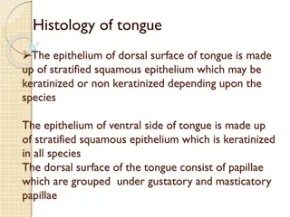
LINGUAL PAPILLAE These are the small, nipple-like structures on the upper surface of the tongue that give it its characteristic rough texture. The four types of papillae on the human tongue have different structures and are accordingly classified as circumvallate (or vallate), fungiform, filiform, and foliate. All except the filiform papillae are associated with taste buds