Understanding Rinderpest: A Cattle Plague Overview
Rinderpest, also known as Cattle Plague, is caused by the Morbillivirus and primarily affects cloven-hooved animals like cattle and buffalo. It is transmitted through contact with infected animals and has distinct clinical signs ranging from fever and mucopurulent secretions to severe hemorrhagic diarrhea. The virus is sensitive to certain disinfectants and has been eradicated globally in 2011. It does not infect humans.
Download Presentation

Please find below an Image/Link to download the presentation.
The content on the website is provided AS IS for your information and personal use only. It may not be sold, licensed, or shared on other websites without obtaining consent from the author. Download presentation by click this link. If you encounter any issues during the download, it is possible that the publisher has removed the file from their server.
E N D
Presentation Transcript
RINDERPEST (Synonym: Cattle Plague) Etiology: Genus: Morbillivirus, within the family Paramyxouiridae. In addition to rinderpest virus (RPV), the genus includes peste des petits ruminants virus (PPRV), which infects sheep and goats; canine distemper virus (CDV), which infects carnivores; and human measles virus (MV). There is one serotype of this virus, but 3 genetically distinct lineages lineage 1(African Lineage ), lineage 2(African Lineages) lineage 3(Asian Lineages ) have been identified. Species Affected: Most cloven-hooved animals (order Artiodactyla) are susceptible Cattle, water buffalo, yaks, African buffalo, giraffes, warthogs and Tragelaphinae (spiral-horned antelope) are particularly susceptible to disease. Cattle are the most important maintenance hosts. Rinderpest is rare among Camelidae; especially in endemic areas. They are dead-end hosts and do not transmit the virus. and
Resistance to physical and chemical action: Temperature: Small amounts of virus resist 56 C/60 minutes or 60 C/30 minutes. pH: Stable between pH 4.0 and 10.0. Disinfectants/chemicals: Susceptible to lipid solvents and most common disinfectants (phenol, cresol, -propiolactone, sodium hydroxide 2%/24 hours used at a rate of 1 litre/m2). Survival: Quickly inactivated in environment (to light, drying and ultraviolet radiation). Can remain viable for long periods in chilled or frozen tissues. Transmission: By direct or close indirect contact between infected and susceptible animals Airborne transmission is limited RPV is sensitive to direct sunlight thus fomites are not a viable means of transmission No evidence of vertical transmission Introduction of RPV into free areas is most commonly by means of infected animals
Sources of virus: Shedding of virus begins 1 2 days before pyrexia in tears, nasal secretions, saliva, urine and faeces. Blood and all tissues are infectious before the appearance of clinical signs During periods of clinical disease, high levels of RPV can be found in expired air, nasal and ocular discharges, saliva, faeces, semen, vaginal discharges, urine and milk. Infection is via the epithelium of the upper or lower respiratory tract. No carrier state. The eradication campaign concluded in 2011 with an international declaration of global freedom from rinderpest. Zoonotic potential : Rinderpest virus has not been reported to infect humans .
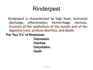
CLINICAL SIGNS: Severity depending on the virulence of the strain and resistance of the infected animal. Infection is initiated in the upper respiratory tract, usually through nasal entry, and is followed by initial replication in the tonsils and local lymph nodes. In Cattle: A peracute form: characterized primarily by high fever and sudden death, is mainly seen in young and newborn animals. The clinical form of the disease can be divided in 3 phases: In the prodromal phase: hyperthermia develops rapidly and it is followed by the mucosal phase, in which ocular and nasal mucopurulent secretions are accompanied by anorexia and depression. The final phase is characterized by severe hemorrhagic diarrhea and prostration, followed by dehydration and death (affected animals usually die 6 12 days after symptoms appear).
Mortality varies with the strain. Convalescence can be prolonged and may be accompanied by secondary infections. Pregnant cows often abort during this period Diarrhea usually starts after the onset of oral necrosis; it is typically profuse and watery, but may contain mucus, blood and shreds of epithelium in the later stages Sheep and goats Variable signs; some pyrexia, anorexia and minor ocular discharge Sometimes diarrhoea Asian RPV strains can be transmitted to cattle by contact with infected small ruminants Pigs Swine is commonly affected Pyrexia, anorexia and prostration
Erosions of buccal mucosa 12 days after fever and diarrhoea at 2 3 days Diarrhoea may last a week leading to dehydration and possible death Lesions: Either areas of necrosis and erosions, or congestion and haemorrhage in the mouth, intestines and upper respiratory tracts Highly engorged or grey discolouration of abomasum (epithelial necrosis of mucous membrane); pyloric region severely affected and shows congestion, petaechiation and oedema of the submucosa Rumen, reticulum and omasum usually unaffected; necrotic plaques are occasionally encountered on the pillars of the rumen Enlarged and oedematous lymph nodes White necrotic foci in Peyer s patches; lymphoid necrosis and sloughing leaves the supporting blackened architecture engorged or
Linear engorgement and blackening of the crests of the folds of the caecum, colon and rectum ( Zebra striping ). Linear haemorrhages on the folds of mucosa of rectum appear like 'Zebra marking' which is pathognomonic in Rinder pest. Typically the carcass of the dead animal is dehydrated, emaciated and soiled DIAGNOSIS: A.Laboratory diagnosis Samples Blood for isolating virus; do not use glycerol as preservative transport media as it inactivates RPV Spleen, prescapular or mesenteric lymph nodes of dead animals chilled to sub-zero temperatures for virus isolation
Histopathology and immunohistochemistry (10% neutral buffered formalin ) Base of the tongue, retropharyngeal lymph node and third eyelid are suitable tissues Ocular and nasal secretions Antigen detection by agar gel immunodiffusion Histopathology and immunohistochemistry Chromatographic strip test: Used for assisting field personnel in investigating suspected outbreaks of rinderpest. Serological tests The competitive enzyme-linked immunosorbent assay VN test
Prevention and Control: A ban on entry of animals from infected areas. Quarantine at border and creation of immune barrier zones is advisable for disease free countries having land borders with enzootic areas. In enzootic states/countries, annual vaccination until no more outbreak are reported for last 5 years and a surveillance to monitor the immune status of animals should be adopted. Control of animal movement and ring vaccination of herd is necessary, if any outbreak occurs. Both killed and attenuated vaccines are used: Attenuated vaccines are good for enzootic areas and killed vaccines of tissue culture origin are recommended for free areas with the danger of introduction through border.
Goat tissue vaccine(GTV) is good for indian plain type Zebu cattle but not safe for buffalo or exotic cattle which have low innate resistance and to be given@ 1ml sc ,provide life long immunity. Lapinized vaccine also used and provide immunity for 2-3years. Avianized vaccine is most safe for exotic breeds and protect them for16 months. Tissue culture vaccine(TCRP)is given @ 1ml sc provide immunity for 10 years. It is best vaccine and has replaced all other vaccines and used for sheep and goat to prevent PPR also. With all these vaccines, calves and lambs born of immune mothers should be vaccinated at 9 and 6 months of age respectively. Measles vaccine is good for young calves deriving maternal antibodies and also for adult cattle, although the degree of immunity is not as strong as with ordinary rinderpest vaccine. Recombinant vaccine is also useful for calves deriving maternal antibody and gives the immunity for 1year and administered by scarification.
Peste des petits ruminants (PPR) (Goat plague, Ovine rinderpest, plague of small ruminants, Kata) Morbillivirus genus in the family Paramyxoviridae, preferentially replicates in lymphoid tissues and epithelial tissue of the GI and respiratory tracts Transmission: By close contact The virus is present in ocular, nasal, and oral secretions as well as feces. Inhalation of aerosols from sneezing and coughing animals. Clinical signs: The incubation period is 4 6 days High fever (41 C/1060F), erosive gastroenteritis, and pneumonia, drymuzzle, oculo-nasal discharges, Watery blood-stained diarrhea stomatitis, conjunctivitis,
Erosions-small pin-point red-greyish areas on the gums, dental pad, palate, lips, inner aspects of the cheeks and upper surface of the tongue. Eye, nose and mouth dischargeswith scabs or nodules around the mouth Death usually occurs 4 6 days after the onset of fever. High morbidity (up to 100%) and up to 90% mortality Post mortem findings: Prominent crusty scabs along the outer lips and severe congestion of lungs due to interstitial pneumonia Diagnosis: Clinical Signs Whole blood in heparin or EDTA. Virus isolation Antigen capture ELISA and reverse transcription-PCR
Competitive ELISA and virus neutralization are the OIE- recommended tests. Treatment: Only supportive treatment to counteract secondary bacterial infection. One course of antibiotics (Ceftriaxone, Gentamycin), 5% dextrose, Multivitamins, Meloxicam can be given in affected animals Control: Farm Disinfection: PPR virus can be killed by most common Disinfectants-Phenols, 2%Sodium hydroxide Vaccination: Live attenuated PPR vaccine
Names: PPR vaccine, Raksha PPR. Availability: 100 and 50 doses with diluent and freeze dried vaccine vials. Dose: 1 ml. Age group: 3 months kids. Route: Subcutaneous route. Immunity: 3 years.


