Understanding Retinal Image Processing and Disorders
Discover the intricacies of retinal image processing, the structure of the retina, and its correlation with the brain. Learn about retinal disorders such as Age-related Macular Degeneration, Diabetic Retinopathy, and Glaucoma. Explore visualization techniques like Fundus Photography and Optical Coherence Tomography imaging for a comprehensive view of the eye's health.

Download Presentation

Please find below an Image/Link to download the presentation.
The content on the website is provided AS IS for your information and personal use only. It may not be sold, licensed, or shared on other websites without obtaining consent from the author. If you encounter any issues during the download, it is possible that the publisher has removed the file from their server.
You are allowed to download the files provided on this website for personal or commercial use, subject to the condition that they are used lawfully. All files are the property of their respective owners.
The content on the website is provided AS IS for your information and personal use only. It may not be sold, licensed, or shared on other websites without obtaining consent from the author.
E N D
Presentation Transcript
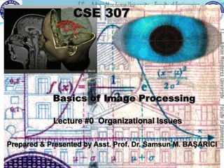
Retinal image processing
The innermost part of the eye Connected with the optic nerve
Structure of Retina Consists of light sensitive tissues of the eyes There are two layers: Pigment epithelium (First Layer) Ensures selective input of substances from choroid into the retina Light sensitive cells (Photoreceptor) These cells help to create visual perception Three types of photoreceptor cells: Rods Cones Ganglion cells Additional structural elements
Creating visual perception Light passes through cornea and brain Light hits a segment of retina Photoreceptor cells of the brain receives the light Retina is closely connected with the brain Photoreceptor cells send electrical neural impulses to the brain The visual cortex of the brain creates visual perception from the electrical neural impulses
Visualization of retina Fundus photography Takes image of the rear of the eye Used to visualize retina, optic disc, blood vessels and macula Image is taken with a microscope attached flash enabled camera
Visualization of retina (2) OCT (Optical coherence Tomography) imaging A non invasive technique to generate cross sectional image of the retina Visualizes different layers of a retina
Retinal disorders Age related Macular Degeneration (AMD) A medical condition which may result in no vision in the center of the eyes because of age Diabetic Retinopathy (DR) A medical condition which cases damage to retina because of diabetes Glaucoma A disease that damages optic nerve. Extra fluid built up in front part of the eye causes this Drusen Drusen is tiny yellow or white material build up in between layers of retina. Excessive amount of drusen may cause AMD And many more!
Correlation with the brain Retina is the part of the central nervous system. Hence, it is very close to the retina. As a result, many brain disorders also tend to affect the retina. Research from the University of Eastern Finland (UEF) shows that retinal changes may be detected earlier than brain changes. Therefore, retinal examination is very crucial for early detection of brain disorders. Brain disorders like Alzheimer, Schizophrenia, perkinson s disease may also affect the brain.
Structural features for anomaly detection in retina Existence of drusen Damage of the ganglion cell layers Reduced thickness of RNFL (Retinal Nerve Fiber Layer) Reduced thickness of blood vessels Cherry red spots on fundus imaging Gathering of different types of fluids
Figure: Normal retina (left) and retinal with anomaly (right) caused by macular degeneration
Screening features for anomaly detection Reaction caused by intense light Eye movement pattern during reading Patterns of following fast moving objects with eyes Patterns of eye movements while watching vehement reactions These are not directly associated with retina, but very useful for brain disease diagnosis
Machine learning for retinal disease diagnosis Machine learning can be used to diagnosis disease from retinal images SVM and Neural Network can be very helpful Deep neural network is an extremely powerful tool for diagnosis Classification model learns from given fundus or OCT images General image processing techniques (blurring, smoothing) can be used but often produce worse result Including handcrafted features may produce better results [1]
Deep learning for retinal disease diagnosis Deep neural networks works very well for large number of datasets Transfer learning from imagenet dataset (with VGG, Inception or Resnet model) can provide fast and fairly accurate results. [2] Ensemble of deep networks with different structure can improve classification accuracy. [3]
Different existing deep learning models for retinal image processing VGG 16 Inception V4 ResNet (Residual Neural Network) Xception NasNet All the models are part of keras tensorflow library. Therefore, implemetation is fairly easy!
References 1. Li, X., Shen, L., Shen, M., & Qiu, C. S. (2019). Integrating Handcrafted and Deep Features for Optical Coherence Tomography Based Retinal Disease Classification. IEEE Access. Kermany, D. S., Goldbaum, M., Cai, W., Valentim, C. C., Liang, H., Baxter, S. L., ... & Dong, J. (2018). Identifying medical diagnoses and treatable diseases by image-based deep learning. Cell, 172(5), 1122-1131. Rasti, R., Rabbani, H., Mehridehnavi, A., & Hajizadeh, F. (2017). Macular OCT classification using a multi-scale convolutional neural network ensemble. IEEE transactions on medical imaging, 37(4), 1024-1034. 2. 3.


")


 and retinal with anomaly")