Understanding Radioactivity: A Comprehensive Overview
Radioactivity is the spontaneous decay of unstable atomic nuclei, emitting alpha, beta, or gamma rays. This phenomenon is regulated by Massachusetts laws, with institutions like Clark University licensed for responsible use. Alpha decay involves emission of helium nuclei, while beta decay releases electrons and antineutrinos. Proper understanding and handling of radioactive materials are crucial for safety. Resources like articles by Lauriston S. Taylor and University of Michigan provide useful insights.
Download Presentation

Please find below an Image/Link to download the presentation.
The content on the website is provided AS IS for your information and personal use only. It may not be sold, licensed, or shared on other websites without obtaining consent from the author. Download presentation by click this link. If you encounter any issues during the download, it is possible that the publisher has removed the file from their server.
E N D
Presentation Transcript
Clark Universitys Radioactive Materials License Clark University is licensed to use radioactive materials by the Commenwealth of Massachusetts (Radiation Control Program), as specified by 105 CMR 120. http://www.mass.gov/dph/rcp/radia.htm Copies of the license, active amendments, and relevant correspondence are maintained in the office of Clark s Radiation Safety Officer, Dr. David L. Thurlow, Sackler S229, X7621, dthurlow@clarku.edu.
Massachusetts 105 CMR 120 Except as otherwise specifically provided, 105 CMR 120.000 apply to all persons who receive, possess, use, transfer, own, or acquire any source of radiation; provided, however, that nothing in 105 CMR 120.000 shall apply to any person to the extent such person is subject to regulation by the U.S. Nuclear Regulatory Commission (NRC). Regulation by the Commonwealth of source material, byproduct material, and special nuclear material in quantities not sufficient to form a critical mass is subject to the provisions of the agreement between the State and the NRC and to 10 CFR Part 150 of the NRC's regulations. The authority for the Department of Public Health to promulgate 105 CMR 120.000 is found in M.G.L. c. 111, 3, 5M, 5N, 5O, 5P. 105 CMR 120.000 shall be known and may be cited as the Massachusetts Regulations for the Control of Radiation (MRCR).
What is radioactivity? Radioactivity can be described as the spontaneous disintegration (decay) of an unstable nucleus of an atom, resulting in the emission of radiation from the nucleus. Instability of an atom's nucleus may result from an excess of either neutrons or protons. There can be many forms of emitted radiation, the most common of which are alpha, beta, and gamma rays. A very useful and easy to read summary article about radioactivity is: WHAT YOU NEED TO KNOW ABOUT RADIATION to protect yourself to protect your family to make reasonable social and political choices By Lauriston S. Taylor http://www.physics.isu.edu/radinf/lstintro.htm Another very useful and easy to read summary of radioativity is found at: http://www.umich.edu/~radinfo/introduction/cover.htm
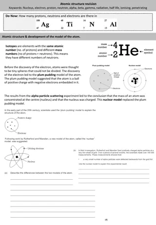
Alpha rays Alpha decay is a radioactive process in which a particle with two neutrons and two protons is ejected from the nucleus of a radioactive atom. The particle is identical to the nucleus of a helium atom. Alpha decay only occurs in very heavy elements such as uranium, thorium and radium. After an atom ejects an alpha particle, a new parent atom is formed which has two less neutrons and two less protons. Because alpha particles contain two protons, they have a positive charge of two. Alpha particles are very heavy and very energetic compared to other common types of radiation. These characteristics allow alpha particles to interact readily with materials they encounter, including air, causing many ionizations in a very short distance. Typical alpha particles will travel no more than a few centimeters in air and are stopped by a sheet of paper.
Beta rays Beta decay is a radioactive process in which an electron is emitted from the nucleus of a radioactive atom, along with an unusual particle called an antineutrino. When a nucleus ejects a beta particle, one of the neutrons in the nucleus is transformed into a proton plus the ejected electron (beta particle). Since the number of protons in the nucleus has changed, a new daughter atom is formed which has one less neutron but one more proton than the parent. Beta particles have a single negative charge and weigh only a small fraction of a neutron or proton. As a result, beta particles interact less readily with material than alpha particles. Depending on the beta particles energy (which depends on the radioactive atom and how much energy the antineutrino carries away), beta particles will travel up to several meters in air, and are stopped by thin layers of metal or plastic. Phosphorus-32 is the most commonly used isotope at Clark, and it emits strong beta particles.
Gamma rays After a decay reaction, the nucleus is often in an excited state. This means that the decay has resulted in producing a nucleus which still has excess energy. This energy is lost by emitting a pulse of electromagnetic radiation called a gamma ray, identical in nature to light or microwaves, but of very high energy. The gamma ray has no mass and no charge. Gamma rays interact with material by colliding with the electrons in the shells of atoms. They lose their energy slowly in material, being able to travel significant distances before stopping. Depending on their initial energy, gamma rays can travel from 1 to hundreds of meters in air and can easily go right through people. It is important to note that most alpha and beta emitters also emit gamma rays as part of their decay process. However, there is no such thing as a pure gamma emitter. Important gamma emitters including Technetium-99m which is used in nuclear medicine, and Cesium-137 which is used for calibration of nuclear instruments.
Other types of radiation and summary of radioactive particles Cosmic radiation: very energetic particles including protons which bombard the earth from outer space. It is more intense at higher altitudes than at sea level where the earth's atmosphere is most dense and gives the greatest protection. Neutrons: uncharged particles which are also very penetrating. On Earth they mostly come from the splitting, or fissioning, of certain atoms inside a nuclear reactor. Water and concrete are the most commonly used shields against neutron radiation from the core of the nuclear reactor. Radiation Radiation Type of Radiation Type of Radiation Mass (AMU) Mass (AMU) Charge Charge Shielding material Shielding material Alpha Alpha Particle 4 +2 Paper, skin, clothes Beta Beta Particle 1/1836 1 Plastic, glass, light metals Gamma Gamma Electromagnetic Wave 0 0 Dense metal, concrete, Earth Neutrons Neutrons Particle 1 0 Water, concrete, polyethylene, oil
Nuclear Equations Nuclear equations describe events that occur in the nucleus. They are balanced; i.e., the sum of atomic masses (superscript; number of neutrons plus protons) on the left must equal the sum of atomic masses on the right. Similarly, the atomic numbers (subscript; number of protons) must balance. 226 4 2 222 86 Alpha rays: Ra(s) He + Rn(g) 88 This describes decay of Radium, found in many rocks and minerals. Radium in basement materials decays to the gas Radon, which itself is radioactive. Radon, if breathed into the lungs, emits radioactive particles and is very likely to cause cancer. 14 14 7 0 -1 Beta rays: C(s) N + e 6 This describes decay of carbon-14. It is used in carbon dating of fossilized materials, and is also commonly used as a tracer for carbon in biochemical experiments. 131 131 54 0 -1 0 0 Gamma rays: I Ne + e + 53 This describes decay of iodine-131. It is commonly used as a tracer for iodine in biochemical and physiological (thyroid) experiments.
Half-life Half-life is the time required for the quantity of a radioactive material to be reduced to one-half its original value. Decay is a random process which follows an exponential curve. The number of radioactive nuclei remaining after time (t) is given by: N(t) = N(0) * e(- t) where N(0) = original number of atoms N(t) = number remaining at time t = decay constant (unique for each isotope; related to probability of decay)
Derivation of half-life equation -dN/dt = N # of atoms decaying at any time is proportional to # of atoms present. dN/N = - dt Separate differentials dN/N = - dt Integrate lnN = - t Evaluate between limits N: N(0), N(t) t: 0, t ln N(t) lnN(0) = - t ln(N(t)/N(0)) = - t Rearrange and take antilog N(t)/N(0) = e(- t) Let N(t) = N(0); i.e., t = t Solve for = ln(2)/t Substitute ; take ln of both sides; rearrange; take antilog of both sides; solve for N(t) N(t)/N(0) = e(-ln(2)t/t ) N(t) = N(0)2-(t/t )
Sources of radioactivity and contributions to the public dose. Natural Sources: Cosmic Terrestrial Internal Inhaled Man-made Sources: Medical Uses Consumers Products Industrial Uses Nuclear Power BACKGROUND RADIATION: Natural and Man-Made Sources Average Annual Dose from Background Radiation= 300 mrem/year
Units of radioactivity When given a certain amount of radioactive material, it is customary to refer to the quantity based on its activity rather than its mass. The activity is simply the number of disintegrations or transformations the quantity of material undergoes in a given period of time. The two most common units of activity are the Curie (Ci) and the Becquerel (Bq). One Curie is equal to 3.7x10¹⁰ disintegrations per second. One Becquerel is equal to one disintegration per second. Because the Ci is so large and the Bq is so small, we often use prefixes to define levels of activity. Examples of these prefixes follow: (m) milli (10 ) 3 ( ) micro (10 ) 6 (n) nano (10 ) 9 (p) pico (10 ) -12 (K) kilo (10 ) 3 (M) mega (10 ) 6 (G) giga (10 ) 9 (T) tera (10 ) 12
Units of exposure and dose The SI unit of exposure (X) is the coulomb/kilogram (C/kg). The traditional unit is the roentgen (R). 1 R = 2.58 x 10⁻⁴ C/kg. R only applies to absorption of gamma rays and x-rays in air. The SI unit of absorbed dose (D, energy absorbed by an object per unit mass) is the Gray (Gy). The traditional unit is the rad. 100 rad = 1 Gy = 1 J/kg. D applies to all radiations at all energies in all absorbers. The SI unit of dose equivalent (H, the absorbed dose multiplied by a quality factor that accounts for the different biological effectiveness of different types of radiation) is the Sievert (Sv). The traditional unit is the rem. 100 rem = 1 Sv.
Quality factor A quality factor (Q) is used to compare the biological damage producing potential of various types of radiation, given equal absorbed doses. This factor is used to convert units of absorbed dose (Grays) to units of absorbed dose equivalents (Sieverts). The quality factors for frequently encountered types of radiation are: Radiation Q Gammas and x-rays Beta particles & electrons Alpha particles & fission fragments 20 Neutrons 1 1 10
Levels of radiation from sources of radiation Beta radiation: For energies above 0.6 MeV, the dose rate to the skin from a uniform deposition of 1 Ci/cm2 of a beta emitter on the skin is about 9 rem/hr. For a beta emitter point source, the dose rate can be calculated using the empirical equation 300 x Ci = rad/hr @ 1 foot, where Ci = source strength in Curies. This calculation neglects any shielding provided by the air, which can be significant. Gamma radiation: An empirical rule is 6 x Ci x n x E = R/hr @ 1 foot, where Ci = source strength in curies, E = energy of the emitted photons in MeV, and n = fraction of decays resulting in photons with an energy of E.
Radiation protection standards 105 CMR 120.211 establishes radiation dose limits for occupationally exposed adults. These limits apply to the sum of the dose received from external exposure and the dose from internally deposited radioactive material. Dose Category Adult Occupational Dose Limit Total Effective Dose Equivalent (TEDE) 5 rem/year* Total Organ Dose Equivalent (TODE) 50 rem/year to any individual organ or tissue except the lens of the eye* Eye Dose Equivalent 15 rem/year* Shallow Dose Equivalent 50 rem/year* Embryo/Fetus Dose 0.5 rem for the entire gestation period *Occupational dose limit for minors is 10% of the adult limit
Biological Effects of radiation Radiation risk from occupational exposure is discussed in the format of frequently asked questions at: http://www.mass.gov/dph/rcp/radia.htm For a full discussion of biological effects, refer to the U.S. Nuclear Regulatory Commission's Guide 8.29, Instruction Concerning Risks from Occupational Radiation Exposure. In general, there are three types of biological effects that can be caused by exposure to radiation: Somatic Effects - Physical effects (immediate or delayed) that occur in the exposed person. Genetic Effects - Abnormalities that may occur in future generations of the person that is exposed. Teratogenic Effects - Effects that may be observed in offspring who were exposed during embryonic development. Course Screens
Biological Effects of radiation (continued) Some factors that can affect the probability and significance of potential effects include: Age - Response to radiation differs with age. For example, children are more sensitive to exposure than (most) adults. Was the exposure acute or chronic? - Acute exposures are delivered over a short time period, whereas a chronic exposure is spread over an extended period of time. Was the exposure external or internal? - An external exposure means that the source of radiation is outside the body, and an internal exposure means the source of radiation was ingested and is giving a dose to the body. What part, and how much of the body? Was the exposure localized, perhaps to extremities (such as your hands), or perhaps a small patch of skin? What kind of radiation? Forms of radiation differ in their penetrating power and ability to cause damage to biological tissues.
Biological Effects of radiation (continued) Alpha particles will be stopped by the dead layers of skin, so they are not an external hazard. However, many alpha emitters or their daughters also emit gammas which are penetrating and therefore may present an external hazard. Internally, alphas can be very damaging because they deposit all of their energy in a very small area. Based on their chemical properties, alpha emitters can be concentrated in specific tissues or organs. Externally, beta particles can deliver a dose to the skin or the tissues of the eye. Many beta emitters also emit gammas. A large activity of a high energy beta emitter can create a significant exposure from bremsstrahlung x-rays produced in shielding material. Internally, betas can be more damaging, especially when concentrated in specific tissues or organs. Externally, the hazard from low energy (< 30 keV) gammas and x-rays is primarily to the skin or the tissues of the eye. Higher energies are more penetrating and therefore a whole body hazard. Internally, gamma emitters can affect not only the tissues or organs in which they are deposited, but also surrounding tissues.
Biological Effects of radiation (continued) Radiation causes atoms and molecules to become ionized or excited. These ionizations and excitations can result in: Production of free radicals. Breakage of chemical bonds. Production of new chemical bonds and cross-linkage between macromolecules. Damage to molecules which regulate vital cell processes (e.g. DNA, RNA, proteins). TISSUE SENSITIVITY: The radiation sensitivity of a tissue varies directly with the rate of proliferation of its cells and inversely with the degree of differentiation. EFFECTS OF ACUTE HIGH RADIATION DOSES: A whole body radiation dose of greater than 25 to 50 rem received in a short time results in the clinical 'acute radiation syndrome.' This syndrome can result in disruption of the functions of the bone marrow system (>25 rem), the gastro-intestinal system (>500 rem), and the central nervous system (>2000 rem). An acute dose over 300 rem can be lethal. EFFECTS OF LOW RADIATION DOSES There is no disease uniquely associated with low radiation doses. Immediate effects are not seen below doses of 25 rem. Latent effects may appear years after a dose is received. The effect of greatest concern is the development of some form of cancer.
Methods of controlling radiation dose Plan, Control, Time, Distance, and Shielding Plan: Design your experiment and safety considerations to reduce exposure to As Low As Reasonably Achievable (ALARA). Control: Utilize proper pipetting techniques and safety equipment, and monitor frequently.
Methods of controlling radiation dose (cont) Time: Minimize the time spent handling or in the vicinity of radiation sources. Distance: Increasing the distance from a radiation source by the use of handling devices will reduce the dose received, since exposure rate decreases as 1/r2, where r is the distance from a point source. 2 Distance 1 Distance 2 Dose 2 Dose 1 = Shielding: Radiation can be absorbed by materials placed between the source of the radiation and the user. The type of shielding that is the most appropriate to use depends on the nature of the penetrating power of the radiation.
Factors to consider when selecting materials and design of shielding Persons outside the shadow cast by the shield are not necessarily protected. A wall or partition may not be a safe shield for people on the other side. Radiation can be "scattered" around corners. The absorption of high energy beta radiation (e.g. P-32 and Sr-90) in high Z materials such as lead and tungsten may result in the production of electromagnetic radiation (bremsstrahlung) which is more penetrating than the beta radiation that produced it. Low Z materials such as plastics and glass minimize the production of bremsstrahlung.
Radiation Detection Instrumentation: gas filled detectors This instrument works on the principle that as radiation passes through air or a specific gas, ionization of the molecules in the air occur. When a high voltage is placed between two areas of the gas filled space, the positive ions will be attracted to the negative side of the detector (the cathode) and the free electrons will travel to the positive side (the anode). These charges are collected by the anode and cathode which then form a very small current in the wires going to the detector. By placing a very sensitive current measuring device between the wires from the cathode and anode, the small current measured and displayed as a signal. The more radiation which enters the chamber, the more current displayed by the instrument. The Geiger-Muller or GM detector used to measure very small amounts of radiation.
Radiation Detection Instrumentation: solid scintillation detectors The second most common type of radiation detecting instrument is the solid scintillation detector. The basic principle behind this instrument is the use of a special material which glows or scintillates when radiation interacts with it. The most common type of material is a type of salt called sodium-iodide. The light produced from the scintillation process is reflected through a clear window where it interacts with device called a photomultiplier tube. The signal pulse is then detected and displayed. Solid scintillation detectors are very sensitive radiation instruments and are used primarily to monitor Gamma rays.
Radiation Detection Instrumentation: liquid scintillation detectors Liquid Scintillation Counters (LSC) must be used for analyzing 3H and other low energy beta emitters such and 35S and 14C. The LSC is used in combination with a scintillation fluid that emits light when radiation interacts with it; the amount of light emitted is proportional to the amount of radiation present. The beta decay electron emitted by the radioactive isotope in the sample excites the solvent molecule, which in turn transfers the energy to the solute, or fluor. The energy emission of the solute (the light photon) is converted into an electrical signal by a photomultiplier tube.
Survey monitoring: G-M detectors Using G-M detector to monitor energetic beta or weak gamma emitters: In this type of meter, an ionization in the detector results in a large output pulse that causes meter and audio responses. All initial ionizing events produce the same size output pulse. Therefore, the meter does not differentiate among types or energies of radiation. Most G-M detectors have a thin mica film 'window' at one end. This window is very fragile. Always use the thin end window for detecting pure beta emitters and low energy photons (e.g. P-32, S-35, C-14, Fe-55, I-125, and x-rays less than 40 keV). The aluminum side wall should be used only for the detection of penetrating x-rays and gamma radiation. Very low energy beta emitters such as H-3 are not detectable since their betas do not have enough energy to penetrate the window. They are best detected by using liquid scintillation counting techniques. C-14 and S-35 emit betas energetic enough to pass through the thin window. However, covering the window with plastic wrap or paraffin film will stop most or all of their betas from entering the detector.
Survey monitoring: G-M detectors (continued) The efficiency of a meter for a specific source of radiation is given by the ratio of the meter count rate to the actual disintegration rate of the source (cpm/dpm). Some examples of approximate G-M efficiencies through the end window at 1 inch from a point source are given below: H-3 C-14, S-35 P-32 I-125 not detectable 0.2% - 0.8% * 3% - 8% 0.01% - 0.03% * Not detectable if the detector window is covered with paraffin film, plastic wrap, or other material.
Survey monitoring: G-M detectors (continued) Because of the randomness of radioactive decay, the meter reading at low count rates often fluctuates widely. For this reason, the audio speaker is sometimes a better indicator of small amounts of radioactivity than the meter reading. At higher count rates, the speaker response is often faster than the meter reading. It is better, therefore, to have the speaker on when using a G-M counter. Very high radiation fields may temporarily overload the detector circuit resulting in a partial or complete loss of meter or audio response. If this happens, remove the meter and yourself from the area and push the reset button or turn the meter off then back on. The meter should resume normal operation. Always turn on a survey meter before entering an area that might have high radiation fields. The meter has a dial with the settings OFF, BATT (battery check), X100, X10, X1, and X0.1. To get a meter reading, multiply the number the needle is pointing at (in CPM) by the setting on the dial.
Survey monitoring: G-M detectors (continued) Check the calibration sticker on the side of the meter; be sure the meter has been checked within the last 12 months. DO NOT USE METERS THAT ARE NOT CALIBRATED. 1. 2. Next, check the batteries. If they are low, replace them with fresh ones. 3. Hold the detector away from any sources of radiation, and determine the background level. Move the dial to the lowest setting and look at the response on the meter. If the needle is off-scale (all the way to the right), move to a higher scale. Anything above that background level is considered to be radioactive. 4. Hold the detector about 1 cm from the surface of the item to be checked. 5. Move the GM pancake probe only 2 inches per second. This gives the detector and meter time to respond. 6. Frequently check hands, clothing, equipment, and floor while using radioactivity. The more often you stop and check things, the quicker you will find contamination before it gets out of control.
Survey monitoring: wipe tests (H-3, C-14, or S-35) 1. Wearing gloves and a lab coat, rub a piece of filter paper along the surface of the item of interest. Rub at least 100 cm2. Repeat for other areas of interest. 2. Carefully place each wipe into a scintillation vial, and add enough scintillation fluid to the vial so that it is at least 80% full. Tightly cap the vial. 3. Put an unused piece of filter paper inside a clean vial filled with scintillation fluid. Use this as a blank control to determine the background radiation level. 4. Place all the vials, including a blank one into the LSC to analyze it. 5. If any wipes are 3 times the background level or higher, there is contamination. The item should be cleaned as soon as possible, and then re-wiped to confirm that the area is clean.
Personnel monitoring equipment The film badge is used to measure whole body dose and shallow dose. It consists of a film packet and a holder. The film will be exposed by radiation. The holder has several filters which help in determining the type and energy of radiation. The badge will detect gamma and x-rays, high energy beta particles, and in certain special cases, neutrons. It does not register radiation from low energy beta emitters such as H-3, C-14, and S-35. The Thermo Luminescent Dosimeter(TLD) ring is used to measure dose to the hand. They are issued to individuals who may use millicurie amounts of a gamma or high energy beta emitter. Incident radiation excites atoms in crystals in the TLD. Upon heating (thermo-), the "trapped" excited electrons give up a photon (- luminescent). This photon signal is amplified by a photo-multiplier tube and the output sent to the dosimeter reader to register the dose. Pocket dosimeters are about the size of a pocket fountain pen, and are used to measure accumulative doses or quantities of gamma & X-ray radiation. A metal clip is used to attach the dosimeter to an individual's pocket or to any available object in an area to be monitored for total radiation exposure. It has a thin wall which permits the penetration and detection of radiation.
Personnel monitoring at Clark Monitoring of an individual s external radiation exposure is required by 105 CMR 120.226(A) if the external occupational dose is likely to exceed 10% of the dose limit appropriate for the individual. Calculations below demonstrate that this is not likely for individuals working with isotopes at Clark. Accordingly, no personnel monitoring for external radiation exposure is required. Consider the most extreme likely scenario: A worker using P-32 holds a vial containing 1 mCi at the thinnest spot on the vial (1.2 rem/hr) continuously for 5 min, once a week for 48 weeks of the year. The estimated dose to that worker s hand is 4.8 rem, which is less than 10% of the Total Organ Dose Equivalent limit of 50 rem. Monitoring of an individual s internal radiation exposure is not required at Clark, because volatile radioisotopes are not used and ingestion of radioisotopes is not otherwise permitted.
Safety Equipment 1. for all experiments involving the use of P-32 or S-35. Shielding: -inch plexiglass shields must be used 2. must be worn by anyone handling isotopes. Personnel protective equipment: Disposable gloves and labcoats 3. test tubes (or other reaction vessels) and stock bottles of isotopes. Storage equipment: Plexiglass containers must be used to hold 4. Handled or generated must be carried out in fume hoods. No such materials or reactions are currently authorized at Clark. Fume hoods: All reactions where volatile radioactive material is 5. quantities greater than 1 mCi should be carried out using remote handling equipment. No such experiments are currently authorized at Clark. Remote handling equipment: Handling radioactive materials in
Operating and Emergency Procedures can be obtained from the RSO and are posted on the Office of Environmental Health and Safety website: http://www.clarku.edu/offices/ehs/ As a general principle, in an emergency involving both personal injury and spill of radioactive materials, adhere to the following priorities as the situation permits: 1. Seek medical attention, informing the responders that radioisotopes are present 2. Stop the source of the spill, secure and post the area, warn others nearby, and notify the Radiation Safety Officer (RSO) 3. In coordination with the RSO, decontaminate the spill, and dispose of all contaminated materials in radioactive waste.
Radioactive Wastes Solid wastes: All solid wastes must be kept in conspicuously marked containers in labs authorized to use radioactive materials. Warning labels must be defaced. When the containers are full, they must be moved to and stored for 10 half-lives in the Radioactive Storage Area, as designated by the RSO. After monitoring to ensure that all radioactivity has decayed to background, the wastes may be disposed of in ordinary trash. Liquid wastes: Liquid wastes may be disposed of into ordinary sewerage using designated sinks within the following limits: All experiments must be conducted so as to generate liquid wastes that can be disposed of in this manner. A log recording the isotope, date, and amounts of disposed material must be maintained near the designated sink.
Training All personnel handling radioisotopes at Clark University must be trained by the RSO (first laboratory session of BCMB 271) and by the faculty Authorized User in whose lab they work. They must also attend an annual Laboratory Safety Training Program (including training related to the safe use and handling of radioisotopes) and pass the associated exam. The Laboratory Safety Training Program and exams may also be taken on-line.
