Understanding Protein Structure for Drug Action in Medicine
Explore the importance of protein structure in drug action, focusing on primary, secondary, and tertiary structures. Learn how proteins such as receptors and enzymes play a crucial role in the mechanisms of drug action.

Download Presentation

Please find below an Image/Link to download the presentation.
The content on the website is provided AS IS for your information and personal use only. It may not be sold, licensed, or shared on other websites without obtaining consent from the author. If you encounter any issues during the download, it is possible that the publisher has removed the file from their server.
You are allowed to download the files provided on this website for personal or commercial use, subject to the condition that they are used lawfully. All files are the property of their respective owners.
The content on the website is provided AS IS for your information and personal use only. It may not be sold, licensed, or shared on other websites without obtaining consent from the author.
E N D
Presentation Transcript
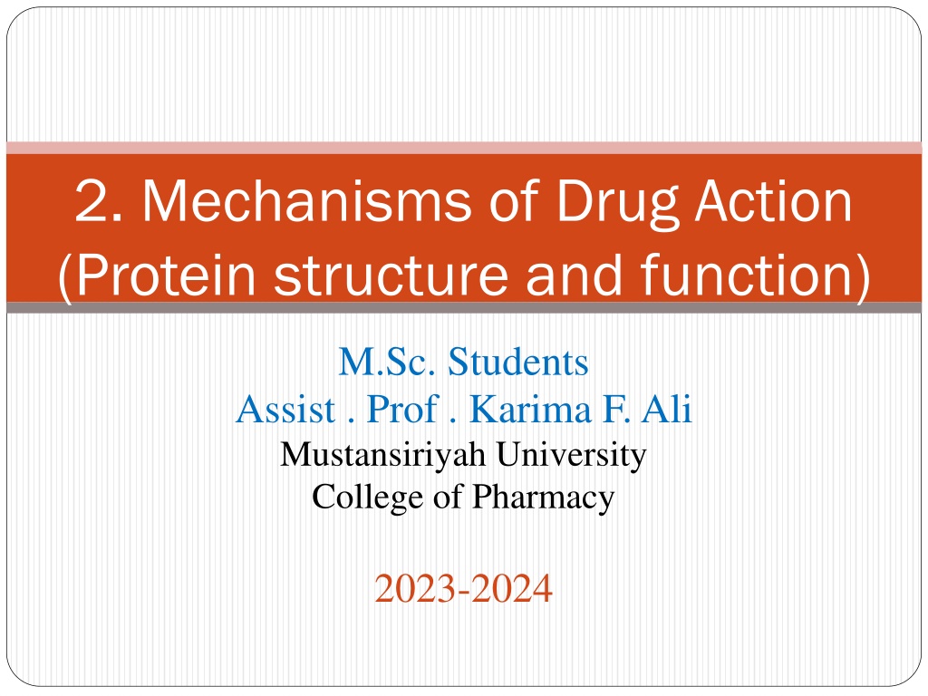
2. Mechanisms of Drug Action (Protein structure and function) M.Sc. Students Assist . Prof . Karima F. Ali Mustansiriyah University College of Pharmacy 2023-2024
Protein structure and function The vast majority of drugs used in medicine are targeted to proteins, such as receptors, enzymes, and transport proteins. Therefore, it is important to understand protein structure in order to understand drug action on proteins. Proteins have four levels of structure: primary, secondary, tertiary, and quaternary.
The primary structure of proteins The primary structure is the order in which the individual amino acids making up the protein are linked together through peptide bonds
The secondary structure of proteins There are three main secondary structures: the -helix, -pleated sheet, and -turn.
The -turn showing hydrogen bonding between the first and third peptide bond. The secondary structure of proteins consists of regions of ordered structure adopted by the protein chain. In structural proteins, such as wool and silk, secondary structures are extensive and determine the overall shape and properties of such proteins. There are also regions of secondary structure in most other proteins.
The tertiary structure of proteins o The tertiary structure is the overall three dimensional shape of a protein. o The tertiary structure of enzymes and receptors is crucial to their function and also to their interaction with drugs o It is important to appreciate the forces that control tertiary structure. Interactions involved in tertiary structure are the same as the intermolecular o Structural proteins are quite ordered in shape o globular proteins, such as enzymes and receptors fold up to form more complex structures. o Globular proteins often contain regions of ordered secondary structure, the extent of which varies from protein to protein. o Some proteins contain species known as cofactors (e.g. metal ions or small organic molecules), which also have an effect on tertiary structure.
Tertiary structure formation as a result of intramolecular interactions.
Relative importance of bonding interactions The relative importance of the bonding interactions in protein tertiary structure to follow the same order as their strengths: covalent, ionic, hydrogen bonding, and, finally, van der Waals. In fact, the opposite is generally true. Usually, the most important bonding interactions are those due to van der Waals interactions and hydrogen bonding, while the least important interactions are those due to covalent and ionic bonding.
First reason: In most proteins there are more possible opportunities for van der Waals and hydrogen bonding interactions than for covalent or ionic bonding. The only amino acid that can form a covalent disulphide bond is cysteine There are many more amino acids that can interact with each other through hydrogen bonding and van der Waal interactions. limited number of amino acids with residues capable of forming ionic bonds Second reason: Water is a highly polar compound that interacts readily with polar, hydrophilic amino acid residues capable of forming hydrogen bonds
The remaining non-polar, hydrophobic amino acid residues cannot interact favorably with water, so the most stable tertiary structure will ensure that most of the hydrophilic groups are on the surface so that they interact with water and that most of the hydrophobic groups are in the centre so that they avoid water and interact with each other The centre of the protein must be hydrophobic and non-polar. As the active site protrudes into the centre of the protein, it tends to be hydrophobic in nature and can provide a non- aqueous environment for the reaction taking place Binding sites are more hydrophobic than the surface and so van derWaals and hydrophobic interactions play an important role in the binding of the ligand. An understanding of these interactions is crucial to the design of effective drugs that will target these binding sites.
The quaternary structure of proteins Only proteins that are made up of a number of protein subunits have quaternary structure. For example, haemoglobin is made up of four protein molecules two identical alpha subunits and two identical beta subunits. Interactions between the exterior surfaces of proteins, ionic bonding can be more important to quaternary structure than it is to tertiary structure. Nevertheless, hydrophobic and van der Waals interactions have a role to play.
Translation and post translational modifications Many proteins are modified following translation ,and these modifications can have wide-ranging effects. For example, the N - terminals of many proteins are acetylated, making these proteins more resistant to degradation. Many of the proteins present on the surface of cells are linked to carbohydrates through asparagine residues. Several proteins are cleaved into smaller proteins or peptides following translation. Active enzymes are sometimes formed by cleaving a larger protein precursor. Often, this serves to protect the cell from the indiscriminate action of an enzyme. Some polypeptide hormones are also produced from the cleavage of protein precursors.
Examples of post-translational modifiations carried out on proteins.
Protein function Structural proteins Structural proteins do not normally act as drug targets. However, the structural protein tubulin is an exception. Tubulin molecules polymerize to form small tubes called microtubules Microtubules have various roles within the cell, including the maintenance of shape, exocytosis , and release of neurotransmitters. They are also involved in the mobility of cells. in the cell s cytoplasm Drugs that target tubulin and inhibit this process are useful anticancer agents
A variety of drugs interfere with this process by either binding to tubulin and inhibiting the polymerization process, or binding to the microtubules to stabilize them and thus inhibit depolymerization. Either way, the balance between polymerization and depolymerization is disrupted, which leads to a toxic effect and the inability of the cell to divide.
Tubulin as a drug target Colchicine is an example of a drug that binds to tubulin and prevents its polymerization. Colchicine is an example of a drug that binds to tubulin and prevents its polymerization. It can be used in the treatment of gout by reducing the mobility of neutrophils into joints. Unfortunately, colchicine has many side effects and it is restricted, therefore, to the treatment of acute attacks of this disease.
The Vinca alkaloids vincristine , vinblastine , vindesine, and vinorelbine bind to tubulin to prevent polymerization and are useful anticancer agents. They are also involved in transport within the cell and cell signaling as well as playing a pivotal role in the movement of chromosomes during mitosis. Colchicine, paclitaxel, and vinca alkaloids are the earliest plant-derived microtubule-targeting agents (MTAs), and paclitaxel and vinca alkaloids are currently important drugs used in the treatment of cancer.
The alkaloids are composed of a catharanthine moiety containing the indole subunit and the vindoline moiety containing the dihydroindole subunit joined by a carbon carbon bond. Vincristine and vinblastine differ only in the group attached to the dihydroindole nitrogen, which is a methyl group in vinblastine and a formyl group in vincristine. Vinorelbine is a semisynthetic material resulting from loss of water across the 3 ,4 bond and first prepared by the use of a modified Polonovski reaction of vinblastine followed by hydrolysis.
Paclitaxel (Taxol) and the semi-synthetic analogue docetaxel are important anticancer agents that inhibit tubulin depolymerization The benzoyl and acetyl substituents, at positions 2 and 4, respectively, play an important role in this binding interaction, as do the side chain and the oxetane ring. These groups dominate the lower or southern half of the molecule (as the structure is normally presented), and so the variations that are possible in this region are restricted when making analogues. In contrast, it is possible to carry out more variations in the northern half of the molecule. This can affect the in vivo efficacy of the molecule, allowing modification of aqueous solubility and pharmacokinetic properties.
Paclitaxel (Taxol), with important binding groups in colour, and docetaxel (Taxotere).
It was possible to replace the aromatic rings of paclitaxel with other hydrophobic groups. Having a suitable acyl group at position 10 has also been found to increase activity against drug-resistant strains of cancers. Such compounds have the ability not only to bind to tubulin, but to inhibit the P-glycoprotein efflux pump. This protein which is present in the cell membrane of cancer cells and can pump drugs out of the cell before they get the chance to work effectively. Further work has demonstrated that acylating the 7-hydroxy group with hydrophobic groups is also effective in blocking efflux. Finally, the addition of a methyl substituent at C-2 has been found to increase activity by inhibiting rotation of the C-2 C-3 bond. The first orally active taxoid structure ortataxel has now been developed and has entered clinical trials. Since the discovery of paclitaxel a variety of other natural products have been found to have a similar mechanism of action and are currently being studied as potential anticancer agents
BMS 188797 and BMS 184476 are two taxoids that have been developed recently and have reached clinical trials. Analogues of paclitaxel.
Structural proteins as drug targets Viral structural proteins as drug targets Viruses consist of a nucleic acid encapsulated within a protein coat called a capsid . If a virus is to multiply within a host cell, this protein coat has to be dismantled in order to release the nucleic acid into the cell. Drugs have been designed which bind to the structural proteins that make up the capsid and which prevent the uncoating process. Enfuvirtide was approved in March 2003 and is an example of an antiviral agent that works in this way.
Enfuvirtide the first member of a new class of fusion inhibitors . It is a polypeptide consisting of 36 amino acids which matches the C -terminal end of the viral protein gp 41 . It works by forming an - helix and binding to a group of three similar -helices belonging to the gp41 protein. This association prevents the process by which the virus enters the host cell. In order to bring about fusion, the gp41 protein anchors the virus to the cell membrane of the host cell. It then undergoes a conformational change where it builds a grouping of six helices using the three already present as the focus for that grouping This pulls the membranes of the virion and the host cell together to permit fusion. By binding to the group of three helices, enfuvirtide blocks formation of the required hexamer and prevents fusion.
( a ) Normal mechanism of fusion. ( b ) Enfuvirtide acting as a fusion inhibitor.
The agents used against flu are ineffective against colds, as these infections are caused by a different kind of virus called a rhinovirus. RNA viruses, containing a positive strand of RNA coated by an icosahedral shell made up of 60 copies of four distinct proteins, VP1 VP4. At the junction between each VP1 and VP3 protein, there is a broad canyon 25 deep, and this is where attachment takes place between the virus and the host cell. On the canyon floor there is a pore which opens into a hydrophobic pocket within the VP1 protein. This pocket is either empty or occupied by a small molecule called a pocket factor.(t is a fatty acid containing seven carbon atoms).
When the virus becomes attached to the host cell, a receptor molecule on the host cell fits into the canyon and induces conformational changes that cause the VP4 protein and the N -terminus of VP1 to move to the exterior of the virus a process called externalization . This is thought to be important to the process by which the virus is uncoated and releases its RNA into the host cell. It is thought that the pocket factor stabilizes the capsid when it is bound, and prevents the conformational changes that are needed to cause infection. Disoxaril inhibits enterovirus replication by binding to the hydrophobic pocket within the VP1 coat protein, thus stabilizing the virion and blocking its uncoating.
Binding of disoxaril (possible hydrogen bonds shown as dashed lines).
A variety of drugs having antiviral activity are thought to mimic the pocket factor by displacing it and binding to the same hydrophobic pocket. The drugs concerned are called capsid-binding agents and are characteristically long-chain hydrophobic molecules. Like the pocket factor, they stabilize the capsid by locking it into a stable conformation and preventing the conformational changes required for uncoating. They also raise the canyon floor and prevent the receptor on the host cell from fitting the canyon
Pleconaril is one such drug that has undergone phase III clinical trials which demonstrate that it has an effect on the common cold. It is an orally active, broad-spectrum agent which can cross the blood brain barrier. The drug may also be useful against the enteroviruses that cause diarrhoea, viral meningitis, conjunctivitis, and encephalitis, as these viruses are similar in structure to the rhinoviruses. The development of pleconaril started when a series of isoxazoles were found to have antiviral activity. This led to the discovery of disoxaril which entered phase I clinical trials, but proved to be too toxic.
X-ray crystallographic studies of VP1-drug complexes involving disoxaril and its analogues showed that: The oxazoline and phenyl rings were roughly coplanar and were located in a hydrophilic region of the pocket near the pore leading into the centre of the virion The hydrophobic isoxazole ring binds into the heart of the hydrophobic pocket and the chain provides sufficient flexibility for the molecule to bend round a corner in the pocket. This also causes conformational changes in the canyon floor. Structure-based drug design was carried out to find safer and more effective antiviral agents. For example, the chain cannot be too short or too long or else there are steric interactions. Placing additional hydrophobic groups on to the phenyl ring improves activity against the HRV2 strain, because increased interactions are possible with a phenylalanine residue at position 116 rather than leucine.
The structure WIN 54954 was developed and entered clinical trials, but results were disappointing because extensive metabolism resulted in 18 different metabolic products due mainly to hydrolysis of the oxazoline ring. Further structure- based drug design led to modifications of the phenyl and oxazoline moieties. This included the introduction of a trifluoromethyl group to block metabolism, resulting in pleconaril, with 70% oral bioavailability.
Transport proteins Transport proteins are present in the cell membrane and act as the cell s smugglers smuggling the important chemical building blocks of amino acids, sugars, and nucleic acid bases across the cell membrane such that the cell can synthesize its proteins, carbohydrates, and nucleic acids. They are also important in transporting important neurotransmitters back into the neuron that released them so that the neurotransmitters only have a limited period of activity. But why is this smuggling operation necessary? Why can t these molecules pass through the membrane by themselves? Quite simply, the molecules concerned are polar structures and cannot pass through the hydrophobic cell membrane
The transport proteins can float freely within the cell membrane because they have hydrophobic residues on their outer surface which interact favourably with the hydrophobic centre of the cell membrane. The portion of the transport protein that is exposed on the outer surface of the cell membrane contains a binding site that can bind a polar molecule, such as an amino acid, stow it away in a hydrophilic pocket, and ferry it across the membrane to release it on the other side Transport proteins are not all identical; there are specific transport proteins for the different molecules that need to be smuggled across the membrane. The binding sites for these transport proteins vary in structure such that they can recognize and bind their specific guest. There are several important drugs which target transport proteins
Transport proteins as drug targets Transport protein accepting a drug that resembles the usual guest. If that drug remains strongly bound to the transport protein, it will prevent the protein from carrying out its normal role. Transport protein is an important target for various drugs which inhibit noradrenaline uptake and thus prolong adrenergic activity. The tricyclic antidepressants, desipramine , imipramine , and amitriptyline work in this fashion in the CNS and were the principal treatment for depression. Amphetamines compete with noradrenaline for the transport proteins responsible for transporting noradrenaline back into the presynaptic neuron. Adrenergic activity is increased in the CNS. Cocaine increases peripheral adrenergic activity by blocking noradrenaline reuptake. In the CNS it inhibits the reuptake of dopamine.
The antidepressant drugs fluoxetine (Prozac), citalopram ,and s- citalopram selectively block the transport protein responsible for the uptake of a neurotransmitter called serotonin from nerve synapses, and are called selective serotonin reuptake inhibitors (SSRIs). Citalopram are chiral molecules which are marketed as racemates. The S -enantiomer of citalopram is more active than the R - enantiomer and is now marketed as escitalopram
Lipids as drug targets The number of drugs that interact with lipids is relatively small and, in general, they all act in the same way by disrupting the lipid structure of cell membranes. It has been proposed that general anaesthetics work by interacting with the lipids of cell membranes to alter the structure and conducting properties of the membranes. Another agent thought to disrupt cell membrane structure is the anticancer agent cephalostatin I , which is thought to span the phospholipid bilayer. It has not yet entered clinical trials and there is a need to develop an efficient synthesis of the compound to obtain sufficient quantities.


.")


 and the semi-synthetic analogue docetaxel")
, with important binding groups in colour,")


 Normal mechanism of fusion.")


, citalopram")