Understanding Osseous Surgery in Periodontology
Explore the fundamentals of osseous surgery in periodontology, including normal alveolar bone morphology, terminology, techniques, and more. Gain insights into the rationale behind osseous resective therapy and specific learning objectives in this field.

Download Presentation

Please find below an Image/Link to download the presentation.
The content on the website is provided AS IS for your information and personal use only. It may not be sold, licensed, or shared on other websites without obtaining consent from the author. If you encounter any issues during the download, it is possible that the publisher has removed the file from their server.
You are allowed to download the files provided on this website for personal or commercial use, subject to the condition that they are used lawfully. All files are the property of their respective owners.
The content on the website is provided AS IS for your information and personal use only. It may not be sold, licensed, or shared on other websites without obtaining consent from the author.
E N D
Presentation Transcript
RUNGTA COLLEGE OF DENTAL SCIENCES AND RESEARCH,BHILAI RESECTIVE OSSEOUS SURGERY PRESENTED BY: Dr Sonika Bodhi Reader Dept. of Periodontology 1
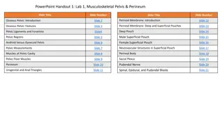
SPECIFIC LEARNING OBJECTIVES Core areas Domain Affective Category Desire to know Introduction Cognitive Nice to know Rationale Cognitive Must to know Normal alveolar bone morphology Cognitive Must to know Terminology Cognitive Must to know Factors in selection of technique Cognitive Must to know Examination , diagnosis and treatment planning Cognitive Must to know Techniques Affectice Nice to know Specific situations 2
CONTENTS Introduction Rationale Normal alveolar bone morphology Terminology Factors in selection of technique Examination , Diagnosis and Treatment Planning Techniques Specific situations Summary References 3
INTRODUCTION Bony deformities .....resulting from periodontal disease...... are not uniform; They may be Horizontal bone loss, Vertical bone loss or combination 4
Horizontal bone loss generally results in a relative thickening of the marginal alveolar bone, because bone tapers as it approaches its most coronal margin. To provide a more physiologic bone pattern, a method for osseous recontouring must be followed. 7
RATIONALE The goal of osseous resective therapy is to reshape the marginal bone to resemble that of the alveolar process undamaged by periodontal disease. The technique is performed in combination with apically positioned flaps, and the procedure eliminates periodontal pocket depth and improves tissue contour to provide a more easily maintainable environment. 8
NORMAL ALVEOLAR BONE MORPHOLOGY 1. The interproximal bone is more coronal in position than the labial or lingual/palatal bone and pyramidal in form. 9
2. The form of the interdental bone is a function of the tooth form and the embrasure width. The more tapered the tooth, the more pyramidal is the bony form. The wider the embrasure, the more flattened is the interdental bone mesiodistally and buccolingually 10
3. The position of the bony margin mimics the contours of the cementoenamel junction. The molar teeth have less scalloping and a more flat profile than bicuspids and incisors Teeth with prominent roots or those displaced to the facial or lingual side may also have fenestrations or dehiscences. 11
TERMINOLOGY OSSEOUS SURGERY .............the procedure by which changes in the alveolar bone can be accomplished to rid it of deformities induced by the periodontal disease process or other related factors, such as exostosis and tooth supraeruption. 13
Additive osseous surgery includes procedures directed at restoring the alveolar bone to its original level. Subtractive osseous surgery is designed to restore the form of preexisting alveolar bone to the level present at the time of surgery or slightly more apical to this level. 14
OSTEOPLASTY: defined as a procedure by which nonsupporting bone is reshaped to achieve a physiologic gingival and osseous contour. OSTECTOMY: is the removal of radicular and interradicular supporting bone to eliminate osseous deformities. 15
Definitive osseous reshaping implies that further osseous reshaping would not improve the overall result. Compromise osseous reshaping indicates a bone pattern that cannot be improved without significant osseous removal that would be detrimental to the overall result. 16
The architecture is said to be positive if the radicular bone is apical to the interdental bone. The bone is said to have negative architecture if the interdental bone is more apical than the radicular bone. Flat architecture is the reduction of the interdental bone to the same height as the radicular bone. 17
Osseous form is considered to be ideal when the bone is consistently more coronal on the interproximal surfaces than on the facial and lingual surfaces. The ideal form of the marginal bone has similar interdental height, with gradual, curved slopes between interdental peaks. 19
FACTORS IN SELECTION OF RESECTIVE OSSEOUS SURGERY early to moderate bone loss (2-3 mm) with moderate-length root trunks that have bony defects with one or two walls. These shallow to moderate bony defects can be effectively managed by osteoplasty and osteoectomy 21
Contraindicated in patients with advanced attachment loss and deep intrabony defects 22
EXAMINATION AND TREATMENT PLANNING Periodontal probing and exploration are key aspects of the examination. Careful probing reveals the presence of pocket depth greater than that of a normal gingival sulcus, the location of the base of the pocket relative to the mucogingival junction and attachment level on adjacent teeth, the number of bony walls, and the presence of furcation defects 23
Transgingival probing, or sounding, under local anesthesia confirms the extent and configuration of the intrabony component of the pocket and of furcation defects 24
OSSEOUS RESECTION TECHNIQUE Instrumentation Rotary instruments .........osteoplasty procedures Hand instruments .......... ostectomy procedures. 26
sequential steps are suggested for resective osseous surgery Vertical grooving Radicular blending Flattening interproximal bone Gradualizing marginal bone 28
VERTICAL GROOVING It is the first step It defines the general thickness and subsequent form of the alveolar housing It also provides continuity from the interproximal surface onto the radicular surface. This step is usually performed with rotary instruments, such as round carbide burs or diamonds 30
The advantages of vertical grooving are most apparent with thick bony margins, shallow crater formations, or other areas that require maximal osteoplasty and minimal ostectomy. Vertical grooving is contraindicated in areas with close roots or thin alveolar housing. 31
RADICULAR BLENDING Second step of the osseous reshaping technique and is an extension of vertical grooving Conceptually, it is an attempt to gradualize the bone over the entire radicular surface to provide the best results from vertical grooving. This provides a smooth, blended surface for good flap adaptation 32
The indications are the same as for vertical grooving (i.e., thick ledges of bone on the radicular surface, where selective surgical resection is desired). Naturally, this step is not necessary if vertical grooving is very minor or if the radicular bone is thin or fenestrated 33
FLATTENING INTERPROXIMAL BONE Flattening of the interdental bone requires the removal of very small amounts of supporting bone. It is indicated when interproximal bone levels vary horizontally. By definition, most of the indications for this step are one-walled interproximal defects or hemiseptal defects 34
The omission of flattening in such cases results in increased pocket depth on the most apical side of the bone loss. This step is typically not necessary with inter- proximal crater formations or flat interproximal defects. 35
GRADUALIZING MARGINAL BONE Bone removal is minimal but necessary to provide a sound, regular base for the gingival tissue to follow. Failure to remove small bony discrepancies on the gingival line angles (widow's peaks) allows the tissue to rise to a higher level than the base of the bone loss in the interdental area. 36
This may make the process of selective recession and subsequent pocket reduction incomplete. This step of the procedure also requires gradualization and blending on the radicular surface. 37
The two ostectomy steps should be performed with great care so as not to produce nicks or grooves on the roots. Not required....When the radicular bone is thin. chisels and curettes are preferred 38
FLAP PLACEMENT AND CLOSURE Flaps may be replaced to their original position, to cover the new bony margin, or they may be apically positioned. Replacing the flap in areas that previously had deep pockets may result initially in greater postoperative pocket depth, although a selective recession may diminish the depth over time. 39
The sutures should be placed with minimal tension to coapt the flaps, prevent their separation, and maintain the position of the flaps. 40
POSTOPERATIVE MAINTENANCE Nonresorbable sutures such as silk are usually removed after 1 week of healing, although some of the newer synthetic materials may be left for up to 3 weeks or longer without adverse consequences 41
Resorbable sutures maintain wound approximation for varying periods of 1 to 3 weeks or more, depending on the type of suture material. 42
At the suture removal appointment the periodontal dressing, if present, is removed, and the surgical site is gently cleansed of debris with a cotton pellet dampened with saline. Nonresorbable sutures are then cut and removed. post-surgical maintenance instructions are given 43
A second postoperative visit is often performed at the second or third week, and the surgical site is lightly debrided for optimal results. Healing should proceed uneventfully, with the attachment of the flap to the underlying bone completed in 14 to 21 days. Maturation and remodeling can continue for up to 6 months. 44
It is usually advisable to wait at least 6 weeks after completion of healing of the last surgical area before beginning dental restorations. 45
SPECIFIC OSSEOUS RESHAPING SITUATIONS Correction of one-walled hemiseptal defects requires that the bone be reduced to the level of the most apical portion of the defect. Therefore, great care should be taken to select the appropriate case. If one-walled defects occur next to an edentulous space, the edentulous ridge is reduced to the level of the osseous defect. 46
Summary Osseous surgery can eliminate and modify defects, as well as gradualize excessive bony ledges, irregular alveolar bone, early furcation involvement, excessive bony exostosis, and circumferential defects. When properly performed, resective osseous surgery achieves a physiologic architecture of marginal alveolar bone conducive to gingival flap adaptation with minimal probing depth. 47
References Newman MG, Takei HH, Klokkevold PR, Carranza FA. Carranza s clinical periodontology, 10th ed. Saunders Elsevier; 2007. Lindhe J, Lang NP and Karring T. Clinical Periodontology and Implant Dentistry. 6th ed. Oxford (UK): Blackwell Publishing Ltd.; 2015. Newman MG, Takei HH, Klokkevold PR, Carranza FA. Carranza s clinical periodontology, 13th ed. Saunders Elsevier; 2018. 48