Understanding NMR Spectroscopy for Structure Identification
NMR spectroscopy is a powerful tool in determining the structure of organic compounds. This summary outlines the process of using 1H NMR spectroscopy to identify an unknown compound, detailing steps such as determining different proton types, analyzing integration data, and interpreting splitting patterns to deduce the compound's structure from its molecular formula and functional groups.
Download Presentation

Please find below an Image/Link to download the presentation.
The content on the website is provided AS IS for your information and personal use only. It may not be sold, licensed, or shared on other websites without obtaining consent from the author. Download presentation by click this link. If you encounter any issues during the download, it is possible that the publisher has removed the file from their server.
E N D
Presentation Transcript
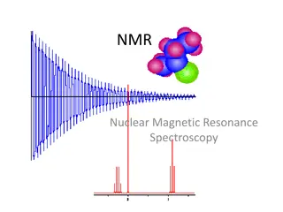
NUCLEAR MAGNETIC RESONANCE spectroscopy NMR spectroscopy Alkhair Adam Khalil, B. Pharm., M. Pharm. Department of Pharmaceutical Chemistry College of Pharmacy Karary University
1H NMR to Identify an Unknown Using Once we know a compound s molecular formula from its mass spectral data and the identity of its functional group from its IR spectrum, we can then use its spectrum to determine its structure. A suggested procedure is illustrated for compound X, whose molecular formula (C4H8O2) and functional group (C=O) were determined. 1H NMR
Step [1] Determine the number of different kinds of protons. The number of NMR signals equals the number of different types of protons. This molecule has three NMR signals ([A], [B], and [C]) and therefore three types of protons (Ha, Hb, and Hc).
Step [2] Use the integration data to determine the number of H atoms giving rise to each signal. Total number of integration units: 14 + 11 + 15 = 40 units Total number of protons = 8 Divide: 40 units/8 protons = 5 units per proton Then, divide each integration value by this answer (5 units per proton) and round to the nearest whole number.
Step [3] Use individual splitting patterns to determine what carbon atoms are bonded to each other. Start with the singlets. Signal [C] is due to a CH3group with no adjacent nonequivalent H atoms. Possible structures include:
Because signal [A] is a triplet, there must be 2H s (CH2group) on the adjacent carbon. Because signal [B] is a quartet, there must be 3H s (CH3group) on the adjacent carbon. This information suggests that X has an ethyl group CH3CH2
To summarize, X contains CH3 , CH3CH2 , and C=O (from the IR). Comparing these atoms with the molecular formula shows that one O atom is missing. Because O atoms do not absorb in a presence can only be inferred by examining the chemical shift of protons near them. O atoms are more electronegative than C, thus deshielding nearby protons, and shifting their absorption downfield. 1H NMR spectrum, their
Step [4] Use chemical shift data to complete the structure. Put the structure together in a manner that preserves the splitting data and is consistent with the reported chemical shifts. In this example, two isomeric structures are possible for X considering the splitting data only:
Chemical shift information distinguishes the two possibilities. The electronegative O atom deshields adjacent H s, shifting them downfield between 3 and 4 ppm. If A is the correct structure, the singlet due to the CH3 group (Hc) should occur downfield, whereas if B is the correct structure, the quartet due to the CH2group (Hb) should occur downfield. Because the NMR of X has a singlet (not a quartet) at 3.7, A is the correct structure.
13C NMR Spectroscopy 13 C NMR spectroscopy is also an important tool for organic structure analysis. The physical basis for 1H NMR. When placed in a magnetic field, B0, can align themselves with or against B0. 13C NMR is the same as for 13 C nuclei
More nuclei are aligned with B0because this arrangement is lower in energy, but these nuclei can be made to spin flip against the applied field by applying RF radiation of the appropriate frequency. 13C NMR spectra, like intensity versus chemical shift, using TMS as the reference signal at 0 ppm. 1H NMR spectra, plot peak 13C occurs in only 1.1% natural abundance, however, so 13C NMR signals are much weaker than 1H NMR signals.
To overcome this limitation, modern spectrometers irradiate samples with many pulses of RF radiation and use mathematical tools to increase signal sensitivity and decrease background noise.
13C NMR spectra are easier to analyze than spectra because signals are not split. Each type of carbon atom appears as a single peak. Because of the low natural abundance of nuclei (1.1%), the chance of two bonded to each other is very small (0.01%), and so no carbon carbon splitting is observed. 1H 13C 13C nuclei being
A protons. This a spectrum, however, by using an instrumental technique that decouples the proton carbon interactions, so that every peak in a spectrum is a singlet. 13C NMR signal can also be split by nearby 1H 13C splitting is usually eliminated from 13C NMR
13CNMR Spectroscopy Features of Two features of most structural information: 1. Number of signals observed 2. Chemical shifts of those signals. 13C NMR spectra provide the
In contrast to what occurs in proton NMR, peak intensity is not proportional to the number of absorbing carbons, so integrated. 13C NMR signals are not
13C NMR: Position of Signals In contrast to the small range of chemical shifts in 1H NMR (0 12 ppm usually), absorptions occur over a much broader range, 0 220 ppm. The chemical shifts of carbon atoms in chemical shifts of protons in 13C NMR 13C NMR depend on the same effects as the 1H NMR:
The sp3hybridized C atoms of alkyl groups are shielded and absorb upfield. Electronegative elements like halogen, nitrogen, and oxygen shift absorptions downfield. The sp2hybridized C atoms of alkenes and benzene rings absorb downfield. Carbonyl carbons are highly deshielded, and absorb farther downfield than other carbon types.
Magnetic Resonance Imaging (MRI) A powerful diagnostic technique. The sample is the patient, who is placed in a large cavity in a magnetic field, and then irradiated with RF energy. Living tissue contains protons (especially the H atoms in H2O) in different concentrations and environments.
These data (due to different environments of protons) are analyzed by a computer that generates a plot that delineates tissues of different proton density. Moreover, because the calcium present in bones is not NMR active, an MRI instrument can see through bones such as the skull and visualize the soft tissue underneath.
Questions ! 39
