Understanding Mechanical Ventilation in Critical Care Environments
Mechanical ventilation, overseen by Dr. Manish Sharma, Pediatric Intensivist at JK Lon Hospital, Jaipur, is essential for treating respiratory failure. It involves using ventilators to deliver gas to the lungs and meet specific goals like correcting hypoxemia and hypercapnia. This process requires understanding basic terminologies, criteria for ventilatory support, types of ventilators, and indications for ventilation.
Download Presentation

Please find below an Image/Link to download the presentation.
The content on the website is provided AS IS for your information and personal use only. It may not be sold, licensed, or shared on other websites without obtaining consent from the author. Download presentation by click this link. If you encounter any issues during the download, it is possible that the publisher has removed the file from their server.
E N D
Presentation Transcript
MECHNICAL VENTILATION DR MANISH SHARMA Additional Superintendent JK Lon Hospital (PAEDIATRIC INTENSIVIST) In Charge Accident/Emergency, JK Lon Hospital, Jaipur SMS MEDICAL COLLEGE , JAIPUR
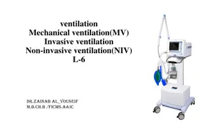
Mechanical Ventilation is ventilation of the lungs by artificial means usually by a ventilator. A ventilator delivers gas to the lungs with either negative or positive pressure.
Indications and Goals for Ventilation Type I or hypoxemic respiratory failure Type II or hypercapnic respiratory failure Goals : Correct hypoxemia PO260mmHg, O2Saturation 90% Correct hypercapnia PCO2~ 40mmHg Reduce work of breathing and hence cardiac workload Provide rest to respiratory muscles and reduce oxygen cost of breathing
Basic terminologies Respiratory cycle (Ti/Te/PIP/PEEP/Vt)
Criteria for institution of ventilatory support: Parameters Ventilation indicated Normal range A- Pulmonary function studies: Respiratory rate (breaths/min). Tidal volume (ml/kg body wt) Vital capacity (ml/kg body wt) Maximum Inspiratory Force (cm HO2) >10- 15 15-20,20-25 < 5 6-8 < 15 65-75 <-20 75-100
Criteria for institution of ventilatory support: Parameters Ventilation indicated Normal range B- Arterial blood Gases PH PaO2 (mmHg) PaCO2 (mmHg) PaO2/FiO2 < 7.25 <60 >50 <100 7.35-7.45 75-100 35-45 >500
Types of Mechanical ventilators: Negative-pressure ventilators Positive-pressure ventilators.
Positive-pressure ventilators Positive-pressure ventilators deliver gas to the patient under positive- pressure, during the inspiratory phase.
Types of Positive-Pressure Ventilators 1- Volume Ventilators. 2- Pressure Ventilators 3- High-Frequency Ventilators
1- Volume Ventilators The volume ventilator is commonly used in critical care settings. The basic principle of this ventilator is that a designated volume of air is delivered with each breath. The amount of pressure required to deliver the set volume depends on :- - Patient s lung compliance - Patient ventilator resistance factors.
Therefore, peak inspiratory pressure (PIP ) must be monitored in volume modes because it varies from breath to breath. With this mode of ventilation, a respiratory rate, inspiratory time, and tidal volume are selected for the mechanical breaths.
2- Pressure Ventilators The use of pressure ventilators is increasing in critical care units. A typical pressure mode delivers a selected gas pressure to the patient early in inspiration, and sustains the pressure throughout the inspiratory phase. By meeting the patient s inspiratory flow demand throughout inspiration, patient effort is reduced and comfort increased.
Although pressure is consistent with these modes, volume is not. Volume will change with changes in resistance or compliance, Therefore, exhaled tidal volume is the variable to monitor closely. With pressure modes, the pressure level to be delivered is selected, and with some mode options (i.e., pressure controlled [PC], described later), rate and inspiratory time are preset as well.
3- High-Frequency Ventilators High-frequency ventilators use small tidal volumes (1 to 3 mL/kg) at frequencies greater than 100 breaths/minute. The high-frequency ventilator accomplishes oxygenation by the diffusion of oxygen and carbon dioxide from high to low gradients of concentration.
This diffusion movement is increased if the kinetic energy of the gas molecules is increased. A high-frequency ventilator would be used to achieve lower peak ventilator pressures, thereby lowering the risk of barotrauma.
outcom es Alarms reset Mode Inspiratory hold Expiratory hold 100% FiO2 Accept Mode Preset variables and alarms
Ventilator mode The way the machine ventilates the patient How much the patient will participate in his own ventilatory pattern. Each mode is different in determining how much work of breathing the patient has to do.
Modes of Mechanical Ventilation A- Volume Modes B- Pressure Modes
A- Volume Modes 1- Assist-control (A/C) 2- Synchronized intermittent mandatory ventilation (SIMV)
1- Assist Control Mode A/C The ventilator provides the patient with a pre-set tidal volume at a pre-set rate . The patient may initiate a breath on his own, but the ventilator assists by delivering a specified tidal volume to the patient. Patient can initiate breaths that are delivered at the preset tidal volume.
The total respiratory rate is determined by the number of spontaneous inspiration initiated by the patient plus the number of breaths set on the ventilator. In A/C mode, a mandatory (or control ) rate is selected. If the patient wishes to breathe faster, he or she can trigger the ventilator and receive a full-volume breath.
Often used as initial mode of ventilation When the patient is too weak to perform the work of breathing (e.g., when emerging from anesthesia). Disadvantages: Hyperventilation,
2- Synchronized Intermittent Mandatory Ventilation (SIMV) The ventilator provides the patient with a pre-set number of breaths/minute at a specified tidal volume and FiO2. In between the ventilator-delivered breaths, the patient is able to breathe spontaneously at his own tidal volume and rate with no assistance from the ventilator. However, unlike the A/C mode, any breaths taken above the set rate are spontaneous breaths taken through the ventilator circuit.
The tidal volume of these breaths can vary drastically from the tidal volume set on the ventilator, because the tidal volume is determined by the patient s spontaneous effort. Adding pressure support during spontaneous breaths can minimize the risk of increased work of breathing. Ventilators breaths are synchronized with the patient spontaneous breathe. ( no fighting)
B- Pressure Modes 1- Pressure-controlled ventilation (PCV) 2- Pressure-support ventilation (PSV) 3- Continuous positive airway pressure (CPAP) 4- Positive end expiratory pressure (PEEP) 5- Noninvasive bilevel positive airway pressure ventilation (BiPAP)
Inverse ratio ventilation (IRV) mode reverses this ratio so that inspiratory time is equal to, or longer than, expiratory time (1:1 to 4:1). Inverse I:E ratios are used in conjunction with pressure control to improve oxygenation by expanding stiff alveoli by using longer distending times, thereby providing more opportunity for gas exchange and preventing alveolar collapse.
4- Continuous Positive Airway Pressure (CPAP) Constant positive airway pressure during spontaneous breathing CPAP allows the nurse to observe the ability of the patient to breathe spontaneously while still on the ventilator. CPAP can be used for intubated and nonintubated patients. It may be used as a weaning mode and for self breathing patients (nasal or mask CPAP)
5- Positive end expiratory pressure (PEEP) Positive pressure applied at the end of expiration during mandatory \ ventilator breath positive end-expiratory pressure with positive-pressure (machine) breaths.
Uses of CPAP & PEEP Prevent atelactasis or collapse of alveoli Treat atelactasis or collapse of alveoli Improve gas exchange & oxygenation Treat hypoxemia refractory to oxygen therapy.(prevent oxygen toxicity Treat pulmonary edema ( pressure help expulsion of fluids from alveoli
Common Ventilator Settings parameters/ controls Fraction of inspired oxygen (FIO2) Tidal Volume (VT) Peak Flow/ Flow Rate Respiratory Rate/ Breath Rate / Frequency ( F) Minute Volume (VE) I:E Ratio (Inspiration to Expiration Ratio) Sigh
Fraction of inspired oxygen (FIO2) The percent of oxygen concentration that the patient is receiving from the ventilator. (Between 21% & 100%) (room air has 21% oxygen content). Initially a patient is placed on a high level of FIO2 (60% or higher). Subsequent changes in FIO2 are based on ABGs and the SaO2.
In adult patients the initial FiO2 may be set at 100% until arterial blood gases can document adequate oxygenation. An FiO2 of 100% for an extended period of time can be dangerous ( oxygen toxicity) but it can protect against hypoxemia For infants, and especially in premature infants, high levels of FiO2 (>60%) should be avoided. Usually the FIO2 is adjusted to maintain an SaO2 of greater than 90% (roughly equivalent to a PaO2 >60 mm Hg). Oxygen toxicity is a concern when an FIO2 of greater than 60% is required for more than 25 hours
Tidal Volume (VT) The volume of air delivered to a patient during a ventilator breath. The amount of air inspired and expired with each breath. Usual volume selected is between 5 to 15 ml/ kg body weight)
Peak Flow/ Flow Rate The speed of delivering air per unit of time, and is expressed in liters per minute. The higher the flow rate, the faster peak airway pressure is reached and the shorter the inspiration; The lower the flow rate, the longer the inspiration.
Respiratory Rate/ Breath Rate / Frequency ( F) The number of breaths the ventilator will deliver/minute (10-16 b/m). Total respiratory rate equals patient rate plus ventilator rate. The nurse double-checks the functioning of the ventilator by observing the patient s respiratory rate.
For adult patients and older children:- With COPD A reduced tidal volume A reduced respiratory rate For infants and younger children:- A small tidal volume Higher respiratory rate
Minute Volume (VE) The volume of expired air in one minute . Respiratory rate times tidal volume equals minute ventilation VE = (VT x F) In special cases, hypoventilation or hyperventilation is desired
I:E Ratio (Inspiration to Expiration Ratio):- The ratio of inspiratory time to expiratory time during a breath (Usually = 1:2)
Sensitivity(trigger Sensitivity) The sensitivity function controls the amount of patient effort needed to initiate an inspiration Increasing the sensitivity (requiring less negative force) decreases the amount of work the patient must do to initiate a ventilator breath. Decreasing the sensitivity increases the amount of negative pressure that the patient needs to initiate inspiration and increases the work of breathing.
Ensuring humidification and thermoregulation All air delivered by the ventilator passes through the water in the humidifier, where it is warmed and saturated. Humidifier temperatures should be kept close to body temperature 35 C- 37 C. In some rare instances (severe hypothermia), the air temperatures can be increased. The humidifier should be checked for adequate water levels
Complications of Mechanical Ventilation:- I- Airway Complications, II- Mechanical complications, III- Physiological Complications, IV- Artificial Airway Complications.
Nursing care of patients on mechanical ventilation Assessment: 1- Assess the patient 2- Assess the artificial airway (tracheostomy or endotracheal tube) 3- Assess the ventilator
Nursing Interventions 1-Maintain airway patency & oxygenation 2- Promote comfort 3- Maintain fluid & electrolytes balance 4- Maintain nutritional state 5- Maintain urinary & bowel elimination 6- Maintain eye , mouth and cleanliness and integrity:- 7- Maintain mobility/ musculoskeletal function:-
Nursing Interventions 8- Maintain safety:- 9- Provide psychological support 10- Facilitate communication 11- Provide psychological support & information to family 12- Responding to ventilator alarms /Troublshooting ventilator alarms 13- Prevent nosocomial infection 14- Documentation
Noninvasive Bilateral Positive Airway Pressure Ventilation (BiPAP) BiPAP is a noninvasive form of mechanical ventilation provided by means of a nasal mask or nasal prongs, or a full-face mask. The system allows the clinician to select two levels of positive-pressure support: An inspiratory pressure support level (referred to as IPAP) An expiratory pressure called EPAP (PEEP/CPAP level).
Criteria for Using of CPAP or BiPAP: Patients who are candidates for CPAP or BiPAP should meet the following general criteria: Awake Able to follow basic commands Protect their airway Not actively vomiting Not having seizures
