Understanding Magnesium Metabolism and Hypomagnesaemia
Magnesium, mainly an intracellular ion, plays a crucial role in enzyme function and cell membrane stability. Its renal handling involves filtration and reabsorption, with hypomagnesaemia often linked to hypocalcaemia. Causes include inadequate intake, excessive losses from GI and urinary sources, and various medical conditions. Clinical features of hypomagnesaemia include tetany, cardiac arrhythmias, and seizures. Treatment involves identifying and addressing underlying causes, with intravenous magnesium chloride as a primary option.

Download Presentation

Please find below an Image/Link to download the presentation.
The content on the website is provided AS IS for your information and personal use only. It may not be sold, licensed, or shared on other websites without obtaining consent from the author. Download presentation by click this link. If you encounter any issues during the download, it is possible that the publisher has removed the file from their server.
E N D
Presentation Transcript
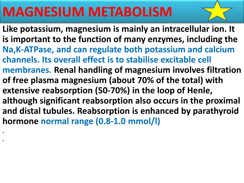
MAGNESIUM METABOLISM Like potassium, magnesium is mainly an intracellular ion. It is important to the function of many enzymes, including the Na,K-ATPase, and can regulate both potassium and calcium channels. Its overall effect is to stabilise excitable cell membranes. Renal handling of magnesium involves filtration of free plasma magnesium (about 70% of the total) with extensive reabsorption (50-70%) in the loop of Henle, although significant reabsorption also occurs in the proximal and distal tubules. Reabsorption is enhanced by parathyroid hormone normal range (0.8-1.0 mmol/l) . .
hypomagnesemia Reduced intake is rarely sufficient alone to cause magnesium depletion, and generally there is excessive loss from either the gastrointestinal tract (notably in chronic diarrhoea) or the kidney (during prolonged use of loop diuretics). Excessive alcohol ingestion can cause magnesium depletion through both gut and renal losses. Some inherited tubular transport disorders result in urinary magnesium wasting, notably Gitelman's syndrome . Hypomagnesaemia is frequently associated with hypocalcaemia, probably because magnesium is required for the normal secretion of PTH in response to a fall in serum calcium,
Clinical features of hypomagnesaemia and hypocalcaemia are similar; there may be tetany, cardiac arrhythmias (notably torsades de pointes, central nervous excitation and seizures, as well as vasoconstriction and hypertension. Magnesium depletion is also associated (through uncertain mechanisms) with hyponatraemia and hypokalaemia, which may mediate some of the clinical manifestations
CAUSES OF HYPOMAGNESAEMIA Mechanism Examples Inadequate intake Starvation, malnutrition (esp. alcoholism), parenteral alimentation Excessive losses Gastrointestinal Prolonged vomiting/nasogastric aspiration, chronic diarrhoea/laxative abuse, malabsorption, small bowel bypass surgery, fistulae Urinary Diuretic therapy (loop, thiazide), alcohol, tubulotoxic drugs (gentamicin, cisplatin), volume expansion (e.g. primary hyperaldosteronism), diabetic ketoacidosis, post-obstructive diuresis, recovery from acute tubular necrosis, inherited tubular transport defect (Bartter's syndrome, Gitelman's syndrome, primary renal magnesium wasting) others Acute pancreatitis, foscarnet therapy, hungry bone syndrome
Treatment 1- involves identification and correction of the cause where possible. 2-Oral magnesium salts have limited effectiveness due to poor absorption and may cause diarrhoea. Where symptoms are present, treatment should be with intravenous magnesium chloride, at a rate not exceeding 0.5 mmol/kg in the first 24 hours. When intravenous access is not available, magnesium sulphate can be given intramuscularly. If hypomagnesaemia is due to diuretic treatment, adjunctive use of a potassium-sparing agent will also reduce magnesium loss into the urine.
HYPERMAGNESAEMIA Aetiology and clinical assessment This is a much less common abnormality than hypomagnesaemia. The cause nearly always involves acute or chronic renal failure, but adrenocortical insufficiency also predisposes to magnesium retention. Any cause of increased intake, either through medications containing magnesium (antacids, laxatives, enemas) or through iatrogenic parenteral therapy, will greatly increase the risk when renal impairment is present.
Clinical features include bradycardia, hypotension, reduced consciousness and respiratory depression. Management Management of hypermagnesaemia involves ceasing all magnesium intake, promoting urinary magnesium excretion using a loop diuretic with intravenous hydration,. Calcium gluconate may be given intravenously to reverse overt cardiac effects. Ultimately, if renal function is minimal, dialysis may be necessary to remove the magnesium load.
. However, when hyponatraemia develops more gradually, cerebral neurons have time to respond by reducing the intracellular osmolality, through reduction in cell potassium and by reduced synthesis of intracellular organic osmolytes . The osmotic gradient favouring water movement into the cells is thus reduced, and patients may present with minimal symptomatology
CAUSES OF HYPONATRAEMIA Volume status Examples Hypovolaemic (sodium deficit with a relatively smaller water deficit) Renal Na losses :Diuretic therapy (especially thiazides) Adrenocortical failure Gastrointestinal Na losses :Vomiting ,Diarrhoea Euvolaemic (water retention alone) Primary polydipsia , Excessive electrolyte-free water infusion ,SIADH* ,Hypothyroidism Hypervolaemic (sodium retention with relatively greater water retention) Congestive cardiac failure Cirrhosis Nephrotic syndrome Chronic renal failure (during free water intake)
MANAGEMENT The treatment for hyponatraemia is critically depon the rate of development and severity, and on the underlying causeendent. In general, if hyponatraemia has developed rapidly (over hours to days), morbidity will be high due to cerebral oedema, and it is generally safe to correct the plasma sodium relatively rapidly. This can include infusion of hypertonic (3%) sodium chloride solutions, especially when the patient is obtunded or convulsing. On the other hand, rapid correction of hyponatraemia which has developed slowly (over weeks to months) can itself be hazardous to the brain. This is because cerebral cells adapt to slowly developing hypo-osmolality by reducing the intracellular osmolality, thus maintaining normal cell volume . Under these conditions, an abrupt increase in extracellular osmolality can lead to water shifting out of the cerebral neurons, abruptly reducing their volume and risking detachment from their myelin sheaths. The resulting 'myelinolysis' can produce permanent structural and functional damage to midbrain structures, and is generally fatal. The rate of correction of the plasma Na concentration in chronic asymptomatic hyponatraemia should not exceed 10 mmol/l/day, and an even slower rate would generally be safer.
Specific treatment measures should be related to the underlying cause. -For hypovolaemic patients, this will involve controlling the source of sodium loss, and administering intravenous saline if clinically warranted. -Patients with dilutional hyponatraemia will generally respond to fluid restriction in the range 600-1000 ml/day, accompanied where possible by withdrawal of the precipitating stimulus (e.g. a drug causing SIADH). Where an inadequate rise in plasma Na results, treatment with demeclocycline (600-900 mg/day) may enhance water excretion, by interfering with collecting duct responsiveness to ADH. An effective alternative for subjects with persistent hyponatraemia due to prolonged SIADH is oral urea therapy (30-45 g/day), which provides a solute load to promote water excretion. - Hypervolaemic patients need optimal treatment of the underlying condition, accompanied by cautious use of diuretics in conjunction with strict fluid restriction. -Potassium-sparing diuretics may be particularly useful in this context where there is significant secondary hyperaldosteronism
HYPERNATRAEMIA Aetiology and clinical assessment hypernatraemia (plasma Na > 150 mmol/l) reflects an inadequacy of the kidney in concentrating the urine in the face of relatively restricted water intake. This can be due to failure to generate an adequate medullary concentration gradient (low GFR states, loop diuretic therapy), but more commonly it is due to failure of the ADH system, either because no ADH is released from the pituitary (central or 'cranial' diabetes insipidus, or because the collecting duct cells are unable to respond to circulating ADH (nephrogenic diabetes insipidus, either inherited or acquired). Patients with hypernatraemia generally have reduced cerebral function, either as a primary problem or as a consequence of the
hypernatraemia itself, which results in dehydration of cerebral neurons and brain shrinkage. In the presence of an intact thirst mechanism and preserved capacity to obtain water, hypernatraemia may not progress very far as water intake proceeds. Where adequate water is not obtained, dizziness, confusion, weakness and ultimately coma and death can result. As for hyponatraemia, the causes of hypernatraemia are best grouped according to the simultaneous disturbance, if any, in total body sodium content . It is important to note the risk of iatrogenic induction of hypernatraemia, and to reiterate that whatever the underlying cause, sustained or severe hypernatraemia must reflect an impaired thirst mechanism or responsiveness to thirst, which would otherwise lead to sufficient water being ingested to prevent this disorder progressing
Congestive cardiac failure Cirrhosis Nephrotic syndrome Chronic renal failure (during free water intake)
Treatment of hypernatraemia depends on both the rate of development and the underlying cause. If there is reason to think that the condition has developed rapidly, cerebral shrinkage may be acute, and correction with appropriate volumes of intravenous hypotonic fluid may be attempted relatively rapidly. However, in older, institutionalised patients it is more likely that the disorder has developed slowly, and extreme caution should be exhibited in lowering the plasma sodium rapidly, to avoid the risk of cerebral oedema in the osmotically adapted cerebral neurons. Where possible, the underlying cause should also be addressed, especially where this involves the simple cessation of salt administration to patients with blunted thirst. Elderly patients are predisposed, in different circumstances, to both hyponatraemia and hypernatraemia, and a high index of suspicion of these electrolyte disturbances is appropriate in aged patients with recent alterations in behaviour
TREATMENT OF HYPERKALAEMIA Mechanism Therapy Stabilise cell membrane potential1 Intravenous calcium gluconate (10 ml of 10% solution) Shift K into cells Inhaled 2agonist, e.g. salbutamol Intravenous glucose (50 ml of 50% solution) and insulin (5 U Actrapid) Intravenous sodium bicarbonate2 (100 ml of 8.4% solution) Intravenous furosemide and normal saline3 Ion-exchange resin (e.g. Resonium) orally or rectally Dialysis Remove K from body