Understanding Hysteroscopes: A Comprehensive Guide
Hysteroscopy is a vital medical procedure for diagnosing intrauterine pathologies. This guide covers the history, components, and types of hysteroscopes, emphasizing rigid and flexible styles. It explains the significance of diameter, lens angles, and instrument options, showcasing images for better understanding. Learn about the evolution of hysteroscopy techniques and instruments to enhance precise diagnosis and treatment.

Download Presentation

Please find below an Image/Link to download the presentation.
The content on the website is provided AS IS for your information and personal use only. It may not be sold, licensed, or shared on other websites without obtaining consent from the author. Download presentation by click this link. If you encounter any issues during the download, it is possible that the publisher has removed the file from their server.
E N D
Presentation Transcript
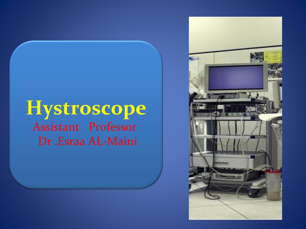
Image result for bettocchi hysteroscope Hystroscope Assistant Professor Dr .Esraa AL-Maini
Related image Hysteroscopy is the process of viewing and operating in the endometrial cavity from a transcervical approach.
-First described in 1969 and done as an office procedure only . -1st optical hysteroscopy was introduced in 1907. -First distension media used was CO2 - In 1980s hysteroscopy replaced blind D&C as a standard procedure for precise diagnosis of intrauterine pathologies
Image result for micro colposcopy hysteroscopy Hystroscope include: -The telescope consists of 3 parts: the eyepiece, barrel, objective lens. -The focal length and angle of the distal tip of the instrument are important for visualization and also fiberoptics of the light source
Image result for micro colposcopy hysteroscopy Angle options include 0 , 12 , 15 , 25 , 30 , and 70 . A 0 hysteroscope provides a panoramic view whereas an angled one allows you to view lateral structures without angling the scope might improve the view of the ostia in an abnormally shaped cavity
Hysteroscopes styles diameter is an important consideration. -Rigid -Flexible
Rigid hysteroscopes most commonly used instruments. Different diameters allows for in-office and complex operating-room procedures. Narrow type:(3-5 mm in diameter), -The 4-mm scope offers the sharpest and clearest view. - It accommodates surgical instruments -Require minimal cervical dilation. -Patients tolerate this instrument well with only paracervical block anesthesia
Related image Related image Conventionally for diagnostic hysteroscopy a 30 degree oblique lens with a diameter of 3 mm and 5 mm detachable external sheath are used.
Related image Minihysteroscopes, introduced recently -Have a 30-degree lens -Diameter of 2 and to 3 mm external sheath. Image quality is similar to conventional hysteroscopes. Microhysteroscopes -Have a diameter less than 2 mm. These are very fragile and have a poor image quality.
Related image Rigid scopes larger than 5 mm in diameter (commonly 8-10 mm) -Require increased cervical dilation for insertion. -Therefore, they are most frequently used in the operating room -With intravenous (IV) sedation or general anesthesia. -Include an outer sheath to introduce and remove media and to provide ports to accommodate large and varied surgical instruments.
flexible hysteroscope -Commonly used for office hysteroscopy. -It tip deflects over a range of 120-160 . -Its most appropriate use is to accommodate the irregularly shaped uterus -To navigate around intrauterine lesions and Allow to visualize the areas that are not visible with the rigid hysteroscope (e.g. uterine retroflexion) - It is also used for diagnostic and operative procedures. 3.7- 5.3 mm diameters with a working channel are use for surgery.
-During insertion, the flexible contour accommodates to the cervix more easily than does a rigid scope of a similarly small diameter. The view was initially described as having a ground-glass quality, which was markedly less desirable than the view obtained with rigid scopes. New, digitally enhanced scopes now offer similar image quality to a rigid hysteroscope lens.
Image result for light source hysteroscope Light source Each hysteroscope is attached to an internal or external light source for illumination at the distal tip. . A xenon light source with a liquid cable is considered the superior option.
Image result for diagnostic sheet hysteroscope DIAGNOSTIC SHEATHS A diagnostic sheath is required to deliver the distention media into the uterine cavity. The telescope fits into the sheath and is secured by means of a watertight seal that locks into place. The sheath is 4 to 5 mm in diameter,.
Image result for operative sheet hysteroscope OPERATIVE SHEATHS Have a larger diameter than diagnostic sheaths. they range from 7 to 10 mm and average 8 mm in diameter. it has channels for 3 - 4 mm telescope , instillation of medium, operating instrument Types : -Standard single operating sheath -Isolated multiple channel operating sheath constant flow of medium leads to very clear operative field.
Image result for resectoscope loop RESECTOSCOPE is a specialized electrosurgical endoscope that consists of an inner sheath and outer sheath. The outer sheath is for medium return. The inner sheath has a common channel for the telescope, medium and electrode. The monopolar or bipolar electrode is fitted to a trigger device. Lens is angled towards the electrode for clear view Electrode The operating tools consist of three basic electrodes: a ball, barrel, and a cutting loop.
Accessory instruments Image result for Accessory instruments FOR hysteroscope The standard accessories are grasping forceps, biopsy forceps, and scissors. be inserted through the operating channels of the scopes Instrument are used with a resectoscope Roller ball, barrel, or ellipsoid - Loop electrode - Scalpel and Vaporizing electrodes Morcellator To cut and remove endometrial polyps or fibroids
Image result for MOMOPOLAR Energy sources and uses Monopolar cautery The resectoscope is often used with a monopolar, use in hypotonic, nonconductive media, such as glycine. It cuts and coagulates tissue by means of contact desiccation with resistive heating. The depth of thermal damage is based on several factors: endometrial thickness; speed, pressure, and duration of contact during motion; and power setting. A thin electrode can cut tissue, whereas one with a large surface area, such as a ball or barrel, is best suited for coagulation. Image result for MOMOPOLAR
Distending media types Gases -Carbon dioxide (CO2) is rapidly absorbed and easily cleared from the body by respiration. -The gas easily flows through narrow channels in small-diameter scopes, making it useful for office-based diagnostic hysteroscopy With CO2, BUT a hysteroscopic insufflator (not safe)is required to regulate flow and limit maximal intrauterine pressure -A flow rate to 40-60 mL/min at a maximum pressure of 100 mm Hg is generally accepted as safe. -Pressures and rates higher than this can result in cardiac arrhythmias, embolism, and arrest.
Liquid Media -The advantage of fluid over gas is the symmetric distention of the uterus with fluid -Its effective ability to flush blood, mucus, bubbles, and small tissue fragments out of the visual field. Both low-viscosity and high-viscosity fluid media can be used for distention. A pressure of 75 mm Hg is usually adequate for uterine distention; rarely is more than 100 mm Hg required, and pressures higher than this can increase the risk of intravasation of medium.
Low viscosity :Ionic ,electrolyte , Isotonic, conductive fluids NS,RL,5%D,10%D,4% and 6% dextran solution Used for diagnostic hysteroscopy and for limited operative procedures. Surgical procedures using mechanical, laser, monopolar ,bipolar VersaPoint system) are safe . Two major disadvantages (1) their miscibility with blood, which obscures visibility with bleeding, leading to the need for increased volumes to clear the operative field 2) their excellent conductivity, which precludes procedures that use standard monopolar electrosurgery
Low viscosity :Non ionic,The hypotonic, nonconductive, fluids 5% mannitol, 3% sorbitol, and 1.5% glycine improve visualization when bleeding occurs. They can be used in diagnostic as well as operative hysteroscopy. risk of volume overload and hyponatremia from intravascular absorption -Combination of 2.8%sorbitol and 0.5% mannitol. -
High viscosity : hyskon(Dextran70) This the only high-viscosity medium available, is a nonelectrolytic, nonconductive fluid that can be used in all types of procedures. It is immiscible with blood and minimally leaks through the cervix and tubes, allowing for excellent visibility during surgical procedures. Allergic reactions and anaphylaxis, fluid overload, disseminated intravascular coagulopathy, and destruction of instruments are adverse effects of this medium
Various delivery systems - The simplest delivery system is a syringe that most often is used with high-viscosity Dextran 70. - Hanging, gravity-fed containers to deliver low-viscosity fluids can be or compressed with a cuff; however, these can be unreliable in estimating intrauterine pressures. - Pumps are available to monitor pressure and volume for low- viscosity media. This design creates laminar flow, which keeps the visual field clear.
Image result for happy mother day Thank you THANK YOU