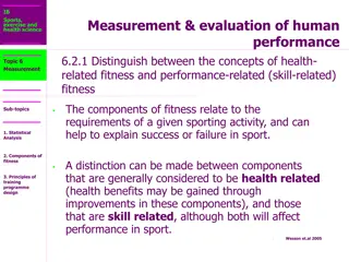
Understanding Hypocalcemia and Related Conditions

Hypocalcemia can be caused by various factors such as hypoparathyroidism, nonparathyroid conditions, and postoperative complications. Symptoms include muscle tetany, paresthesia, and seizures. Conditions like pseudohypoparathyroidism and congenital hypoparathyroidism also contribute to calcium imbalances, affecting bone health. Proper diagnosis and treatment involving calcium and vitamin D are essential for managing these conditions.

Download Presentation

Please find below an Image/Link to download the presentation.
The content on the website is provided AS IS for your information and personal use only. It may not be sold, licensed, or shared on other websites without obtaining consent from the author. Download presentation by click this link. If you encounter any issues during the download, it is possible that the publisher has removed the file from their server.
E N D
Presentation Transcript
Causes of Hypocalcaemia Hypoparathyroid Nonparathyroid PTH Resistance Postoperative Vitamin D deficiency Pseudo- hypoparathyroidism Idiopathic Malabsorption Hyperproduction of calcitonin (medullary thyroid cancer). Post radiation Liver disease Kidney disease Vitamin D resistance
Hypoparathyroidism Commonly occur accidentally after surgical removal of the thyroid gland Latent or overt tetany. Characterized by hypersensitivity (Low threshold) nerves and muscles. of Can be demonstrated by two signs: Chvostek s sign: Tapping the facial nerve as it emerge from the parotid gland In front of the ear Contraction of the facial muscles. Trousseau s sign: Arresting blood flow to the forearm for few minutes Flexion of the wrist, thumb and metacarpophalangeal joints.
Hypocalcaemia Symptoms: Chvosteks and Trousseau s signs Neuromuscular excitability Tetany Paresthesia Seizures
Tetnus influx of Na++ ions at motor neurons and interneurons conduction of reflex muscle contraction causing: Impulses 1- Spasm of larynx death. and bronchus asphyxia and 2- Muscle cramps. 3- Coronarospasm infarction (cardiotetanus) angina
Hypofunction Hyperfunction hypocalciemia hyperphosphatemia hypophosphaturia tetanus - hypercalciemia hypophosphatemia hyperphosphaturia osteoporosis Accumulation of lcium in tissues - - - - - - -
Pseudohypoparathyroidism Symptoms and signs Characteristic physical appearance: short stature, round face, short thick neck, obesity, shortening of the metacarpals Autosomal dominant Hypocalcemia Hyperphosphatemia Resistance to parathyroid hormone The patients have normal parathyroid glands, but they fail to respond to parathyroid hormone or PTH injections Symptoms begin in children of about 8 years Tetany and seizures Hypoplasia of dentin or enamel and delay or absence eruption occurs in 50% of people with the disorder of Treatment: vitamin D and calcium
Pseudohypoparathyroidism Short stature, enamel hypoplasia
Congenital Hypoparathyroidism Hypoplasia of the teeth, shortened roots, and retarded eruption
Case Study A 27 years old man presents to his physician 3 weeks after his thyroid surgically removed for a thyroid cancer. However, since he went home from the hospital, he noticed painful, involuntary muscular cramping. He also felt numbness and tingling around his mouth & in his hands and feet. His parents said that he was irritable for the last 2 weeks. He is on levothyroxine medication. On examination He has a well-healingthyroidectomyscar & no palpablemassesin the thyroid bed. Bloodpressure cuff inflatedabove the systolicpressure induces involuntary muscular contracture in the ipsilateralhand after 60 seconds (Trousseau`s sign) Tapping on the face interior to the ears cause twitchingin the ipsilateralcorner of the mouth (Chevostek`s sign) Lab Investigations: Calcium: 5.6 mg/dl (N: 8.5 10.2) Albumin: 4.1 g/dl (N: 3.5) 4.8) PTH:< 1 pg/ml (N:11 - 54)
DIAGNOSIS The parathyroid glands were removed thyroidectomy during PTH undetectable Hypocalcemia Clinical Manifestations of hypocalcemia (increased reflexes & muscular cramping)
Causes of Hypercalcemia Common Uncommon Malignant disease, e.g. some lung cancers Renal failure Hyperparathyroidism Sarcoidosis Vitamin D toxicity (excessive intake) Multiple myeloma
Hypercalciemia The quantity of c lcium in blood more than 2.6 mmol/L Causes: Hyperparathyroidism and Malignant neoplasms account for majority of hypercalcemia Neoplasms most frequently associated with hypercalcemia: Breast cancer, lung cancer and multiple myeloma Most hypercalcemias in malignancy are caused by humoral hypercalcemia of malignancy ( PTHrP). Single adenomas of the parathyroid gland account for 75% of primary hyperparathyroidism associated with hypercalcemia.
Parathyroid Hormone related Peptide (PTHrP) Can activate the PTH receptor Plays a physiological role in lactation, possibly as a hormone for the mobilization and/or transfer of calcium to the milk May be important in fetal development May play a role in the development of hypercalcemia of malignancy - Some lung cancers are associated with hypercalcemia - Other cancers can be associated with hypercalcemia
Clinical Manifestations of Hypercalcemia Stones Bones Abdominal moans Psychic groans Neuromuscular Cardiovascular Other
Clinical Manifestations of Hypercalcemia Renal stones Nephrolithiasis Nephrogenic DI: polydipsia and polyuria Dehydration Nephrocalcinosis
Clinical Manifestations of Hypercalcemia Skeleton bones Bone pain, arthralgias Osteoporosis of cortical bone such as wrist In primary hyperparathyroidism: Subperiosteal resorption, leading to osteitis fibrosa cystica with bone cysts and brown tumors of the long bones
Clinical Manifestations of Hypercalcemia Gastrointestinal abdominal moans Nausea, vomiting Anorexia weight loss Constipation Abdominal pain Pancreatitis Peptic ulcer disease
Clinical Manifestations of Hypercalcemia Psychic groans Impaired concentration and memory Confusion, stupor, coma Lethargy and Fatigue
Clinical Manifestations of Hypercalcemia Neuromuscular Reduced neuromuscular excitability and muscle weakness Easy fatigability and muscle weakness more common in hyperparathyroidism than other hypercalcemic conditions Clinical features of hyperparathyroid myopathy: Proximal muscle weakness, wasting and mild nonspecific myopathic features on electromyogram and muscle biopsy
Clinical Manifestations of Hypercalcemia Cardiovascular Shortened QT interval on electrocardiogram Cardiac arrhythmias Vascular calcification
Clinical Manifestations of Hypercalcemia Other Itching Keratitis Conjunctivitis Corneal calcification, band keratopathy Carpal tunnel syndrome has occasionally been associated with hyperparathyroidism
Metabolic Diseases of Bones RICKETS Normal formation of the collagen matrix BUT Incomplete mineralization (poor calcification) Soft Bones CLINICALLY: Bone Deformity (Rickets) OSTEOMALACIA Demineralization (poor calcification) of preexisting bones CLINICALLY: More Susceptibility to Fracture
Vitamin D deficiency: Rickets Inadequate intake and absence of sunlight The most prominent clinical effect of Vitamin D deficiency in adults is osteomalacia, or the defective mineralization of bone matrix. Vitamin D deficiency in children produces rickets leading to (bow-legged) due to weight bearing on the legs. A deficiency of renal 1 -hydroxylase produces vitamin D-resistant rickets Sex linked gene on the X chromosome Teeth may be hypoplastic and eruption may be retarded
Images of Rickets Wrist expansion: cupping and fraying of hypertrophied metaphyseal plate Rachitic Rosary Bone demineralization and deformity
nal R ste In Chronic Renal Failure Low activity of Renal 1 -hydroxylase Decreased ability to form the active form of vitamin D (1, 25 DHCC will be low) Treatment: 1,25 DHCC (Calcitriol)
Laboratory Investigations for the Diagnosis of Rickets & Osteomalacia Investigations to confirm the diagnosis of rickets: Blood levels of 25-hydroxycholecalciferol (25 HCC) Blood calcium, (hypocalcemia ) Blood Alkaline phosphatase (ALP )
Osteoporosis Most prevalent metabolic bone disease in adults It means reduction in bone mass. i.e. bone matrix composition is normal, but it is reduced. Typically silent (without symptoms) until it leads to fracture at a minimal trauma. Most affected: vertebral compression (may be asymptomatic) & hip fractures (requires surgery in most cases). Post-menopausal women lose more bone mass than men (primary osteoporosis)
Sequelae of Osteoporosis
Secondary Osteoporosis Risk Factors Risk Factors for osteoporosis: Advanced age (esp. in females) Certain Drugs Family history of osteoporosis or Immobilization Smoking Excess alcohol intake Cushing s syndrome Long term glucocorticoids therapy Hyperparathyroidism Hyperthyroidism Vitamin D disorders Certain malignancies fractures


")