Understanding Giardia: Morphology, Life Cycle, and Pathogenesis
Giardia is a protozoan parasite that causes chronic diarrhea in various hosts, including humans and animals. This parasite has unique morphology with trophozoite and cyst stages. Its life cycle involves direct reproduction through binary fission and transmission through contaminated food or water. Giardia's pathogenesis leads to clinical signs like giardiosis, resulting in diarrhea, dysentery, and fat digestion deficiency. Understanding these aspects is crucial for effective prevention and treatment strategies.
Download Presentation

Please find below an Image/Link to download the presentation.
The content on the website is provided AS IS for your information and personal use only. It may not be sold, licensed, or shared on other websites without obtaining consent from the author. Download presentation by click this link. If you encounter any issues during the download, it is possible that the publisher has removed the file from their server.
E N D
Presentation Transcript
Hexamita and Giardia Bihar Animal Sciences University | Dr. AJIT KUMAR Dr. AJIT KUMAR HoD HoD Department of Veterinary Parasitology Department of Veterinary Parasitology Bihar Veterinary College Bihar Veterinary College Bihar Animal Sciences University Bihar Animal Sciences University Patna Patna- -800014 800014 Image source: Google image
Family: Family: Hexamitidae Hexamitidae Both have two similar nuclei. Genus: Hexamita and Giardia
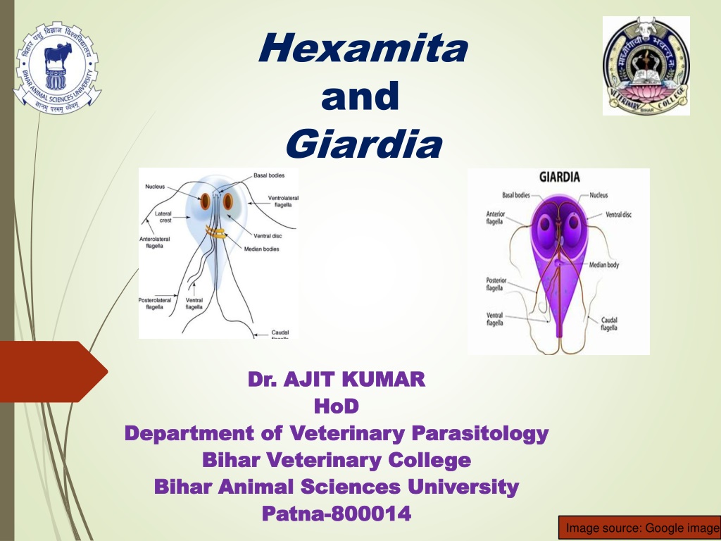
Giardia Giardia Morphology: o Two developmental stages - trophozoite and cyst. Trophozoite : o Pyriform to elliptical in shape and symmetrical. o Anterior end is rounded and posterior pointed. bilaterally end is Image source: Google image
Giardia Giardia Morphology: o Trophozoite : o A large adhesive disc is present on the ventral side whereas dorsal convex. o It has two anterior nucleus, two median axostyle and eight flagella arranged in two pairs side Image source: Google image
Genus: Genus: Giardia Giardia Cyst: Cysts are oval or elliptical in shape with 2 or 4 nuclei and a number of fibrillar remnants of the trophozoite organelles. Image source: Google image
Genus: Genus: Giardia Giardia Species Species Hosts Hosts Location Location Giardia intestinalis or Giardia lamblia Man, monkey and pig Upper digestive tract G. canis Dog G. bovis Ox G. cati Cat G. caprae Goat
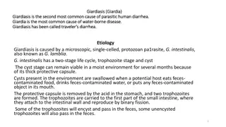
Genus: Genus: Giardia Giardia Life-cycle : Direct Reproduction :- By binary fission Transmission: through contaminated food or water. the ingestion of cysts
Life-cycle of Giardia Image source: Google image
Giardia Giardia Pathogenesis: o Chronic diarrhoea in man especially children. o Interference in fat digestion which results deficiency in the fat soluble vitamins. Clinical signs : o Giardiosis (beaver fever) results diarrhoea, dysentery and steatorrhoea (fats comes with stool) and deficiency of fat soluble vitamin.
Giardia Giardia Diagnosis: o Microscopic examination oval or elliptical shaped cyst contain 2 or 4 nuclei. o Trophozoite stage may be found in diarrhoeic stool. faecal reveals Trophozoites Image source: Google image Cyst
Giardia Giardia Treatment: o Metroniodazole,Tinidazole, Fenbendazole, etc. Chloroquine Control: o Good hygiene and proper sanitation.
Hexamita meleagridis Morphology Morphology Pyriform shaped, two o similar nuceli, out of eight flagella, six flagella anteriorly directed in two groups of three in each and two caudal flagella, two axostyles but adhesive disc absent.
Hexamita meleagridis (Spironucleus Spironucleus meleagridis meleagridis) ) Host and location o It occurs in the duodenum and small intestine of young turkey, peafowl, quail, partridge, duck and also chicken. Image source: Google image
Hexamita meleagridis Transmission Transmission o Organisms transmitted through contaminated feed and drinking water.
Hexamita meleagridis Pathogenesis Pathogenesis o Young turkey up to two months of age are most susceptible whereas old birds act carrier. as symptomless o It causes catarrhal enteritis in the small intestine and intestinal contents become thin, watery and foamy. Myriad of Hexamita are occur in the bulbous area of caeca.
Hexamita meleagridis Clinical Clinical signs o Hexamitosis disease is known as Infectious catarrhal enteritis ion turkey poults. signs o Symptoms include foamy and watery diarrhoea, loss nervousness and death of affected birds. of body weight, Image source: Google image
Hexamita meleagridis Diagnosis Diagnosis o Examination of intestinal contents or scraping of small intestine of sacrificed birds to microscopic detection of living Hexamita organisms. Treatment Treatment o Nithiazide @ 0.02 percent Furazolidone @ 0.01 percent in feed / Oxyteracycline drugs are used in treatment. Control Control o Adoption of hygienic measures in drinking water/
