Overview of Flagellates and Their Clinical Significance
Flagellates are single-celled organisms with whip-like flagella that can cause parasitic infections in humans. Key species like Trichomonas vaginalis, Trichomonas hominis, and Giardia lamblia are discussed, highlighting their clinical implications, diagnostic methods, and treatment options.

Download Presentation

Please find below an Image/Link to download the presentation.
The content on the website is provided AS IS for your information and personal use only. It may not be sold, licensed, or shared on other websites without obtaining consent from the author.If you encounter any issues during the download, it is possible that the publisher has removed the file from their server.
You are allowed to download the files provided on this website for personal or commercial use, subject to the condition that they are used lawfully. All files are the property of their respective owners.
The content on the website is provided AS IS for your information and personal use only. It may not be sold, licensed, or shared on other websites without obtaining consent from the author.
E N D
Presentation Transcript
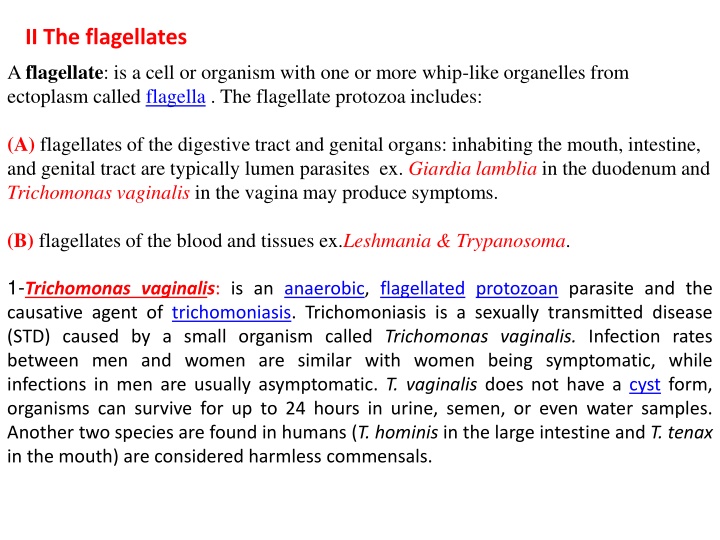
II The flagellates A flagellate: is a cell or organism with one or more whip-like organelles from ectoplasm called flagella . The flagellate protozoa includes: (A) flagellates of the digestive tract and genital organs: inhabiting the mouth, intestine, and genital tract are typically lumen parasites ex. Giardialamblia in the duodenum and Trichomonasvaginalis in the vagina may produce symptoms. (B) flagellates of the blood and tissues ex.Leshmania & Trypanosoma. 1-Trichomonas vaginalis: is an anaerobic, flagellated protozoan parasite and the causative agent of trichomoniasis. Trichomoniasis is a sexually transmitted disease (STD) caused by a small organism called Trichomonas vaginalis. Infection rates between men and women are similar with women being symptomatic, while infections in men are usually asymptomatic. T. vaginalis does not have a cyst form, organisms can survive for up to 24 hours in urine, semen, or even water samples. Another two species are found in humans (T. hominis in the large intestine and T. tenax in the mouth) are considered harmless commensals.
Laboratory Diagnosis 1- Classically, with a cervical smear, infected women have a transparent "halo" around their superficial cell nucleus. It is unreliably detected by studying a genital discharge or with a cervical smear because of their low sensitivity. T. vaginalis was traditionally diagnosed via a wet mount, in which "corkscrew" motility was observed. 2-Currently, the most common method of diagnosis is via culture ex. Diamond' s medium with a sensitivity range of 75-95%. 3-Newer methods, such as rapid antigen testing and transcription-mediated amplification, PCR, using specific primers. Treatment : for both pregnant and non-pregnant patients usually utilizes metronidazole (Flagyl), but with caution especially in early stages of pregnancy 2000 mg by mouth once. Sexual partners, even if asymptomatic, should be treated concurrently.
2-Trichomonas hominis This flagellate is cosmopolitan in its distribution. It is thought to be non-pathogenic although it has been associated with diarrhoeic stools. Laboratory Diagnosis: In a fresh stool, the flagellates move very rapidly in a jerky, non-directional manner.its swim with a characteristic wobbly movement, which makes them unmistakable during diagnosis.
3- Giardia lamblia is a flagellated protozoan parasite that colonizes and reproduces in the small intestine, causing giardiasis. The parasite attaches to the epithelium by a ventral adhesive disc, and reproduces via binary fission. Giardiasis remains confined to the lumen of the small intestine. Giardia infection can occur through ingestion of dormant contaminated water, food, or by the fecal-oral route through poor hygiene practices cysts in CYST TROPHOZOITE
http://fcmdsc.files.wordpress.com/2010/04/giardia-trph1.jpg TROPHOZOITE
Laboratory Diagnosis 1-Trophozoites are found by examination of saline wet preparations of fresh, diarrhoeic stool, duodenal or jejunal aspirate or in a permanently stained faecal preparation. 2-Cysts can be found by examination of the deposit of a formol-ether concentrate of a stool preparation. The oval cysts with thick walls serve as characteristic features for these organisms. The flagella disintegrate and form a central streak which becomes visible when stained with iodine or MIF (merthiolate-iodine-formaldehyde). 4-Chilomastix mesnili is cosmopolitan in distribution although found more frequently in warm climates. It is thought to be non-pathogenic although the trophozoite has been associated with diarrhoeic stool. This is the largest flagellate found in the large intestine.
Laboratory Diagnosis A-The characteristic lemon shaped cysts can be seen in a formol-ether concentrate. B- Motile organisms can be seen in a wet preparation of a fresh stool TROPHOZOITE CYST
TROPHOZOITE CYST