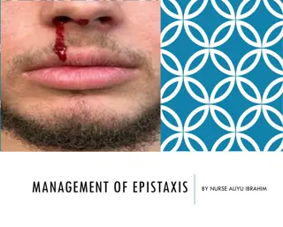
Understanding Epistaxis: Causes, Management, and Prevention
Epistaxis, commonly known as nosebleed, is a prevalent issue affecting a significant portion of the population. It can result from various factors such as trauma, infections, structural abnormalities, environmental conditions, medications, and systemic issues like cardiovascular diseases and coagulopathies. Emergency physicians can successfully treat most cases, with only a fraction needing referral to ENT specialists for further evaluation and care. Recognizing the causes and risk factors of epistaxis is crucial for effective management and prevention.

Download Presentation

Please find below an Image/Link to download the presentation.
The content on the website is provided AS IS for your information and personal use only. It may not be sold, licensed, or shared on other websites without obtaining consent from the author. Download presentation by click this link. If you encounter any issues during the download, it is possible that the publisher has removed the file from their server.
E N D
Presentation Transcript
EPISTAXIS EPISTAXIS Assist prof Dr. FALIH AL Dr. FALIH AL- -ANBAKY ANBAKY Consultant otolaryngologist Consultant otolaryngologist
EPISTAXIS Bleeding from the nose anteriorly or Nasopharynx posteriorly. Relatively common occurrence of hemorrhage from the nose, usually noticed when the blood drains out through the nostrils. There are two types: anterior )nommoc dna , posterior nommoc ssel ) .Sometimes in more severe cases, the blood can come up the nasolacrimal duct and out from the eye. Fresh blood and clotted blood can also flow down into the stomach and cause nausea and vomiting . Although the sight of large amounts of blood can be alarming and may warrant medical attention, nosebleeds are rarely fatal , tsom eht( ( ][
Epistaxis: Epistaxis is a frequent complaint 60% of the population will suffer from a nose bleed during their lifetime, and 6% will require medical attention. Majority of epistaxis occurs between the ages of 2-10 and 50-80 years old. Epistaxis results from an interaction of factors that damage the nasal mucosal lining, affect the vessel walls, or alter the coagulability of the blood. Emergency physicians have a 90% success rate at treating epistaxis in emergency department, and only have to refer 10% to ENT for further assessment and management
Causes of Epistaxis: Despite multiple causes for epistaxis, literature shows that in 85% of cases no causes are found (idiopathic=spontanous). Local causes 1-Trauma Nose picking facial trauma --Blunt trauma (usually a blow to the face such as a punch, sometimes accompanying a nasal fracture. Foreign bodies 2-Nasal or sinus infections 3-Nasal septum deviation, septal spur,Septal perforation 4-Nasal ,Nasopharyngeal Tumour 5-Environmental: Dry cold conditions (presentations increase during winter) Prolonged inhalation of dry air (Oxygen)
Local causes 6-Iatrogenic: Surgery: septoplasty ,endoscopic sinus surgery rhinoplasty, etc Nasogastric tube insertion N asotracheal intubation 7-Medication: Topical corticosteroids and antihistamines Solvent inhalation or (huffing ) Snorting cocaine ( sniffer of cocaine) ischemia necrosisi. Anticoagulants: Aspirin, warfarin, platelet inhibitors
SYSTEMIC CAUSES Cardiovascular (Heart failure) Coagulopathies: Inherited coagulopathies, haemophilia A & B,christmas disease. Splenomegaly Thrombocytopenia , Platelet disorders Haematologigal malignancy like leukemia Liver diseases( cirrhosis ,failure.) Renal failure(ureamia). Chronic alcohol abuse .Vitamin K,C deffeciency AIDS
Systemic causes Vascular Abnormalities: Sclerotic vessels-elderly hyertension. Hereditary haemorrhagic telangiectasia (Osler-Weber-Rendu syndrom) : von Willebrand`s disease Ateriovenous malformation Neoplasm Aneurysms Hypertension: Controversial topic and is often misunderstood as acause of epistaxis . Hypertension is rarely a direct cause of epistaxis Epistaxis is however more common in hypertensive patients this is postulated to be caused from long standing hypertension causing vascular fragility of the blood vessels. Epistaxis in patients presenting to ED, will generally have an associated anxiety that will increase blood pressure. .
Pathophysiology ; Nosebleeds are due to the rupture of a blood vessel within the richly perfused nasal mucosa. Rupture may be spontaneous or initiated by trauma. An increase in blood pressure (e.g. due to general hypertension) tends to increase the duration of spontaneous epistaxis.[ Anticoagulantmedication and disorders of blood clotting can promote and prolong bleeding. Spontaneous epistaxis is more common in the elderly as the nasal mucosa (lining) becomes dry and thin and blood pressure tends to be higher. The elderly are also more prone to prolonged nose bleeds as their blood vessels are less able to constrict and control the bleeding.
Sites The vast majority of nose bleeds occur in the anterior (front) part of the nose from the nasal septum. This area is richly endowed with blood vessels (Kiesselbach's plexus). This region is also known as Little's area. And from retrocollumellar vein. posterior bleeding is usually due to bleeding from Woodruff's plexus, a venous plexus situated in the posterior part of inferior meatus. From sphenopalatine or posterior ethmoid artries. Posterior bleeds are often prolonged and difficult to control. They can be associated with bleeding from both nostrils and with a greater flow of blood into themouth.
http://3.bp.blogspot.com/_u337ThvlyDw/SYtf3ZAEwoI/AAAAAAAABuo/W_7-P1zEozI/s400/Epistaxis+Management.gifhttp://3.bp.blogspot.com/_u337ThvlyDw/SYtf3ZAEwoI/AAAAAAAABuo/W_7-P1zEozI/s400/Epistaxis+Management.gif
Assessment of the patient presenting with Epistaxis: History: Obtain the following: Laterality, duration, frequency Severity, estimated blood loss Any contributing or inciting factors Family history of bleeding disorder Past medical history Current medications (aspirin warfarin ,heparin nsaid,etc)
Physical Examination: Look for vital signs (pulse ,bd pressure Focus on trying to identify if the bleed is coming anteriorly or posteriorly. Suctioning or blowing of the nose to clear away clots, and application of topical vasoconstrictors or anaesthetics will help visualisation. Anterior rhinoscopy by nasal speculum to visualize site of bleeding. A posterior source of bleeding is suggested by failure to visualise an anterior source, or bleeding from both nares, and the visualisation of blood in the posterior pharynx. IF bleeding is severe or recurrent and no site is seen, fiberoptic endoscopy may be necessary. The general examination should look for signs of bleeding disorders, including petechiae, purpura, and perioral and oral mucosal telangiectasias as well as any intranasal masses ,
Investigations: CBC, BT,CT PT ,PTT ,PLATELATE COUNT. Patients taking warfarin should have an INR checked. Coagulation studies are only of benefit in patients with a known coagulopathy or chronic liver disease, and should not be routine in patients presenting with epistaxis. Other bloods test should only be ordered if past medical history warrants further investigation (renal failure = urea and creatinin, chronic alcohol abuse = LFTs. Radiological investigations have little role ,CT scan is indicated if neoplasm suspected
Emergency Department Management: Prehospital Care: Good effective first aid should stop 90-95% of nose bleeds. Provide a calm and quite area for the patient to decrease anxiety Patient position forward, which ever provides the most comfort and prevents the patient from swallowing or aspirating any blood. Tip: Fresh blood is irritating to the stomach and will cause nausea and vomiting. pinching the anterior aspect of the nose for 15-20mins, which provides tamponade to the anterior septal vessels. local application of a vasoconstrictive agent has been shown to reduce the bleeding time in benign cases of epistaxis.( adrenalin xylometazolin ,phenylphrin,, by spray or cotton pledget. Some authors advocate placing ice pack to the forehead or nape of the neck with belief it produces a reflex vasoconstriction.
Treatment at ENT department Cleaning the nose by: Suction by an angled Fraser sucker, 10-12 French gauge. or Nose blowing. Cautery: Chemical: Silver nitrate sticks, or Trichloroacetic acid by wiping the tip of the silver nitrate stick over littles area until it becomes discoloured and grey. The area should dry as possible foreffectiveness of silver nitrate sticks, The sticks should be applied for 4-5secs until a grey residue or eschar develops. Only one septum should be cauterised using silver nitrate, as bilateral can cause sepal perforation Generally effective in mild anterior bleeds, however there is a risk of rebleeding. Electrocautery: Genrally performed by ENT specialist after effective topical anaesthetic needs to be provide first. The red-hot electrocautery loop is passed over the mucosal blood vessels. Topical antibiotics and/or petroleum jelly can be used postoperativley.
Packing: Packing: Anterior packing is required when the bleeding fails to stop with vasoconstrictors and cautery. Types of anterior nasal packs :: Traditional Vaseline gauze packing: generally not used these days, it is ribbon gauze soaked in vasalin or other gel. Compressed sponge/tampon: Merocel is a dehydrated polyvinyl polymer sponge, formed into flat tampons of various sizes. These are inserted into the nasal cavity, and then rehydrate by blood or saline, causing then to expand up the three time their original size, filling the nasal cavity. Anterior epistaxis ballons: Rapid Rhino consist of an outer layer of carboxycellulose that promotes platelet aggregation, with an inflatable balloon that compresses the nasal cavity upon inflation tamponading the bleeding site. Rapid Rhino have been shown to be as effective as nasal tampons and allow for superior patient comfort on insertion and removal. Absorbable materials:, carboxymethycellulose sponges (surgecil) and calcium alginate dressings and wicks. These dressing can be left in place for between 1-5 days, but remember the longer the packing is left insitu the increase risk of developing toxic- shock syndrome
Posterior Packing/ Ballon/ Catheters: Posterior nasal bleeds can be difficult to manage related to the relatively inaccessible site of bleeding and generally don t respond the above standard medical treatment and packing. Analgesia will be required here. 1-The posterior gauze pack consists of 4-inch gauze squares folded, rolled, tied into a tight bundle with 2 strands of heavy silk suture, and coated with antibiotic ointment. Remain4-5days. 2-Double balloon catheters consist off of a posterior and anterior balloon, are relatively easy to insert, although cost may limit their use. Generally used in difficult posterior epistaxis. Saline is preferred over air to inflate balloon as air can leak out causing deflation. Avoid over-inflating ballon (discomfort, rupture of the ballon, or pressure necrosis of the nasal mucosa). 3-A foley catheter can be used 10-14 French G. Posterior pack \ballon= need admession, (decrease pao2),put on antibiotic cover 7dys.
Typical contents of an epistaxis tray. Top row: nasal decongestant sprays and local anesthetic, silver nitrate cautery sticks, bayonet forceps, nasal speculum, Frazier suction tip, posterior double balloon system and syringe for balloon inflation. Bottom row: Packing materials, including nonadherent gauze impregnated with petroleum jelly and 3 percent bismuth tribromophenate (Xeroform), Merocel, Gelfoam, and suction cautery
Rarely if these measures failed to control bleeding . Examination under general anesthesia, use of nasoendoscopy: Cauterization if possible like sphenopalatine artery .if not do: Ligation of selective(anterior or posterior ethmoidal artery) or internal maxillary artery. Embolizatin of artery to stop or to reduce bleeding.
Bleeding disorders In Rendu-Osler-Weber syndrome, A split-thickness skin graft (septal dermatoplasty) reduces the number of nosebleeds and allows the anemia to be corrected. Laser (Nd:YAG) photocoagulation can be done in the operating room. Selective embolization also is very effective, particularly in patients who cannot tolerate general anesthesia or for whom surgical intervention has not been successful. New endoscopic sinus devices have made transnasal surgery more effective. Blood may be swallowed in large amounts and, in patients with liver disease, should be eliminated promptly with enemas and cathartics to prevent hepatic encephalopathy. The GI tract should be sterilized with nonabsorbable antibiotics (eg, neomycin 1 g po qid) to prevent the breakdown of blood and the absorption of ammonia.