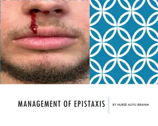
Understanding Epistaxis: Causes, Assessment, and Management

Epistaxis, or nosebleed, can occur in individuals of all ages and has various causes, including local factors like trauma and systemic conditions like hypertension. Clinical assessment is crucial to manage epistaxis effectively, involving checking vital signs and blood tests. Proper management techniques include applying pressure, nasal packing, and addressing underlying conditions. Learn more about the prevention and treatment of epistaxis in this comprehensive guide.

Download Presentation

Please find below an Image/Link to download the presentation.
The content on the website is provided AS IS for your information and personal use only. It may not be sold, licensed, or shared on other websites without obtaining consent from the author. Download presentation by click this link. If you encounter any issues during the download, it is possible that the publisher has removed the file from their server.
E N D
Presentation Transcript
EPISTAXIS PREPARED BY DR.RAFID YASEEN ENT LECTURER.
Introduction Epistaxis : haemorrhage from the nose, affect all ages without sex predilection. -Anterior epistaxis is more common in the child or young adult. -Posterior bleeding is more often in the older adult with hypertension or atherosclerosis. - The incidence is higher the winter months when URTIs are more frequent. -
CAUSES: Local : 1- idiopathic. 2- traumatic(fractures,FB,nose picking). 3- inflammatory(rhinitis, sinus diseases). 4- neoplastic(tumours of the nose, sinuses,and nasopharynx). 5- Environmental (high altitude, air conditioning). 6- Endocrine ( menstruation, pregnancy). 7- Iatrogenic (surgery , steroid nasal spray).
Systemic causes: 1- anticougulants( warfarin, aspirin) 2- Diseases of the blood ( haemophilia, leukaemia). 3- familial haemorrhagic telangiectasia ( Osler-Rendu-Weber) 4- Hypertension. 5- raised venous pressure( whooping cough, Pneumonia, congestive heart failure).
Clinical assessment: A clinical assessment is made by recording the pulse , BP., Signs of shock should be looked for : 1- pallor. 2- tachycardia. 3- sweating. * In cases of severe nosebleed , an IV. Line should be inserted.
Continue. Blood should be taken for cross matching. Checking CBC, coagulation profile. Arrest the bleeding: The nostrils should be pinched together and respiration continued through the mouth . The patient should sit upright to lower the blood pressure and lean forward slightly over a container. Compressing the bony bridge of the nose is of no value.
Proper management : 1- Pressure on the nostrils( can be supported by ice-cold packs and sucking ice cubes). 2- local cautery (chemical or electrocautery). 3- anterior nasal packing (paraffin gauze, BIPP , merocel ) 4- packing of the postnasal space (gauze, Foley s catheter , Brighton balloon).
Continue 5- patient reassurance sometimes with sedation ( iv diazepiam) will relieve patient s anxiety. 6- If nasal packing is required , systemic antibiotics should be given to prevent otitis media , and rhinosinusitis. 7- Examination under GA., it may be necessary if the above measures fail to better identification of the bleeding site and more effective cautery and packing.
Surgical options: 1- submucosal resection ( SMR). Indications: a- bleeding from septal spur. b- to improve the access for inspection, cautery , packing. c- interruption the blood supply of the little s area.
Continue. 2- Endoscopic SPA ligation , clipping , cautery. This type of treatment has success up to 90-100%. 3- anterior ethmoid artery clipping: It is required for uncontrolled bleeding from the upper part of the nose, above middle turbinate. 4- maxillary artery ligation , clipping. 5- external carotid artery ligation. 6- angiograghy and embolization. With gel sponge or beads sometimes associated with complications (0.1%) like CVA.
Familial haemorrhagic :telangiectasia FHT: it is familial autosomal dominant disorder, in which patients develop prominent telangiectasia recognized as red spots on the lips and mucous membranes of the mouth especially the tongue as well as the face and in the nose. Pathology: these nasal telangiectasia consist of dilated venules located just below the basement membrane .they are lined with endothelium without elastic tissue and appear spiderlike, punctate, nodules.
Continue. - the condition may be complicated by the presence of bleeding lesions in the gut , lungs , and genitourinary tract. Treatment options: 1- oestrogens therapy. 2- radiotherapy. 3- diathermy, laser(argon, KTP,YAG) 4- Septal dermoplasty. 5- microembolization 6- YOUNG s operation.