Understanding Computational Vision and Color Perception
Explore the role of MST in heading computation, the influence of eye movements on MSTd cells, and how light wavelengths and color perception are intertwined. Discover the complexities of neural processing in color perception and how it shapes our visual experiences.

Download Presentation

Please find below an Image/Link to download the presentation.
The content on the website is provided AS IS for your information and personal use only. It may not be sold, licensed, or shared on other websites without obtaining consent from the author. Download presentation by click this link. If you encounter any issues during the download, it is possible that the publisher has removed the file from their server.
E N D
Presentation Transcript
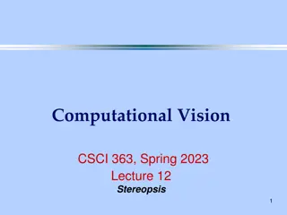
Computational Vision CSCI 363, Spring 2023 Lecture 23 Heading, Color
Does MST compute heading? Prediction: If MST is involved in heading computation, one would expect to find cells tuned to a particular position for the center of expansion. Duffy and Wurtz (1995) tested this prediction.
Do MSTd cells use eye- movement information? Psychophysical experiments showed that humans can make use of eye movement information to compute heading. Some MSTd cells have responses that are modulated by eye movements. Do eye movements affect the responses of MSTd cells to compensate for rotation? This was tested in an experiment by Bradley et al (1996). They recorded from MSTd cells while showing flow fields that consisted of an expansion plus a rotation. The rotation was generated by real or simulated eye movements.
Simulated eye movement condition
Results No eye movement Eye movement in preferred direction. Eye movement in anti-preferred direction. This cell seems to take into account eye movements. The effect was not consistent among all cells tested.
Wavelengths and Color 1. Light is composed of photons with different wavelengths. 2. Humans are sensitive to a small range of wavelengths: 400 - 700 nm.
Light Spectra White light is a mixture of all colors: A variety of wavelengths of light come from different light sources. These are reflected in the spectra of the light. The light reflected off surfaces can be characterized by spectra as well.
Color Perception is not equivalent to wavelength When light hits a surface, some is reflected and some is absorbed and some may be transmitted, depending on the surface properties. Some of the reflected light may reach the eye and is focused on the retina. Neural processing of the light that hits the retina determines our perception of color. Perception of color is not an inherent object property. It depends on neural processing. Different people may perceive the same wavelengths (or spectra) differently (e.g colorblind people).
Color Space Human color perception is defined in terms of 3D color space. The 3 dimensions of this space are Hue, Saturation and Brightness. brightness goes from bottom (dark) to top (light). 2 sides of color space (or spindle).
Hue and Saturation A slice through the color spindle shows a color circle of a given brightness. Hue is the direction from the center of the circle. It varies as you move around the circumference of the circle. Saturation is the distance from the center of the circle. The most vibrant colors (high saturation) are at the outside of the circle. The grayest colors (low saturation) are at the inside of the circle.
Another view of Hue, Saturation and Brightness In a simple case, if the spectrum forms a Gausian distribution, then: Hue is given by the mean of the Gaussian. Saturation is given by the variance (width) of the Gaussian. Brightness is given by the area under the Gaussian curve.
Trichromatic Color Theory Observation: You can match any color in color space with a linear combination of 3 appropriately chosen (no co-linear) colors. Color Matching "Subtract" a color by adding it into the test color.
Three types of cone photoreceptors The retina has three types of cone photoreceptors. Each has a different pigment molecule that absorbs best at a given wavelength. S (Short wavelength), "Blue"=>440 nm M (Middle), "Green" => 530 nm L (Long), "Red" => 560 nm A single wavelength stimulus will stimulate each type of cone by some amount. A mixed color match is a set of stimuli that stimulate the cones by the same amounts as the single wavelength.
1 cone cannot detect color 1 cone type: Cannot tell change in brightness from change in Hue. 2 cone types: Can distinguish some colors. 3 cone types: Can distinguish more colors.
Color perception requires comparison of responses A given wavelength of light will cause a set of responses in the three receptor types. If the intensity changes, the ratio of responses between the photoreceptors will stay the same (but all will increase or decrease). If the wavelength (or spectrum) changes, the ratio of responses between the photoreceptors will change. Next Lecture: Why are certain colors complementary? (E.g. Red/Green or Blue/Yellow).
Color Opponency Observation: Color appears to be organized in terms of 3 sets of complementary colors. (This was first established by Hering) Red/Green Blue/Yellow White/Black Evidence: 1) We don't see a Reddish-Green when mix Red and Green light. Instead, we see less red and green and more yellow. 2) Color adaptation: If you stare at a Green surface for a while, then look at a white background, it will appear red.
Color Adaptation Stare at the white dot
Color Opponent Cells Retinal Ganglion Cells and LGN cells have center-surround receptive fields that show color opponency. B+ (G+R)- (R+G)- (G+R)+ B- (G+R)+ (R+G)+ (G+R)- Concentric Broadband Co-extensive single-opponent Concentric single-opponent
Types of Single-opponent cells Concentric single-opponent: Signal Red vs. Green. Also useful for edge detection. Concentric broad-band: Useful for overall luminance changes (responsive to white light changes). Useful for edge detection. Co-extensive single-opponent: Signal Blue vs. Yellow. Blue cones are probably not involved in edge-detection because of chromatic aberration. The image of the blue wavelength light is more distorted on the retina than of red or green wavelength light.
Color Contrast Observation: The appearance of a color depends on the surrounding colors. There is only one shade of pink in this picture.
Color Constancy Some surfaces appear the same color, even when the ambient lighting changes. The wavelength of the blue squares on the left matches the wavelength of the yellow squares on the right!
Color Constancy This is the same picture as before, but the surrounding squares have been masked out to show the "true" color of the blue and yellow squares.
Responses of double-opponent cells R+G- center R-G+ surround In V1, many color sensitive cells are double-opponent (found in the blobs). Small red or green spot G-R+ G+R- Red or green annulus G+R- G-R+ Large red or green spot B- (G+R)+ B+ (G+R)- B+ (G+R)- B- (G+R)+ Spot inside annulus
Retinex Theory The perception of a color depends on the colors of all objects in the scene. Edwin Land came up with Retinex Theory to predict what appearance colors would have for a given scene. Retinex theory integrates colors across space by measuring the ratios of color changes across color borders. Because we can only measure the relative luminance of surfaces, the retinex theory uses the brightest region in the scene as an anchor. It sets this region to white, and all other regions are computed relative to the anchor.
Color Blindness A small proportion of the population is missing one of the types of cones. Another small proportion has an anomalous pigment. Trichromats: 3 pigments: Normal color vision Dichromats: 2 pigments: Red/Green color blind (1% of males and 0.01% of females) Missing red cones: protanopia Missing green cones: deuteranopia Blue/Yellow color blind (0.02% of males, 0.01% of females) Color anomalies: One of the pigments has a different absorption spectrum.
Colorblindness test Can you see the numbers?