Understanding Acid-Base Disorders in Critical Care
Explore the intricate details of acid-base disorders, including metabolic acidosis with anion gap, high and normal anion gap metabolic acidosis, causes of lactic acidosis, and important considerations like hypoalbuminemia. Learn about the delta ratio and how to interpret it in the context of acid-base imbalances.
Download Presentation

Please find below an Image/Link to download the presentation.
The content on the website is provided AS IS for your information and personal use only. It may not be sold, licensed, or shared on other websites without obtaining consent from the author. Download presentation by click this link. If you encounter any issues during the download, it is possible that the publisher has removed the file from their server.
E N D
Presentation Transcript
ACID BASE DISORDERS Nikki Yeo Critical Care Clinical Fellow Royal Papworth Hospital
Metabolic Acidosis: Anion Gap [Na+] - [Cl-] - [HCO3-] Reference range 8 12 (+/- 4) mmol/L Sometimes [K+] is included: [Na+] + [K+] - [Cl-] - [HCO3-] * Relative to the three other ions, [K+] is low and typically does not change much so omitting it from the equation does not have much clinical significance.
HAGMA and NAGMA High Anion Gap Metabolic Acidosis (HAGMA): Gain of anions (endogenous or exogenous) Normal Anion Gap Metabolic Acidosis (NAGMA) Hyperchloraemia Bicarbonate loss
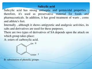
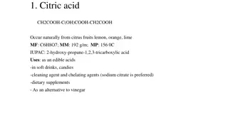
HAGMA NAGMA Renal failure Iatrogenic: Saline Parenteral nutrition Carbonic anhydrase inhibitors Ketoacidosis: Diabetic Alcoholic Starvation Renal losses: Renal tubular acidosis Uretoenterostomy Lactic acidosis GI losses: Diarrhoea Small bowel/pancreatic drainage Toxins: Methanol Ethylene glycol Salicylates Metformin Pyroglutamic acid
Lactic Acidosis Type A: Type B: Imbalance between oxygen supply and demand Altered metabolism Reduced supply -Reduced tissue oxygen delivery: hypoxaemia, anaemia -Impaired tissue utilisation: CO poisoning -Hypoperfusion: Shock B 1: Underlying disease Leukaemia, lymphoma Thiamine deficiency, infection,pancreatitis Failures: renal, liver Increased demand: anaerobic muscle activities Seizures Sprinting B 2: Drugs Beta agonists salicylates Cyanide Ethanol, methanol B 3: Inborn errors in metabolism
Other Considerations Hypoalbuminaemia: Albumin is a an anion Hypoalbuminaemia decreases the AG => For every 10 g/L below normal, add 2.5 to anion gap
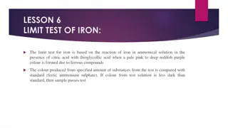
Delta Ratio determine if there is a 1:1 relationship between increase anion gap and decrease in HCO3- Delta ratio = ___increase in anion gap__ decrease in HCO3- < 0.4: associated hyperchloraemia NAGMA 0.4-0.8: consider HAGMA and NAGMA 1-2: uncomplicated HAGMA >2: pre-existing metabolic alkalosis or compensation to chronic respiratory acidosis
Causes of Low Anion Gap Increased Unmeasured Cations Decreased Anion Artefactual Hyperchloraemia Hypercalcaemia Hypoalbuminaemia Iodism Hypermagnesaemia Bromism Lithium intoxication Hypertriglyceridaemia Multiple myeloma *Table reproduced from Toxicology Handbook
Base Excess and Standard Base Excess Base excess definition: Dose of acid or base required to return the pH of a blood sample to 7.40 Measured at standard conditions: 37 C and 40mmHg (5.3 kPa) PaCO2 isolates the metabolic disturbance from the respiratory Standard base excess definition: Dose of acid or base required to return the pH of an anaemic blood sample Calculated for a Hb of 50g/L Haemoglobin buffers both the intravascular and the extravascular fluid => SBE assesses the buffering of the whole extracellular fluid, not just the haemoglobin-rich intravascular fluid
Causes of Metabolic Alkalosis Chronic hypercapnia Volume contraction GI losses Vomiting NG losses (chloride loss) Hypochloraemia Hypokalaemia Renal Losses Diuretics Primary hyperaldosteronism Cushing s syndrome Bartter s syndrome Administration of bases Antacids
Summary of Acid Base Assessment Step 1: Acidaemia (pH < 7.35) Alkalaemia (pH > 7.45) Step 2: Respiratory acidosis or alkalosis Metabolic acidosis or alkalosis Step 3: AG if metabolic acidosis present
Step 4: Check degree of compensation Metabolic acidosis Expected PaCO2 (in mmHg) = (1.5 x HCO3-) + 8 Or, For every 1mmol/L decrease in HCO3- , PaCO2 should decrease by 1.3mmHg Metabolic alkalosis Expected PaCO2 (in mmHg) = (0.7 x HCO3-) + 20 Or, For every 1mmol/L increase in HCO3-, PaCO2 should increase by 0.6mmHg * Conversion of mmHg to kPa 7.5
Step 4 cont: Respiratory acidosis For every 10mmHg (1.3 kPa) increase in PaCO2, HCO3- should increased by 1 mmol/L (acute) or 4 mmol/L (chronic) Respiratory alkalosis For every 10mmHg decrease (1.3 kPa) in PaCO2, HCO3- should decrease by 2 mmol/L (acute) or 5mmol/L (chronic) Step 5: Determine the delta ratio
Question 1 62 year old lady with history of multiple bowel surgeries and severe rheumatoid arthritis presented to ED with abdominal pain and diarrhoea.
Step 1: Acidaemia or alkalaemia Step 2: respiratory acidosis/alkalosis metabolic acidosis/alkalosis Step 3: AG ([Na]-[Cl]-[HCO3-] Step 4: Compensation Step 5: Delta ratio (ref 8-12)
Step 1: Acidaemia Step 2: metabolic acidosis Step 3:AG=133-113-4 = 16 (HAGMA) Step 4: Compensation Expected pCO2= (1.5 x 4) + 8 = 14 mmHg (1.9 kPa) => respiratory alkalosis Step 5: Delta ratio 16-12/ 24-4 = 0.2 (associated hyperchloraemic NAGMA ) (ref range 8-12)
Question 2 A 60-year-old male was admitted after an argument with his partner who found him, 2 hours later, unconscious in his workshop, having likely ingested an unknown substance with empty liquid bottles around him. Describe the significant abnormalities in the results below:
Question 2 Step 1: Acidaemia or alkalaemia Step 2: respiratory acidosis/alkalosis metabolic acidosis/alkalosis Step 3: AG ([Na]-[Cl]-[HCO3-] (ref range 8-12) Step 4: Compensation Step 5: Delta ratio
Question 2 Step 1: Acidaemia Step 2: Metabolic acidosis ?Respiratory acidosis Step 3: AG 141-99-10=32 (ref 8-12) Step 4: Compensation Expected pCO2 (1.5 x 10)+8= 23 Respiratory acidosis/incomplete compensation Step 5: (32-12)/ (24-10) = 1.4 (HAGMA) Osmolar gap
Question 3 72 year old man presented to ED with abdominal pain, nausea and vomiting. PMH: T2DM and AF
Step 1: Acidaemia or alkalaemia Step 2: respiratory acidosis/alkalosis metabolic acidosis/alkalosis Step 3: AG ([Na]-[Cl]-[HCO3-] Step 4: Compensation Step 5: Delta ratio (ref 8-12)
Step 1: Acidaemia Step 2: metabolic acidosis ?respiratory acidosis Step 3: AG ([Na]-[Cl]-[HCO3-] = 36 with profound lactic acidosis Step 4: Compensation Expected pCO2= (1.5x7) + 8 = 19.9 mmHg = 2.7 kPa Step 5: Delta ratio (36-12)/(24-7) = 1.4
Question 4 23 year old female admitted with severe asthma
Step 1: Acidaemia or alkalaemia Step 2: respiratory acidosis/alkalosis metabolic acidosis/alkalosis Step 3: AG ([Na]-[Cl]-[HCO3-] Step 4: Compensation Step 5: Delta ratio (ref 8-12)
Step 1: Severe acidaemia Step 2: respiratory acidosis metabolic acidosis Step 3: AG (139-108-14 ) = 17 with lactic acidosis Step 4: Compensation Expected HCO3- 24+ 3 [(71- 40) = 31] Expected pCO2 = (1.5x 14 )+ 8 = 29 mmHg = 3.9 kPa Step 5: Delta ratio (17-12)/(24-14) = 5/10 = 0.5 (HAGMA and NAGMA) (ref 8-12)
Question 5 35 year old female presented to ED with poorly controlled hypertension, paraesthesia and weakness. Her blood results are as follow:
Step 1: Acidaemia or alkalaemia Step 2: respiratory acidosis/alkalosis metabolic acidosis/alkalosis Step 3: AG ([Na]-[Cl]-[HCO3-] Step 4: Compensation Step 5: Delta ratio (ref 8-12)
Step 1: Alkalaemia (severe hypokalaemia) Step 2: metabolic alkalosis Step 3: AG ([Na]-[Cl]-[HCO3-] Step 4: Compensation pCO2 = (0.8x 40) +20 = 42 mmHg = 5.6 kPa Step 5: Delta ratio (ref 8-12)
References Al-Jaghbeer M, Kellum JA. Acid base disturbances in intensive care patient: etiology, pathophysiology and treatment. Nephrology Dialysis Transplantation 2015; 30(7): 1104-1111. Murray L, Daly F, Little M, Cadogan M. Acid Base Disorders. Toxicology Handbook. 2nd Ed. Elsevier Australia, 2011: 658-685. Derangedphysiology.com UpToDate.com Litfl.com
