the Pancreas Anatomy and Investigations
Situated in the retroperitoneum, the pancreas plays a vital role in digestion and hormonal regulation. Learn about its anatomy, vasculature, investigative methods, and congenital abnormalities affecting this essential organ.

Uploaded on Mar 05, 2025 | 0 Views
Download Presentation

Please find below an Image/Link to download the presentation.
The content on the website is provided AS IS for your information and personal use only. It may not be sold, licensed, or shared on other websites without obtaining consent from the author.If you encounter any issues during the download, it is possible that the publisher has removed the file from their server.
You are allowed to download the files provided on this website for personal or commercial use, subject to the condition that they are used lawfully. All files are the property of their respective owners.
The content on the website is provided AS IS for your information and personal use only. It may not be sold, licensed, or shared on other websites without obtaining consent from the author.
E N D
Presentation Transcript
The pancreas Dr. Ali Jaffer Upper GI surgeon
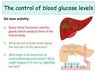
Anatomy The pancreas is situated in the retroperitoneum. It is divided into a head, which occupies 30% of the gland by mass, and a body and tail, which together constitute 70%. The head lies within the curve of the duodenum, overlying the body of the second lumbar vertebra and the vena cava. The aorta and the superior mesenteric vessels lie behind the neck of the gland. Coming off the side of the pancreatic head and passing to the left and behind the superior mesenteric vein is the uncinate process of the pancreas. Behind the neck of the pancreas, near its upper border, the superior mesenteric vein joins the splenic vein to form the portal vein. The tip of the pancreatic tail extends up to the splenic hilum.
Anatomy Vasculature Arteries: Splenic a., superior pancreaticoduodenal a , inferior pancreaticoduodenal a. Veins: Splenic vein, superior mesenteric vein Lympahatics: The pancreas is drained by lymphatic vessels that follow the arterial supply. They empty into the pancreaticosplenal nodes and the pyloric nodes, which in turn drain into the superior mesenteric and coeliac lymph nodes.
INVESTIGATIONS Estimation of pancreatic enzymes in body fluids serum amylase, lipase Pancreatic function tests The nitroblue tetrazolium para-aminobenzoic acid (NBT PABA) The pancreolauryl test Faecal elastase Imaging investigations a. Ultrasonography Ultrasonography is the initial investigation of choice in patients with jaundice to determine whether or not the bile duct is dilated, the coexistence of gallstones or gross disease within the liver such as metastases. It may also define the presence or absence of a mass in the pancreas . However, obesity and overlying bowel gas often make interpretation of the pancreas itself unsatisfactory. b. Computed tomography Most significant pathologies within the pancreas can be diagnosed on high-quality CT scans. c. Magnetic resonance imaging Magnetic resonance cholangiography and pancreatography (MRCP) may well replace diagnostic endoscopic cholangiography and pancreatography (ERCP) as it is non-invasive and less expensive
Invstigation d. Endoscopic retrograde cholangiopancreatography ERCP is performed using a side-viewing fibreoptic duodenoscope. The ampulla of Vater is intubated, and contrast is injected into the biliary and pancreatic ducts to display the anatomy radiologically. Diagnostic and theraputic e. Endoscopic ultrasound This is particularly useful in identifying small tumours that may not show up well on CT or MRI, and in demonstrating the relationship of a pancreatic tumour to major vessels nearby. Transduodenal or transgastric fine needle aspiration (FNA) or Trucut biopsy performed under EUS guidance avoids spillage of tumour cells into the peritoneal cavity.
CONGENITAL ABNORMALITIES Pancreas divisum occurs when the embryological ventral and dorsal parts of the pancreas fail to fuse. The dorsal pancreatic duct becomes the main pancreatic duct and drains most of the pancreas through the minor or accessory papilla. The incidence of pancreas divisum ranges from 5% to10% .Pancreas divisum found incidentally in an asymptomatic person does intervention. The incidence of pancreas divisum ranges from 25 50% in patients with recurrent acute pancreatitis, chronic pancreatitis and pancreatic pain. The minor papilla is substantially smaller than the major papilla (and many of these patients probably have papillary stenosis). A large volume of secretions flowing through a narrow papilla probably leads to incomplete drainage, which obstructive pain or pancreatitis. not warrant any may then cause Idiopathic recurrent pancreatitis, pancreas divisum should be excluded. The diagnosis can be arrived at by MRCP, EUS or ERCP, augmented by injection of secretin if necessary. Endoscopic sphincterotomy and stenting of the minor papilla may relieve the symptoms. Surgical intervention can take the form of sphincteroplasty, pancreatojejunostomy resection of the pancreatic head. or even
CONGENITAL ABNORMALITIES Annular pancreas This is the result of failure of complete rotation of the ventral pancreatic bud during development, so that a ring of pancreatic tissue surrounds the second or third part of the duodenum. It is most often seen in association with congenital duodenal stenosis or atresia and is therefore more prevalent in children with Down syndrome. The usual treatment is bypass (duodenoduodenostomy). The disease may occur in later life as one of the causes of pancreatitis. Ectopic pancreas Islands of ectopic pancreatic tissue can be found in the submucosa in parts of the stomach, duodenum or small intestine (including Meckel s diverticulum), the gall bladder, adjoining the pancreas, in the hilum of the spleen and within the liver. Ectopic pancreas may also be found in the wall of an alimentary tract duplication cyst.
INJURIES TO THE PANCREAS External injury Presentation and management The pancreas, its somewhat protected location in the retroperitoneum, is not frequently damaged in blunt abdominal trauma. If there is damage to the pancreas, it is often concomitant with injuries to other viscera, especially the liver, the spleen and the duodenum. Pancreatic injuries may range from a contusion or laceration of the parenchyma without duct disruption to major parenchymal destruction with duct disruption (sometimes complete transection) and, rarely, massive destruction of the pancreatic head. Blunt pancreatic trauma usually presents with epigastric Pain with the progressive development of more severe pain due to the sequelae of leakage of pancreatic fluid into the surrounding tissues. A rise in serum amylase occurs in most cases. A CT scan of the pancreas will delineate the damage that has occurred to the pancreas. If there is doubt about duct disruption, an urgent ERCP should be sought. MRCP may also provide the answer.
INJURIES TO THE PANCREAS Support with intravenous fluids and a nil by mouth regimen should be instituted while these investigations are performed. It is preferable to manage conservatively at first, investigate and, once the extent of the damage has been ascertained, undertake appropriate action. Operation is indicated if there is disruption of the main pancreatic duct; in almost all other cases, the patient will recover with conservative management. Penetrating trauma to the upper abdomen or the back carries a higher chance of pancreatic injury. In penetrating injuries, especially if other organs are injured and the patient s condition is unstable, there is a greater need to perform an urgent surgical exploration. Sequlae 1. Death, in acute phase as a result of bleeding from associated injuries. 2. Persistent drain output in 1/3 of patients 3. Duct stricture resulting in recurrent episodes of pancreatitis. 4. Pancreatic pseudocyst.
INJURIES TO THE PANCREAS Iatrogenic injury This can occur in several ways: Injury to the tail of the pancreas during splenectomy, resulting in a pancreatic fistula. Injury to the pancreatic head and the accessory pancreatic duct (Santorini), which is the main duct in 7% of patients, during Billroth II gastrectomy Enucleation of islet cell tumours of the pancreas can result in fistulae. Duodenal or ampullary bleeding following sphincterotomy. Pancreatic fistula Management of pancreatic fistulae Tests Measure amylase level in fluid Determine the anatomy of the fistula Check if the main pancreatic duct is blocked or disrupted Measures Correct fluid and electrolyte imbalances Protect the skin Drain adequately Parenteral or nasojejunal feeding Octreotide to suppress secretion Relieve pancreatic duct obstruction if possible (ERCP andstent) Treat underlying cause
Acute pancreatitis The underlying mechanism of injury in pancreatitis is thought to be premature activation of pancreatic enzymes within the pancreas, leading to a process of autodigestion. Once cellular injury has been initiated, the inflammatory process can lead to pancreatic oedema, haemorrhage and, eventually, necrosis. As inflammatory mediators are released into the circulation, systemic complications can arise, such as haemodynamic instability, bacteraemia (due to translocation of gut flora), acute respiratory distress syndrome and pleural effusions, gastrointestinal haemorrhage, renal failure and disseminated intravascular coagulation (DIC). The majority of patients will have a mild attack of pancreatitis, the mortality from which is around 1%. Severe acute pancreatitis is seen in 5 10% of patients, and is characterised by pancreatic necrosis, a severe systemic inflammatory response and often multi-organ failure. In those who have a severe attack of pancreatitis, the mortality varies from 20 to 50%. Acute pancreatitis has an early phase that usually lasts a week. It is characterised by a systemic inflammatory response syndrome (SIRS) which if severe can lead to transient or persistent organ failure (deemed persistent if it lasts for over 48 hours). About one-third of deaths occur in the early phase of the attack, from multiple organ failure. The late phase is seen typically in those who suffer a severe attack, and can run from weeks to months. It is characterised by persistent systemic signs of inflammation, and/or local complications, particularly fluid collections and peripancreatic sepsis. Deaths occurring after the first week of onset are often due to septic complications.
Acute pancreatitis Possible causes of acute pancreatitis Gallstones Alcoholism 50-70% Post ERCP 1-3 % Abdominal trauma Following biliary, upper gastrointestinal or cardiothoracic surgery Ampullary tumour Drugs (corticosteroids, azathioprine, asparaginase, valproic acid, thiazides, oestrogens) Hyperparathyroidism Hypercalcaemia Pancreas divisum Autoimmune pancreatitis Hereditary pancreatitis Viral infections (mumps, coxsackie B) Malnutrition Scorpion bite Idiopathic
Acute pancreatitis Clinical presentation Pain characteristically develops quickly, reaching maximum intensity within minutes and persists for hours or even days. The pain is frequently severe, constant and refractory to the usual doses of analgesics. epigastrium but may be localised to either upper quadrant or felt diffusely throughout the abdomen. There is radiation to the back in about 50% of patients, and some patients may gain relief by sitting or leaning forwards. Nausea, repeated vomiting and retching. The retching may persist despite the stomach being kept empty by nasogastric aspiration. Hiccoughs may be due to gastric distension or irritation of the diaphragm. On examination, the patient may be well or gravely ill. Tachypnoea , tachycardia is usual, and hypotension . The body temperature is often normal or even subnormal Mild icterus can be caused by biliary obstruction in gallstone pancreatitis. Bleeding into the fascial planes can produce bluish discolouration of the flanks (Grey Turner s sign) or umbilicus (Cullen s sign). Neither sign is pathognomonic of acute pancreatitis; Cullen s sign was first described in association with rupture of an ectopic pregnancy. Subcutaneous fat necrosis may produce small, red, tender nodules on the skin of the legs. Abdominal examination may reveal distension due to ileus. There is usually muscle guarding in the upper abdomen. Apleural effusion is present in 10 20% of patients. Pulmonary oedema and pneumonitis
Acute pancreatitis Investigations A serum amylase level three times above normal serum levels lipase contrast- enhanced CT. ASSESSMENT OF SEVERITY Severity stratification assessments should be performed in patients at 24 hours, 48 hours and 7 days after admission. The Ranson and Glasgow scoring systems are specific for acute pancreatitis, and a score of 3 or more at 48 hours indicates a severe attack Regardless of the system used, persisting organ failure indicates a severe attack. A serum C-reactive protein level >150 mg/L at 48 hours after the onset of symptoms is also an indicator of severity. Mild acute pancreatitis: no organ failure; no local or systemic complications. Moderately severe acute pancreatitis: organ failure that resolves within 48 hours (transient organ failure); and/or local or systemic complications without persistent organ failure. Severe acute pancreatitis: persistent organ failure (>48 hours); single organ failure; multiple organ failure.
Acute pancreatitis ASSESSMENT OF SEVERITY
Acute pancreatitis PSEUDOCYST A pseudocyst is a collection of amylase-rich fluid enclosed in a well-defined wall of fibrous or granulation tissue. Formation of a pseudocyst requires 4 weeks or more from the onset of acute pancreatitis. A pseudocyst is usually identified on ultrasound or a CT scan. EUS and aspiration of the cyst fluid. The fluid should be sent for measurement of carcinoembryonic antigen (CEA) levels, amylase levels and cytology. Fluid from a pseudocyst typically has a low CEA level, and levels above 400 ng/mL are suggestive of a mucinous neoplasm. Pseudocyst fluid usually has a high amylase level, but that is not diagnostic, as a tumour that communicates with the duct system may yield similar findings. Cytology typically reveals inflammatory cells in pseudocyst fluid. Pseudocysts will resolve spontaneously in most instances, but complications can develop . Pseudocysts that are thick-walled or large (over 6 cm in diameter), have lasted for a long time (over 12 weeks), or have arisen in the context of chronic pancreatitis are less likely to resolve spontaneously. Therapeutic interventions are advised only if the pseudocyst causes symptoms, if complications develop, or if a distinction has to be made between a pseudocyst and a tumour.
Acute pancreatitis PSEUDOCYST Percutaneous drainage to the exterior under radiological guidance should be avoided. It carries a very high likelihood of recurrence. Moreover, it is not advisable unless one is absolutely certain that the cyst is not neoplastic and that it has no communication with the pancreatic duct (or else a pancreaticocutaneous fistula will develop). Endoscopic drainage usually involves puncture of the cyst through the stomach or duodenal wall under EUS guidance, and placement of a tube drain with one end in the cyst cavity and the other end in the gastric lumen. Surgical drainage involves internally draining the cyst into the gastric or jejunal lumen
Acute pancreatitis STERILE AND INFECTED PANCREATIC NECROSIS Refers to a diffuse or focal area of non-viable parenchyma. Acute necrotic collection (ANC). This is typically an intra or extrapancreatic collection containing fluid and necrotic material, with no definable wall. Gradually, over a period of over 4 weeks, this may develop a well-defined inflammatory capsule, and evolve into what is termed walled-off necrosis (WON). Collections associated with necrotising pancreatitis are sterile to begin with but often become subsequently infected, probably due to translocation of gut bacteria. Infected necrosis is associated with a mortality rate of up to 50%. Sterile necrotic material should not be drained or interfered with. But if the patient shows signs of sepsis, then one should determine whether the collection is infected Dx: aspiration under Ct scan guidance. (the needle should never pass through hollow viscera) Rx: if the aspirated is purulent, the patient should be kept on antibiotics according to sensitivity and drain inserted for drainage. The drain should be with larger size when the aspirate is thick content. if sepsis persist this need pancreatic necrosectomy Patients with peripancreatic sepsis are ill for long periods of time, and may require management in an intensive care unit. Nutritional support is essential. The parenteral and nasojejunal approaches are more popular (on the assumption that they rest the pancreas).
Chronic pancreatitis Chronic pancreatitis is a progressive inflammatory disease in which there is irreversible destruction of pancreatic tissue. Its clinical course is characterised by severe pain and, in the later stages, exocrine and endocrine pancreatic insufficiency. Aetiology and pathology Alcoholism 60-70% Duct obstruction due to trauma, acute pancreatitis or pancreatic cancer Congenital anomalies Hereditary pancreatitis Cystic fibrosis Hyperlipidemia Hypercalcemia Autoimmune pancreatistis (IgG4 phenomena) Idioathic *** At the onset of the disease, the pancreas may appear normal. Later, the pancreas enlarges and becomes hard as a result of fibrosis. The ducts become distorted and dilated with areas of both stricture formation and ectasia. Calcified stones form within the ducts. The ducts may become occluded with a gelatinous proteinaceous fluid and debris, and inflammatory cysts may form. Histologically, the lesions affect the lobules, producing ductular metaplasia and atrophy of acini, hyperplasia of duct epithelium and interlobular fibrosis.
Chronic pancreatitis Clinical features Pain; the site of the pain is depend on the site of the disease. Head of pancreas; the pain felt in the epigastric and right subcostal region, whereas if the disease is limited to the body and tail, the pain felt in the left subcostal region and back. Nausea is common, vomiting may develop. Weight loss The patient s lifestyle is gradually destroyed by pain, analgesic dependence, weight loss and inability to work. Loss of exocrine function leads to steatorrhoea in more than 30% of patients with chronic pancreatitis. Loss of endocrine function and the development of diabetes Infection
Chronic pancreatitis Investigations Only in the early stages of the disease will there be a rise in serum amylase. Tests of pancreatic function merely confirm the presence of pancreatic insufficiency or that more than 70% of the gland has been destroyed. Pancreatic calcifications may be seen on abdominal X-ray CT or MRI scan will show the outline of the gland, the main area of damage and the possibilities for surgical correction, An MRCP will identify the presence of biliary obstruction and the state of the pancreatic duct ERCP is the most accurate way of elucidating the anatomy of the duct EUS
Chronic pancreatitis Medical treatment of chronic pancreatitis Treat the addiction Help the patient to stop alcohol consumption and tobacco smoking Involve a dependency counsellor or a psychologist Alleviate abdominal pain Eliminate obstructive factors (duodenum, bile duct, pancreatic duct) Escalate analgesia in a stepwise fashion Refer to a pain management specialist For intractable pain, consider CT/EUS-guided coeliac axis block Nutritional and pharmacological measures Diet: low in fat and high in protein and carbohydrates Pancreatic enzyme supplementation with meals Correct malabsorption of the fat-soluble vitamins and vitaminB12 Micronutrient therapy with methionine, vitamins C & E, selenium (may reduce pain and slow disease progression) Steroids (only in autoimmune pancreatitis, for relief of symptoms) Medium-chain triglycerides in patients with severe fat malabsorption (they are directly absorbed by the small intestine without the need for digestion) Reducing gastric secretions may help Treat diabetes mellitus
Chronic pancreatitis Endoscopic, radiological or surgical interventions are indicated mainly to relieve obstruction of the pancreatic duct, bile duct or the duodenum, or in dealing with complications (e.g. pseudocyst, abscess, fistula, ascites or variceal haemorrhage). Prognosis Chronic pancreatitis is a difficult condition to manage. Patients often suffer a gradual decline in their professional, social and personal lives. The pain may abate after a surgical or percutaneous intervention, but tends to return over a period of time. In a proportion of patients, the inflammation may gradually burn out over a period of years, with disappearance of the pain, leaving only the exocrine and endocrine insufficiencies. Development of pancreatic cancer is a risk in those who have had the disease for more than 20 years. New symptoms or a change in the pattern of symptoms should be investigated and malignancy excluded.
CARCINOMA OF THE PANCREAS Risk factors for the development ofpancreatic cancer. Demographic factors Age (peak incidence 65 75 years) Male gender Black ethnicity Environment/lifestyle Cigarette smoking Genetic factors /medical conditions Family history Germline BRCA2 mutations in some rare high-risk families Hereditary pancreatitis (50- to 70-fold increased risk) Chronic pancreatitis (5- to 15-fold increased risk) Lynch syndrome (HNPCC) Ataxia telangiectasia Peutz Jeghers syndrome Familial breast ovarian cancer syndrome Familial atypical multiple mole melanoma Familial adenomatous polyposis risk of ampullary/duodenal carcinoma Diabetes mellitus
CARCINOMA OF THE PANCREAS Pathology of pancreatic tumours 85% ductal adenocarcinoma Intense desmoplastic reaction (extensive fibrosis), difficult to differentiate from ch pancreatitis Spread; locally nerve sheath, blood vessels and lymphatic liver and peritoneal spread Cystic tumours ; serous and mucinous Serous : benign, old female, small and multiple Mucinous : potential for malignant transformation it is either Mucinous cystic neoplasm (MCNs) or intraductal papillary mucinous neoplasms (IPMNs) Adenomas of ampulla of Vater; premalignant lesion, may harbour invasive adenocarcinoma, it is common in patients with FAP, need endoscopic surveillance Ampullary adenocarcinoma Ampullary neuroendocrine tumours
CARCINOMA OF THE PANCREAS Clinical features Painless jaundice, pruritus, dark urine, pale stool, steatorrhoea Vague symptoms, weight loss , poor appitite Recent onset DM over 50 with non specific symptms Attacks of pancreatitis Body and tail of pancreas tumours usually preset late as a big tumour with backache Examination; jaundice, weight loss, palpable liver and gall bladder, Courvoisier sign, sings of advance disease (acites, supraclavicular LN, pelvic metastasis etc) Investigation blood test; abnormal liver function test, anemia, tumour markers CA19-9 Ultrasound; dilated CBD, pancreatic mass liver metastasis, peritoneal mass Contrast enhanced CT scan; site of tumour, relation to the arteries vein, duodenum, stomach, bowel, peritoneum lymphnodes MRI ERCP; mainly therapeutic ( stenting) EUS, tissue to confirm the Dx (ch. Pancreatitis, Mucinous tumours) Diagnostic laparoscopy
CARCINOMA OF THE PANCREAS Management 85% unrespectable at time of diagnosis palliative treatment Operable surgical resection 1. head of pancreas and periampullary tumours: pancreaticoduodenectomy whipple s operation ( resection of gastric antrum, duodenum, head of pancreas, CBD& gall bladder) or pylorus preserving pancreaticodudenectomy 2. body and tail of pancreas ; distal pancreatectomy and splenectomy 3. total pancreatectomy; a multifocal tumour (e.g. a main duct IPMN), or the body and tail of the gland are too inflamed or too friable to achieve a safe anastomosis with the bowel.
CARCINOMA OF THE PANCREAS Palliation of pancreatic cancer Relieve jaundice and treat biliary sepsis Surgical biliary bypass Stent placed at ERCP or percutaneous transhepaticcholangiography Improve gastric emptying Surgical gastroenterostomy Duodenal stent Pain relief Stepwise escalation of analgesia Coeliac plexus block Transthoracic splanchnicectomy Symptom relief and quality of life Encourage normal activities Enzyme replacement for steatorrhoea Treat diabetes Consider chemotherapy
Questions Which of the following statements are true? A Pancreatic injury is common following blunt abdominal trauma. B Pancreatic injury is often accompanied by damage to the liver, spleen and duodenum. C The serum amylase is raised in most cases of pancreatic injury. D A CECT scan will delineate the damage. E In doubtful cases, urgent ERCP is helpful. BCDE Which of the following statements is false? A All patients with pancreatic trauma should undergo an exploratory laparotomy. B Pancreatic duct disruption requires surgical exploration. C Severe injury to the duodenum and the head of the pancreas requires pancreatoduodenectomy. D After conservative management for pancreatic injury, duct stricture and pseudocyst may occur as complications. E During splenectomy, iatrogenic injury to the pancreatic tail can occur. A
Which of the following statements are true with regard to complications in acute pancreatitis? A Patients with severe acute pancreatitis require a CECT scan to detect pancreatic necrosis. B In severe acute pancreatitis, a laparotomy must be done in all cases of pancreatic necrosis. C Aneurysm of the superior mesenteric artery can occur. D The vast majority of patients with peripancreatic sepsis can be treated conservatively. E Pleural effusion is seen in 10 20 per cent of patients. ACDE