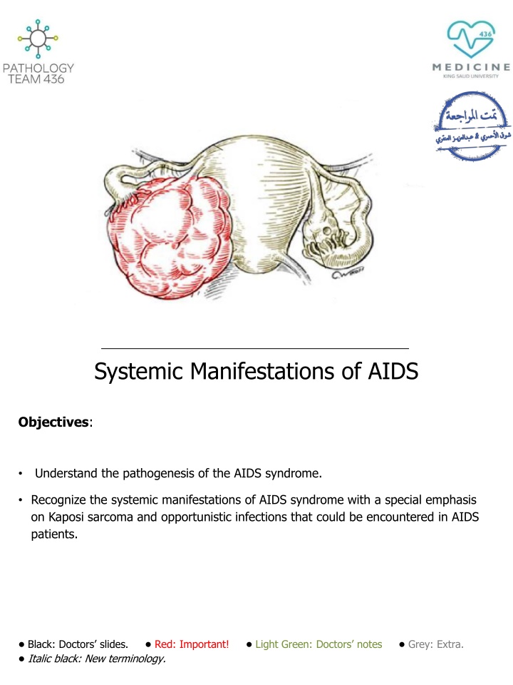
Systemic Manifestations of AIDS
Human Immunodeficiency Virus (HIV) is a retrovirus causing Acquired Immunodeficiency Syndrome (AIDS). HIV leads to immune system destruction, making individuals vulnerable to opportunistic infections and tumors. Common sexually transmitted diseases like syphilis, chlamydia, and gonorrhea are discussed alongside HIV pathogenesis. Systemic manifestations of AIDS, including Kaposi sarcoma and opportunistic infections, are emphasized. The structure of HIV, modes of transmission, and societal impact are also covered.

Download Presentation

Please find below an Image/Link to download the presentation.
The content on the website is provided AS IS for your information and personal use only. It may not be sold, licensed, or shared on other websites without obtaining consent from the author.If you encounter any issues during the download, it is possible that the publisher has removed the file from their server.
You are allowed to download the files provided on this website for personal or commercial use, subject to the condition that they are used lawfully. All files are the property of their respective owners.
The content on the website is provided AS IS for your information and personal use only. It may not be sold, licensed, or shared on other websites without obtaining consent from the author.
E N D
Presentation Transcript
Systemic Manifestations of AIDS Objectives: Understand the pathogenesis of the AIDS syndrome. Recognize the systemic manifestations of AIDS syndrome with a special emphasis on Kaposi sarcoma and opportunistic infections that could be encountered in AIDS patients. Black: Doctors slides. Red: Important! Light Green: Doctors notes Grey: Extra. Italic black: New terminology.
Dr.Ammar Alrikabis notes The most common sexually transmitted diseases (also called venereal diseases) are: 1. Syphilis: it is a sexually transmitted disease caused by the bacteria Treponema pallidum (responsive to penicillin). It starts with a primary infection which is characterized by painless, shallow ulcers ( ) which disappear spontaneously (within 2 to 6 weeks) or with treatment, then the patient gets a serology +ve test for syphilis. The most common serological test used for the identification of the disease is venereal disease research laboratory (VDRL). In primary syphilis, we can take a sample from the ulcer and stain it with IF to see the treponema pallidum under dark field microscopy. Secondary syphilis follows primary syphilis (if no/inadequate treatment was used) after months or years, the patient presents with rash and erythematous papules (which can also be found in plenty of other diseases) but what specializes syphilis is that it affects the palms and soles of foot. A biopsy from the skin shows inflammatory cells including a lot of plasma cells. The diagnosis can be confirmed by VDRL. We have two types of condylomas - condyloma - - which are caused by HPV infection (the malignant type), also called condyloma accumenato. 2) Flat condyloma which are caused by secondary syphilis, also called condylomata lata. Tertiary syphilis comes after secondary syphilis (but is rare now), it affects the CNS. - : 1) Exophytic 2. Chlamydia: only lives intracellularly, causes atypical pneumonia in the lungs, trachoma in the eyes and inflammation in the urethra (non-specific urethritis), vagina and cervix. In non-specific urethritis there is a yellowish discharge which DOES NOT contain bacteria, this way we can differentiate chlamydia from gonorrhea (gonorrhea contains bacteria in the discharge). Chlamydia is treated by tetracycline and erythromycin. 3. Gonorrhea: causes urethritis with brownish discharge, the organism can live outside or inside the host cell. It is treated with ampicillin or penicillin (with a very high dose). Inadequate treatment leads to epididymitis chronic inflammation fibrosis blocking of epididymis infertility. 4. AIDS: explained in the upcoming slides.
Human Immunodeficiency Virus (Introduction): Human immunodeficiency virus (HIV) is the causative agent for AIDS. HIV is caused by a retrovirus of the lentivirus family that contains only RNA. It was known until the early 1980s, but since then it has spread around the world to infect millions of people. All HIV infected persons are at risk of illness and death from the development of opportunistic infections, tumors and the inevitable manifestations of AIDS. The most common type of HIV infections is known as HIV-1 and is the type that has led to the worldwide AIDS epidemic. Responsible for most of the tumor associated with HIV. There s also an HIV-2 that is much less common. The result of HIV infections is the destruction of the immune system. HIV Structure: The mature virus consists of an electron dense core containing the viral genome consisting of two short strands of RNA (ribonucleic acid). It also contains enzymes like: 1. Reverse transcriptase. 2. Protease. 3. Ribonuclease 4. Integrase. All are encased by an outer lipid envelope. This picture is different than that in the lecture, but this one has more details
Understand the pathogenesis of the AIDS syndrome. Pathogenesis of HIV Infection: The HIV virion expresses a cell surface protein/antigen called gp120. They enable the virus to attach itself to different types of cells. Gp120 aids in the binding of the virus to the target cells. Once the virus enters the human body it attaches itself to the target cell via the CD4 receptors on the surface of the target cell and therefore gains entry into the target cell. Gp120 on the surface of the virus is responsible for tropism/attraction to CD4 receptors on the surface of the host cell, this function helps in entry of HIV into the host cell. In addition, gp120 also binds to two co-receptors CXCR4 and CCR5 on the host cell surface, they also assist in the entry of the virus into the host cell. The T-lymphocytes have surface CD4 receptors (CD4+ T-lymphocytes) to which HIV can attach to promote entry into the cell. HIV is shown crossing the mucosa of the genital tract to infect CD4+ T-lymphocytes. The probability of infection depends on both the number of the infective HIV virions in the body fluid which contacts the host as well as the number of cells with CD4 receptors available at the site of contact. That s why it infects/spreads by blood not through any other organ. o o Retroviruses are unable to replicate outside the living host cells because they only contain RNA and do not contain DNA. Therefore, once HIV infects a cell, it must use its reverse transcriptase enzyme to transcribe/convert its RNA to host cell proviral DNA for replication. The enzyme reverse transcriptase in the HIV helps in the reverse transcription (i.e. conversion) of RNA to proviral DNA, the proviral HIV DNA is then inserted into host cell genomic DNA by the integrase enzyme. Once the HIV proviral DNA is within the infected cell s genome, the HIV provirus is replicated by the host cell to produce additional HIV virions which are released by surface budding. Alternatively, the infected cells can undergo lysis with release of new HIV virions which can then infect additional cells.
A Langerhans cell in the epithelium is shown in red in this diagram. Infects the damaged skin not the intact one. HIV viral particles are seen adjacent to the cell surface in the electron micrograph. Doctor s explanation of the pathogenesis (as a summary) 1. Once the virus is attached through gp120 to CD4 cells, it will insert its RNA into the cytoplasm to the nucleus. It will use its enzyme reverse transcriptase to convert RNA to proviral DNA. Then it uses its second enzyme integrase to integrate the viral DNA into the host s DNA so the viral DNA becomes part of the host s DNA. Then it will use the host s proteins, enzymes or glycoproteins to replicate within the cell. Then the new millions of virions will leave the cell by either surface budding or lysis of the cell. 2. 3. 4. 5.
HIV Infection: Macrophages and Langerhans cells are both important as reservoirs and vectors for the spread of HIV in the body including the CNS. Both macrophages and Langerhans cells can be infected by HIV but are not destroyed themselves (they don t undergo lysis, this way they work as reservoirs). HIV can then be carried via these cells elsewhere in the body. Once the infection extends to the lymph nodes, the HIV virions are trapped in the processes of follicular dendritic cells (FDC s), where they provide a reservoir and infect CD4+ T-lymphocytes that are passing through the lymph nodes. The FDC s themselves become infected, but are not destroyed. The target cells are: are all hematopoietic cells (except for RBCs and granulocytes). 1. Blood monocytes. 2. Tissue macrophages. 3. T-lymphocytes and B-lymphocytes. 4. Natural killer (NK) lymphocytes. 5. Dendritic cells (i.e. Langerhans cells of epithelia and follicular dendritic cells in lymph nodes). 6. Hematopoietic stem cells. 7. Endothelial cells. 8. Macroglial cells in the brain. 9. Gastrointestinal epithelial cells. In addition, HIV has the ability to mutate easily. This high mutation rate leads to the emergence of HIV variants within the infected person s cells that are more toxic and can resist drug therapy. Over time, different tissues of the body may harbor differing HIV variants. Why didn t they invent a vaccination or treatment for HIV? o Because the virus itself has a lot of mutations making new versions of it. So by the time you try to invent a vaccine or a drug for one version, there s already 50 new other versions made within the same host, so there s no effective treatment, single drug therapy nor vaccination to be used.
Modes of Transmission of HIV: HIV can be present in a variety of body fluids and secretions. They include: 1. Genital secretions. The highest amount is present there. 2. Blood. 3. Breast milk. So keep in mind that a breastfeeding mother can transmit the disease. 4. 5. 6. 7. Saliva. Urine. Tears. Sweat. However, these four are of no major clinical importance, as transmission of HIV through these fluids does not routinely occur because of the low concentration of HIV in these fluids. HIV is primarily spread as a sexually transmitted disease. Transmission of HIV can occur from male to male, male to female, and female to male. Female to female transmission remains extremely rare. HIV can be transmitted through parenteral routes. IV drug users sharing infected needles, less common practice like the use of instruments such as tattoo needles that are not properly disinfected also carry a potential risk. Health care workers with percutaneous exposures (needle puncture) to HIV- containing blood. Persons receiving multiple blood transfusions (e.g. hemophiliacs). Screening of blood products for HIV has significantly reduced HIV transmission by this means. HIV infection can also be acquired as a congenital infection either perinatally or in infancy. Mothers with HIV infection can pass the virus by: 1. Transplacentally (i.e. in uterus). 2. At the time of delivery through the birth canal. 3. Through breast milk. HIV infection is not spread by casual contact in public places, households, or in the workplace. HIV is not spread by insect vectors. There is no vaccine to prevent HIV infection.
Diagnosis of HIV: Western blot which is a type of PCR technique. ELISA, if positive, should be confirmed by: Dx of HIV IF Test for HIV antibodies is done with a rapid test using an enzyme-linked immunosorbent assay (ELISA) technique. If rapid test is positive, then the next step is to confirm HIV infection with western blot or immunofluorescence assay (IFA). The average HIV-infected person may take up to several weeks to become seropositive, and then may live up to 8 or 10 years, on average, before the development of the clinical signs and symptoms of AIDS. Clinical presentation: Primary HIV infection may go unnoticed in at least half of cases, produce a mild disease (malaise, muscle weakness and common cold symptoms) which quickly subsides, or produce an acute HIV infection, followed by a long clinical latent period lasting for years. Primary acute HIV infections may include fever, generalized lymphadenopathy, pharyngitis, rash, arthralgia and diarrhea. These symptoms diminish over 1 to 2 months.
Recognize the systemic manifestations of AIDS syndrome with a special emphasis on Kaposi sarcoma and opportunistic infections that could be encountered in AIDS patients. Pathogenesis of AIDS/clinical AIDS: The primary target of HIV is the immune system, which is gradually destroyed. Clinically, HIV infection may appear latent for years, during this period there is ongoing immune system destruction but still enough of the immune system remains intact to provide immunity and prevent most infections. Eventually, when a significant number of CD4+ T-lymphocytes have been destroyed and when production of new CD4+ cells cannot match destruction, failure of the immune system leads to the appearance of clinical AIDS. When the immune system can t compensate anymore for its loss of immune cells. The progression to clinical AIDS is also marked by the appearance of syncytia- forming (SI) variants of HIV in about half of HIV infected patients. These SI variants are associated with more rapid CD4+ cell decline. The development of signs and symptoms of AIDS correlates with the CD4+ lymphocyte count, when the CD4+ lymphocyte count drops below 200/microliter, then the stage of clinical AIDS has been reached. This is the point at which the characteristic opportunistic infections and neoplasms of aids appear. The CD4+ T-cells to CD8+ ratio is also greatly reduced, often to less than 1.0. Acquired Immunodeficiency Syndrome (AIDS): The stage of clinical AIDS is reached years after the initial infection and is marked by the development of one or more of the typical opportunistic infections or neoplasms common to AIDS. Symptoms of AIDS could be persistent diarrhea, lymphadenopathy, unexplained fever, early dementia, neurological symptoms, early encephalopathy and weakness. A patient could have HIV but never get AIDS for a long time, it is the reduction in immunity that determines the development to AIDS. Following are some of the more common complications seen in AIDS: 1. Infections e.g. pneumocystis jeroveci, CMV, mycobacteria and fungi. 2. Neoplasms. 3. Miscellaneous (e.g. lymphoid) interstitial pneumonitis which is a condition involving the lung that can be seen in AIDS in children.
Infections Pneumocystis Jeroveci (P.carinii) Pneumocystis? Jiroveci Pneumocystis? Jiroveci H&E stain The most frequent opportunistic infection and it commonly produces a pulmonary infection. The diagnosis is made histologically by finding the organisms in cytologic (bronchoalveolar lavage) or biopsy (transbrnochial biopsy) material from lung. In the lung, there is soap bubble like intra-alveolar exudate and the organism appears as cyst like structures that are positive with silver stain. o Silver Stain Silver Stain Silver stain H&E Stain H&E Stain o o REPR 224 Inflammatory cells within the space of alveoli, but are not enough/effective to defeat the organism. comma shaped. (Hence the name) REPR 224 10/04/2018 10/04/2018 29 29 Cytomegalovirus (CMV) CMV Infection Cyto: cell. Megalo: big. Virus.: Big inclusions within the cell. It causes unexplained pneumonia and serious diseases in the brain and gastrointestinal tract. It is also a common cause for retinitis and blindness in persons with AIDS. o The cells and nuclei are enlarged. The cells show an owl eye shape, large basophilic intranuclear inclusions. Multiple cytoplasmic inclusions. o o REPR 224 10/04/2018 30 Mycobacterial infections Mycobacterium tuberculosis. Mycobacterium avium complex (MAC). Definitive diagnosis of mycobacterial disease is made by culture and PCR. o o o Others Toxoplasmosis caused by Toxoplasma gondii is a protozoan parasite that most often leads to infection of the brain with AIDS. Herpes simplex infection in the mucosa. Aspergillosis especially in the lung. Cryptosporidium and Microsporidium produce voluminous watery diarrhea in patients with AIDS. Viral HIV encephalitis. Syphilis (primary, secondary and tertiary). o o o o o o
Neoplasms: Kaposi s sarcoma (KS) produces reddish purple patches or nodules over the skin and can be diagnosed with skin biopsy. Visceral organs can also be involved with KS. It is a sarcoma of the blood vessels which is common in homosexuals. It is associated with HHV-8 human herpes virus type 8- (so if a patient comes with the same features of KS but is NOT affected with HHV-8 we don t call it KS!) and on histology, it shows malignant spindle cells of vascular origin. HHV-8 affects the endothelium of blood vessels, gets integrated into their nuclei and leads to cells proliferation cancer. Any significant reduction in the immunity can lead to KS, examples: post-renal transplant, post-chemotherapy, AIDS and post-bone marrow transplant. Malignant lymphomas are commonly B-cell non-Hodgkins lymphoma. They are typically of high grade and often in the brain. They are very aggressive and respond poorly to therapy. Clinically: Microscopically: Extra:
Summary The HIV virion expresses a cell surface protein/antigen called gp120. Once the virus enters the human body it attaches itself to the target cell via the CD4 receptors. The T-lymphocytes have surface CD4 receptors (CD4+T lymphocytes). Macrophages and Langerhans cells are both important as reservoirs and vectors for the spread of HIV in the body including the CNS. Modes of Transmission of HIV: HIV can be present in a variety of body fluids and secretions. HIV concentration is high in genital secretions and has low concentration in saliva, urine etc. that s why they re not have clinical importance. The progression to clinical AIDS is also marked by the appearance of syncytia-forming (SI) variants of HIV. These SI variants are associated with more rapid CD4+ cell decline Destroy T-cells . Pneumocystis jiroveci (P. carinii) is the most frequent opportunistic infection seen in AIDS. It commonly produces a pulmonary infection. The diagnosis of Pneumocystis jiroveci is made histologically. the lung, there is soap bubble like intra-alveolar exudate and the organism appears as cyst like structures that are positive with silver stain GMS . Owl's eye appearance of inclusion bodies is highly specific for cytomegalovirus (CMV) infection. Kaposi's sarcoma (KS) produces reddish purple patches or nodules over the skin and can be diagnosed with skin biopsy. Visceral organs & lymph nodes can also be involved. KS is associated with HHV-8 Human Herpes virus-8 .
Questions Q1) Which of the following is a cause of death in HIV infected persons? A. Opportunistic infections. B. Tumors. C. AIDS. D. All of the above. Ans: D Q2) Which of the following enzymes does HIV contain? A. Reverse transcriptase. B. Protease. C. Integrase. D. All of the above. Ans: D Q3) HIV can spread by all of the following except? A. Blood. B. By mother. C. Contact by touch. D. Sexual intercourse. ANS: C Q4) What is the infection that can cause blindness and retinitis in patients with AIDS? A. CMV. B. Fungal infection. C. Aspergillosis. D. Cryptosporidium. ANS: A Q5) A patient with AIDS came to the hospital suffering from reddish purple nodules over his skin, on examination, he was infected with HHV-8 and his blood vessels show spindle cells. Which of the following is the diagnosis? A. Malignant lymphoma. B. Syphilis. C. Kaposi s Sarcoma. D. HSV ANS: C
Questions Q6) Which of the following cells in the mucosal surfaces are most instrumental in transmitting HIV to CD4+ T-lymphocytes? A. CD8+ cells. B. Natural killer cells. C. Neutrophils. D. Dendritic cells. ANS: D Q7) HIV can not be carried in macrophages nor Langerhans cells since it destroys them. A. True. B. False. ANS: B Q8) Which of these is not a target cell for HIV infections? A. Hematopoietic stem cells. B. Dendritic cells. C. Skin stem cells. D. B and T lymphocytes. ANS: C Q9) which of the following is an antigen expressed on the surface of HIV virion? A. Gp120. B. CD4. C. Gp210. D. CCR5. ANS: A
! . : : Editing File Email: pathology436@gmail.com Twitter: @pathology436 References: Doctor s slides + notes, Robbins basic pathology 10th edition.


:")