Surgery of the Vermiform Appendix & acute abdomen
Surgery of the Vermiform Appendix
& acute abdomen
By
Dr. Hosam Elsrogy
Prf. of General Surgery
Sohag University
2018
1
Items
•
Introduction
•
Surgical anatomy
•
Inflammations of appendix
acute
chronic
recurrent
•
Tumours of appendix
Carcinoid (argentaffinoma)
1ry adenocarcinoma
•
Causes of acute abdomen
2
Introduction
•
Appendix is a vestigial organ.
•
Surgical importance-------inflammations------
clinical syndrome of acute appendicitis (AA).
•
AA is the most common cause of acute
abdomen in young adults.
•
Appedecectomy (appendectomy) is the
frequently performed urgent abdominal op.
•
Diagnosis of AA is essentially clinical.
3
Surgical anatomy
•
Vermiform appendix is present only in human
& certain anthropoid apes.
•
Blind muscular tube at distal end of caecum.
•
4 layers--------- mucosa, submucosa,
musculosa & serosa.
•
At birth, it is short & broad at its junction with
caecum.
•
By age of 2 ys------ typical tubular structure
(due to differential growth of the caecum)
4
Surgical anatomy; positions of
appendix
•
Retrocaecal -------- 74%
•
Pelvic -------- 21%
•
Postileal -------- 5%
•
Paracaecal -------- 2%
•
Subcaecal -------- 1.5%
•
Preileal --------- 1% (typical C/P)
•
Subhepatic --------- very rare
•
Lt. sided --------- situs inversus viscerum
5
6
Surgical anatomy (cont.)
•
Position of base of the appendix is constant -------
- at the confluence of the 3 tinea coli.
•
Mesoappendix arises from lower surface of
mesentery of terminal ileum.
•
Bl. Supply -------- appendicular a. (from lower
division of ileocolic a.), it is an end a.-----
thrombosis ----- gangrenous appendicitis.
•
Accessory appendicular a. in most people.
•
Lymph drainage ----- 4-6 lymph channels ----
ileocaecal LNs
7
8
Microscopic anatomy
•
Length ------- 7.5-10 cm
•
Irregular lumen, multiple folds of m.m lined by
col. cell intestinal mucosa of colonic type.
•
Crypts are present but not numerous----- at
base of crypts ------ Argentaffin Cs
(Kultschitzsky Cs) ------- carcinoid tumour.
•
App. is the most frequent site for carcinoid ts.
•
Submucosa ------ lymphatic aggregations or
follicles especially in young adults ----- AA.
9
Acute appendicitis - Incidence
•
One person in 6-7 develops AA at some time.
•
Age:
rare in infants
Common in childhood & early adult life
Peak incidence → teens & early 20s
↓ ↓ after middle age
•
Sex:
Equal before puberty
More in males at teenagers & young adults (3:2)
After that age → equal
10
AA- Incidence (cont.)
•
AA is the commonest abdominal surgical
emergency.
•
AA is a disease of civilisation.
•
Relatively uncommon in developing rural
communities.
•
Incidence ↑ in developing countries adopting
a more-refined western-type diet
11
AA- Surgical pathology
•
Aetiology & predisposing factors:
Infective agents
Obstructive agents
•
Infective agents:
•
Bacterial proliferation within the appendix
•
Mixed intestestinal organisms (aerobic & anaerobic)
e.g., coliforms, entercocci, bacteroides & others.
•
Infection either:
1ry → lymphoid hyperplasia.
2ry → bacteria → wall of appendix through epithelial
erosion caused by pressure of an obstructing agent.
12
AA- Surgical pathology (cont.)
•
Obstructing agents:
1.
Faecolith (inspissated faecal material, Ca
phosphates, bacteria, epithelial debris)
2.
F.B. (vegetable seeds, date stones)
3.
Fibrotic stricture (previous resolved AA)
4.
Tumour (caecal Ca in older ages, carcinoid)
5.
Intestinal parasites (thread; round & pinworms)
6.
Lymphoid hyperplasia in submucosa ; viral
causes
13
AA- Surgical pathology (cont.)
•
Clinicopathological types of AA:
1.
Acute appendicitis
2.
Acute appendicitis with inflammatory mass
3.
Acute appendicitis with generalised
peritonitis
4.
Mucocele of the appendix
14
AA- Surgical pathology (cont.)
•
Early AA → mucosal inflam. & lymphoid
hyperplasia with patent lumen.
•
If obstruction occurs → cont. mucous sec. +
inflam. exudate (pus) → ↑ intraluminal pr. →
obstruct lymphatic drainage → oedema &
mucosal ulceration → bact. translocation into
submucosa.
•
Fate: either
Resolution (spontaneous or Antibiotic)
Progress
15
AA- Surgical pathology (cont.)
•
Progression → further distension→ venous
obstruction → ischaemia of app. wall →
bacterial invasion through muscularis propria
& submucosa → AA (red, turgid appendix).
•
Finally ischaemic necrosis → gangrenous app.
Common close to the tip of appendix due to ↓
blood supply or
At the site of obstruction due to pressure necrosis
16
AA- Surgical pathology (cont.)
•
Fate of gangrenous appendicitis:
1)
Free bacterial contamination in peritoneal cavity
→generalised peritonitis → intense peritoneal
reaction with fluid outpouring → initially clear, late
purulent → serosal surface of bowel is injected &
flaked with clotted lymph.
2)
Rapid localisation by defence mechanism (greater
omentum & coils of small bowel) → phlegmonous
mass or paracaecal abscess if suppuration occurs.
•
Rarely, resolution of app inflam → distended mucous-
filled app → mucocele of the appendix.
17
AA with generalised peritonitis
•
It is the great threat of acute appendicitis
•
Causes of peritonitis:
1)
Free migration of bacteria through ischaemic
appendicular wall
2)
Frank perforation of gangrenous appendix
3)
Delayed perforation of appendicular abscess
18
Risk factors for perforation of
appendix
•
Extreme of age (ill-developed omentum)
•
Immunosuppression
•
Diabetes mellitus
•
Faecolith obstruction
•
Pelvic appendix (lying free in pelvis; 21%)
•
Previous abdominal surgery
•
pregnancy
19
AA- Clinical features
•
Periumbilical colic (poorly localised, visceral)
•
Pain shifts to RIF (intense, constant & localised
somatic pain due to parietal peritoneal irritation)
•
Anorexia (constant especially in children)
•
Nausea
•
Vomiting (1 or 2 episodes after onset of pain)
•
History of previous similar discomfort which
setteled spontaneously
20
AA- Clinical features (cont.)
•
Typical features are present in 50%.
•
Atypical features→ poorly localised somatic or
visceral pain especially in:
Elderly → no RIF pain
Pelvic appendicitis
→ no somatic pain in ant. abd. wall
→ suprapubic discomfort & tenesmus
→ tenderness on PR examination
21
AA- Clinical features (cont.)
•
Typically 2 clinical syndromes of AA:
•
Acute catarrhal (non obstructive) appendicitis
•
Acute obstructive appendicitis
→ acute course
→ abrupt onset
→ generalised abdominal pain from start
→ temp. may be normal & vomiting is common
→ mimic acute intestinal obstruction
22
AA- Signs
•
General
•
Local
•
Special
•
General signs:
1)
Look → unwell, coated tongue & foul breath
2)
Pyrexia
→ low grade (37.2-37.7˚C)
→ absent in 20% of cases
→ In children if > 38.5˚C → other cause
e.g., mesenteric adenitis
1)
Tackycardia → 80-90 beats/ min, absent in 20%
23
AA- Signs
•
Local signs:
1.
Localised tenderness in RIF;McBurney’s point
2.
Muscle guarding over RIF
3.
Rebound tenderness(coughing or percussion)
4.
Limitation of resp. movement in lower abd.
5.
Pointing sign
6.
Tender PR exam on Rt side in pelvic app.
7.
Cutaneous hyperaesthesia in RIF
24
AA- Signs
•
Special signs:
1.
Rovsing’s sign (on deep pressure on LIF)
2.
Blumberg’s sign (crossed or rebound tenderness
on sudden release of deep pressure on LIF)
3.
Psoas sign (pain on extension of Rt. hip)
4.
Obturator (Cope’s) sign (pain on flexion &
internal rotation of Rt. Hip in pelvic app.)
5.
Straight leg raising sign
25
AA- Special features
•
Retrocaecal appendix:
Absent rigidity
Lack localised deep tendern. at RIF (silent app)
Deep tenderness at loin
Rigidity of quadratus lumborum
+ve psoas spasm
26
AA- Special features
•
Pelvic appendix:
Early diarrhoea
Complete absence of abdominal rigidity
Lack of tenderness at McBurney’s point
Deep tendern. above & to Rt. of symphysis pubis
Tendern. in retrovesical or Douglas pouch on PR
Psoas spasm & obturator internus muscles
Frequency of micturition
27
AA- Special features
•
Postileal appendix:
Pain may not shift (missed appendix)
Diarrhoea is present
Ill-defined tenderness or may be present
immediately to the Rt. of the umbilicus
28
AA- Special features
•
Appendicitis in infants & young children:
Rare under 36 months of age
Often delayed diagnosis:
Patient unable to give history
High incidence of perforation
Diffuse peritonitis develop rapidly due to ill-
developed omentum
29
AA- Special features
•
Appendicitis in older children:
Vomiting is always
Complete aversion to food
No sleep during attack
Absent bowel sounds in early stages
•
Appendicitis in elderly:
High incidence of gangrene & perforation
Little signs due to lax abdominal walls or obesity
May simulate subacute intestinal obstruction
Higher mortality rates
30
AA- Special features
•
AA in obese patients:
↓ all local signs with delay in diagnosis
Technically difficult operation
Consider a midline abdominal incision
•
AA in pregnancy:
AA is the most common extrauterine abd condition
Frequency is one in 1500-2000 pregnancies
Delay in presentation & early nonspecific symptoms
High appendix late in pregnancy → flank or back pain
confused with pyelonephritis
Foetal loss occurs in 3-5% & 35% if perforation occurs
31
D.D. of acute appendicitis & acute
abdomen
•
Children:
1)
Acute gastroenteritis
2)
Mesenteric lymphadenitis
3)
Meckel’s diverticulitis
4)
Intussusception
5)
Henoch-Schönlein purpura
6)
Lobar pneumonia & pleurisy
32
D.D. of acute appendicitis & acute
abdomen
•
Adults:
1)
Regional enteritis (Crohn’s disease)
2)
Ureteric colic
3)
Perforated ulcer
4)
Torsion testis
5)
Pancreatitis
6)
Rectus sheath haematoma
33
D.D. of acute appendicitis & acute
abdomen
•
Adult females:
1)
Salpingitis
2)
Mittelschmerz
3)
torsion/rupture of an ovarian cyst
4)
Ectopic pregnsncy
5)
Pyelonephritis
6)
Endometriosis
34
D.D. of acute appendicitis & acute
abdomen
•
Elderly:
1)
Sigmoid diverticulitis
2)
Intestinal obstruction
3)
Colonic carcinoma
4)
Torsion appendix epiploicae
5)
Mesenteric infarction
6)
Aortic aneurysm
35
D.D. of acute appendicitis & acute
abdomen
•
Rare D.D.:
1)
Preherpetic pain of Rt 10
th
&11
th
dorsal
nerves
2)
Tabetic crises
3)
Spinal conditions
4)
Porphyria & D.M.
5)
Cyclical vomiting of infants or young children
6)
Typhlitis or leukaemic ileocaecal syndrome
36
preoperative investigations in AA
•
Routine:
Full blood count
urinlysis
•
Selected cases:
Pregnancy test
Urea & electrolytes
Supine AXR
Ultrasound abdomen/pelvis
37
Ttt of acute AA
•
appendicectomy:
•
Conventional
McBurney’s incision
Lanz incision
Lower midline abdominal incision
Rutherford Morrison’s incision
Retrograde appendectomy
Drainage of peritoneal cavity
•
laparoscopic
38
Problems encountered during
appendicectomy
•
A normal appendix:
Careful exclusion of other possible diagnosis esp Crohn’s
disease, Meckel’s diverticulitis, tubal or ovarian causes
Remove appendix
•
The appendix cannot be found:
Mobilise the caecum& trace tinea coli
Absent appendix
•
An appendix abscess is found & appendix could not
removed easily:
Local peritoneal toilet & drainage of abscess
I.v antibiotics
Very rarely. Caecectomy or partial Rt hemicolectomy
39
AA complicating Crohn’s disease
•
If caecal wall is healthy at base of appendix→
appendicectomy.
•
If appendix is involved in CD→ conservative
approach→ I.V corticosteroids & ststemic AB
40
41
Abscesses complicating AA
•
Appendix abscess
Pus within the phlegmonous appendix mass
Failure of resolution of appendix mass
Continued spiking pyrexia
Percutaneous drainage under US or CT guide
Laparotomy through a midline incision
Extraperitoneal drainage
•
Pelvic abscess
Spiking pyrexia several days after appendicitis or discharge
Pelvic discomfort, loose stool or tenesmus
PR→ boggy mass in pelvis, ant. to rectum
Pelvic US or CT
Transrectal drainage
42
Ttt of appendix mass
•
The standard conservative Ochsner-Sherren regimen
•
Careful record of patient’s condition
•
Regular exam of abdomen
•
Mark limits of the mass on the abdominal wall
•
Nasogastric tube
•
Intravenous fluids
•
Systemic antibiotic therapy
•
Record temp & pulse every 4 hrs
•
Clinical improvement within 24-48 hrs
•
Remove appendix usually after an interval of 6-8 weeks
43
Criteria for stopping conservative ttt of
appendix mass
•
A rising pulse rate
•
Increasing or spreading abdominal pain
•
Increasing size of the mass
•
Vomiting or copious gastric aspirate
44
Neoplasms of appendix
•
Carcinoid tumour (argentaffinoma)
Arise in argentaffin tissue (Kulschitzsky Cs of crypts of
Lieberkuhn)
Most commonly found in appendix
Found once in every 300-400 appendices
10 times more common than any other neoplasms
Subacute or recurrent appendicitis
Frequently involves distal third of appendox
Rarely gives rise to metastases
Small tumours < 2 cm→ appendicectomy
Rt hemicolectomy if involved caecal wall, tumour is 2 cm or
more or involved LN
45
46
Thank you
47
48
Dr. Asem Elsani 2009
49
Dr. Asem Elsani 2009
50
51
52
53
54
This publication by Dr. Hosam Elsrogy, a Professor of General Surgery at Sohag University, delves into the intricacies of performing surgery on the vermiform appendix and managing acute abdomen conditions. A comprehensive guide for medical professionals and students interested in this field of surgery. It covers essential insights, techniques, and approaches relevant to these surgical procedures, aiding in improving medical knowledge and practice.

Download Presentation

Please find below an Image/Link to download the presentation.
The content on the website is provided AS IS for your information and personal use only. It may not be sold, licensed, or shared on other websites without obtaining consent from the author.If you encounter any issues during the download, it is possible that the publisher has removed the file from their server.
You are allowed to download the files provided on this website for personal or commercial use, subject to the condition that they are used lawfully. All files are the property of their respective owners.
The content on the website is provided AS IS for your information and personal use only. It may not be sold, licensed, or shared on other websites without obtaining consent from the author.
E N D
Presentation Transcript
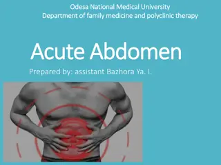
Surgery of the Vermiform Appendix & acute abdomen By Dr. Hosam Elsrogy Prf. of General Surgery Sohag University 2018 1
Items Introduction Surgical anatomy Inflammations of appendix acute chronic recurrent Tumours of appendix Carcinoid (argentaffinoma) 1ry adenocarcinoma Causes of acute abdomen 2
Introduction Appendix is a vestigial organ. Surgical importance-------inflammations------ clinical syndrome of acute appendicitis (AA). AA is the most common cause of acute abdomen in young adults. Appedecectomy (appendectomy) is the frequently performed urgent abdominal op. Diagnosis of AA is essentially clinical. 3
Surgical anatomy Vermiform appendix is present only in human & certain anthropoid apes. Blind muscular tube at distal end of caecum. 4 layers--------- mucosa, submucosa, musculosa & serosa. At birth, it is short & broad at its junction with caecum. By age of 2 ys------ typical tubular structure (due to differential growth of the caecum) 4
Surgical anatomy; positions of appendix Retrocaecal -------- 74% Pelvic -------- 21% Postileal -------- 5% Paracaecal -------- 2% Subcaecal -------- 1.5% Preileal --------- 1% (typical C/P) Subhepatic --------- very rare Lt. sided --------- situs inversus viscerum 5
Surgical anatomy (cont.) Position of base of the appendix is constant ------- - at the confluence of the 3 tinea coli. Mesoappendix arises from lower surface of mesentery of terminal ileum. Bl. Supply -------- appendicular a. (from lower division of ileocolic a.), it is an end a.----- thrombosis ----- gangrenous appendicitis. Accessory appendicular a. in most people. Lymph drainage ----- 4-6 lymph channels ---- ileocaecal LNs 7
Microscopic anatomy Length ------- 7.5-10 cm Irregular lumen, multiple folds of m.m lined by col. cell intestinal mucosa of colonic type. Crypts are present but not numerous----- at base of crypts ------ Argentaffin Cs (Kultschitzsky Cs) ------- carcinoid tumour. App. is the most frequent site for carcinoid ts. Submucosa ------ lymphatic aggregations or follicles especially in young adults ----- AA. 9
Acute appendicitis - Incidence One person in 6-7 develops AA at some time. Age: rare in infants Common in childhood & early adult life Peak incidence teens & early 20s after middle age Sex: Equal before puberty More in males at teenagers & young adults (3:2) After that age equal 10
AA- Incidence (cont.) AA is the commonest abdominal surgical emergency. AA is a disease of civilisation. Relatively uncommon in developing rural communities. Incidence in developing countries adopting a more-refined western-type diet 11
AA- Surgical pathology Aetiology & predisposing factors: Infective agents Obstructive agents Infective agents: Bacterial proliferation within the appendix Mixed intestestinal organisms (aerobic & anaerobic) e.g., coliforms, entercocci, bacteroides & others. Infection either: 1ry lymphoid hyperplasia. 2ry bacteria wall of appendix through epithelial erosion caused by pressure of an obstructing agent. 12
AA- Surgical pathology (cont.) Obstructing agents: 1. Faecolith (inspissated faecal material, Ca phosphates, bacteria, epithelial debris) 2. F.B. (vegetable seeds, date stones) 3. Fibrotic stricture (previous resolved AA) 4. Tumour (caecal Ca in older ages, carcinoid) 5. Intestinal parasites (thread; round & pinworms) 6. Lymphoid hyperplasia in submucosa ; viral causes 13
AA- Surgical pathology (cont.) Clinicopathological types of AA: 1. Acute appendicitis 2. Acute appendicitis with inflammatory mass 3. Acute appendicitis with generalised peritonitis 4. Mucocele of the appendix 14
AA- Surgical pathology (cont.) Early AA mucosal inflam. & lymphoid hyperplasia with patent lumen. If obstruction occurs cont. mucous sec. + inflam. exudate (pus) intraluminal pr. obstruct lymphatic drainage oedema & mucosal ulceration bact. translocation into submucosa. Fate: either Resolution (spontaneous or Antibiotic) Progress 15
AA- Surgical pathology (cont.) Progression further distension venous obstruction ischaemia of app. wall bacterial invasion through muscularis propria & submucosa AA (red, turgid appendix). Finally ischaemic necrosis gangrenous app. Common close to the tip of appendix due to blood supply or At the site of obstruction due to pressure necrosis 16
AA- Surgical pathology (cont.) Fate of gangrenous appendicitis: 1) Free bacterial contamination in peritoneal cavity generalised peritonitis intense peritoneal reaction with fluid outpouring initially clear, late purulent serosal surface of bowel is injected & flaked with clotted lymph. 2) Rapid localisation by defence mechanism (greater omentum & coils of small bowel) phlegmonous mass or paracaecal abscess if suppuration occurs. Rarely, resolution of app inflam distended mucous- filled app mucocele of the appendix. 17
AA with generalised peritonitis It is the great threat of acute appendicitis Causes of peritonitis: 1) Free migration of bacteria through ischaemic appendicular wall 2) Frank perforation of gangrenous appendix 3) Delayed perforation of appendicular abscess 18
Risk factors for perforation of appendix Extreme of age (ill-developed omentum) Immunosuppression Diabetes mellitus Faecolith obstruction Pelvic appendix (lying free in pelvis; 21%) Previous abdominal surgery pregnancy 19
AA- Clinical features Periumbilical colic (poorly localised, visceral) Pain shifts to RIF (intense, constant & localised somatic pain due to parietal peritoneal irritation) Anorexia (constant especially in children) Nausea Vomiting (1 or 2 episodes after onset of pain) History of previous similar discomfort which setteled spontaneously 20
AA- Clinical features (cont.) Typical features are present in 50%. Atypical features poorly localised somatic or visceral pain especially in: Elderly no RIF pain Pelvic appendicitis no somatic pain in ant. abd. wall suprapubic discomfort & tenesmus tenderness on PR examination 21
AA- Clinical features (cont.) Typically 2 clinical syndromes of AA: Acute catarrhal (non obstructive) appendicitis Acute obstructive appendicitis acute course abrupt onset generalised abdominal pain from start temp. may be normal & vomiting is common mimic acute intestinal obstruction 22
AA- Signs General Local Special General signs: 1) Look unwell, coated tongue & foul breath 2) Pyrexia low grade (37.2-37.7 C) absent in 20% of cases In children if > 38.5 C other cause e.g., mesenteric adenitis 1) Tackycardia 80-90 beats/ min, absent in 20% 23
AA- Signs Local signs: 1. Localised tenderness in RIF;McBurney s point 2. Muscle guarding over RIF 3. Rebound tenderness(coughing or percussion) 4. Limitation of resp. movement in lower abd. 5. Pointing sign 6. Tender PR exam on Rt side in pelvic app. 7. Cutaneous hyperaesthesia in RIF 24
AA- Signs Special signs: 1. Rovsing s sign (on deep pressure on LIF) 2. Blumberg s sign (crossed or rebound tenderness on sudden release of deep pressure on LIF) 3. Psoas sign (pain on extension of Rt. hip) 4. Obturator (Cope s) sign (pain on flexion & internal rotation of Rt. Hip in pelvic app.) 5. Straight leg raising sign 25
AA- Special features Retrocaecal appendix: Absent rigidity Lack localised deep tendern. at RIF (silent app) Deep tenderness at loin Rigidity of quadratus lumborum +ve psoas spasm 26
AA- Special features Pelvic appendix: Early diarrhoea Complete absence of abdominal rigidity Lack of tenderness at McBurney s point Deep tendern. above & to Rt. of symphysis pubis Tendern. in retrovesical or Douglas pouch on PR Psoas spasm & obturator internus muscles Frequency of micturition 27
AA- Special features Postileal appendix: Pain may not shift (missed appendix) Diarrhoea is present Ill-defined tenderness or may be present immediately to the Rt. of the umbilicus 28
AA- Special features Appendicitis in infants & young children: Rare under 36 months of age Often delayed diagnosis: Patient unable to give history High incidence of perforation Diffuse peritonitis develop rapidly due to ill- developed omentum 29
AA- Special features Appendicitis in older children: Vomiting is always Complete aversion to food No sleep during attack Absent bowel sounds in early stages Appendicitis in elderly: High incidence of gangrene & perforation Little signs due to lax abdominal walls or obesity May simulate subacute intestinal obstruction Higher mortality rates 30
AA- Special features AA in obese patients: all local signs with delay in diagnosis Technically difficult operation Consider a midline abdominal incision AA in pregnancy: AA is the most common extrauterine abd condition Frequency is one in 1500-2000 pregnancies Delay in presentation & early nonspecific symptoms High appendix late in pregnancy flank or back pain confused with pyelonephritis Foetal loss occurs in 3-5% & 35% if perforation occurs 31
D.D. of acute appendicitis & acute abdomen Children: 1) Acute gastroenteritis 2) Mesenteric lymphadenitis 3) Meckel s diverticulitis 4) Intussusception 5) Henoch-Sch nlein purpura 6) Lobar pneumonia & pleurisy 32
D.D. of acute appendicitis & acute abdomen Adults: 1) Regional enteritis (Crohn s disease) 2) Ureteric colic 3) Perforated ulcer 4) Torsion testis 5) Pancreatitis 6) Rectus sheath haematoma 33
D.D. of acute appendicitis & acute abdomen Adult females: 1) Salpingitis 2) Mittelschmerz 3) torsion/rupture of an ovarian cyst 4) Ectopic pregnsncy 5) Pyelonephritis 6) Endometriosis 34
D.D. of acute appendicitis & acute abdomen Elderly: 1) Sigmoid diverticulitis 2) Intestinal obstruction 3) Colonic carcinoma 4) Torsion appendix epiploicae 5) Mesenteric infarction 6) Aortic aneurysm 35
D.D. of acute appendicitis & acute abdomen Rare D.D.: 1) Preherpetic pain of Rt 10th&11th dorsal nerves 2) Tabetic crises 3) Spinal conditions 4) Porphyria & D.M. 5) Cyclical vomiting of infants or young children 6) Typhlitis or leukaemic ileocaecal syndrome 36
preoperative investigations in AA Routine: Full blood count urinlysis Selected cases: Pregnancy test Urea & electrolytes Supine AXR Ultrasound abdomen/pelvis 37
Ttt of acute AA appendicectomy: Conventional McBurney s incision Lanz incision Lower midline abdominal incision Rutherford Morrison s incision Retrograde appendectomy Drainage of peritoneal cavity laparoscopic 38
Problems encountered during appendicectomy A normal appendix: Careful exclusion of other possible diagnosis esp Crohn s disease, Meckel s diverticulitis, tubal or ovarian causes Remove appendix The appendix cannot be found: Mobilise the caecum& trace tinea coli Absent appendix An appendix abscess is found & appendix could not removed easily: Local peritoneal toilet & drainage of abscess I.v antibiotics Very rarely. Caecectomy or partial Rt hemicolectomy 39
AA complicating Crohns disease If caecal wall is healthy at base of appendix appendicectomy. If appendix is involved in CD conservative approach I.V corticosteroids & ststemic AB 40
Abscesses complicating AA Appendix abscess Pus within the phlegmonous appendix mass Failure of resolution of appendix mass Continued spiking pyrexia Percutaneous drainage under US or CT guide Laparotomy through a midline incision Extraperitoneal drainage Pelvic abscess Spiking pyrexia several days after appendicitis or discharge Pelvic discomfort, loose stool or tenesmus PR boggy mass in pelvis, ant. to rectum Pelvic US or CT Transrectal drainage 42
Ttt of appendix mass The standard conservative Ochsner-Sherren regimen Careful record of patient s condition Regular exam of abdomen Mark limits of the mass on the abdominal wall Nasogastric tube Intravenous fluids Systemic antibiotic therapy Record temp & pulse every 4 hrs Clinical improvement within 24-48 hrs Remove appendix usually after an interval of 6-8 weeks 43
Criteria for stopping conservative ttt of appendix mass A rising pulse rate Increasing or spreading abdominal pain Increasing size of the mass Vomiting or copious gastric aspirate 44
Neoplasms of appendix Carcinoid tumour (argentaffinoma) Arise in argentaffin tissue (Kulschitzsky Cs of crypts of Lieberkuhn) Most commonly found in appendix Found once in every 300-400 appendices 10 times more common than any other neoplasms Subacute or recurrent appendicitis Frequently involves distal third of appendox Rarely gives rise to metastases Small tumours < 2 cm appendicectomy Rt hemicolectomy if involved caecal wall, tumour is 2 cm or more or involved LN 45
Thank you 47


")


")

")
")
")
")
")


")
")