Supplementary Figures for ACPA IgG Galactosylation Study in Pregnant Rheumatoid Arthritis Patients
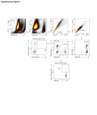
Time curves illustrating sialylation, bisection, fucosylation, and DAS28-CRP disease activity in pregnant rheumatoid arthritis patients based on ACPA IgG galactosylation. The figures show changes in glycosylation patterns across IgG subclasses and disease activity in ACPA-positive and ACPA-negative patients during pregnancy and post-delivery.

Download Presentation

Please find below an Image/Link to download the presentation.
The content on the website is provided AS IS for your information and personal use only. It may not be sold, licensed, or shared on other websites without obtaining consent from the author.If you encounter any issues during the download, it is possible that the publisher has removed the file from their server.
You are allowed to download the files provided on this website for personal or commercial use, subject to the condition that they are used lawfully. All files are the property of their respective owners.
The content on the website is provided AS IS for your information and personal use only. It may not be sold, licensed, or shared on other websites without obtaining consent from the author.
E N D
Presentation Transcript
SUPPLEMENTAL FIGURES to the manuscript entitled ACPA IgG galactosylation associates with disease activity in pregnant rheumatoid arthritis patients by Bondt et al. Supplemental Figure S1 Time curves sialylation Supplemental Figure S2 Time curves bisection Supplemental Figure S3 Time curves fucosylation Supplemental Figure S4 Time curves DAS28-CRP
Sialylation IgG1 IgG2/3 15 15 % % 10 10 5 5 IgG4 15 % 10 5 Supplemental Figure S1 Sialylation time curves for IgG1, IgG2/3 and IgG4. The ACPA glycosylation (red line, circle markers) is depicted together with total IgG of ACPA+ individuals (yellow line, triangle markers), ACPA- patients (grey line, square markers) and healthy controls (blue line, cross markers). Error bars represent standard errors.
Bisection IgG1 IgG2/3 15 15 % % 10 10 5 5 Supplemental Figure S2 Bisection time curves for IgG1 and IgG2/3. The ACPA glycosylation (red line, circle markers) is depicted together with total IgG of ACPA+ individuals (yellow line, triangle markers), ACPA- patients (grey line, square markers) and healthy controls (blue line, cross markers). Error bars represent standard errors.
Fucosylation IgG1 98 96 % 94 92 90 Supplemental Figure S3 Fucosylation time curves for IgG1. The ACPA glycosylation (red line, circle markers) is depicted together with total IgG of ACPA+ individuals (yellow line, triangle markers), ACPA- patients (grey line, square markers) and healthy controls (blue line, cross markers). Error bars represent standard errors.
DAS28-CRP 3.8 DAS28-CRP 3.2 2.6 PC TM1 TM2 TM3 PP1 PP2 PP3 ACPA+ ACPA- Supplemental Figure S4 Disease activity in ACPA positive and negative patients. The mean disease activity during the course of pregnancy and after delivery is depicted for the patients which are ACPA-positive (orange line, triangle markers) and ACPA-negative (dark green line, square markers) patients. Error bars represent standard errors.