Structure and Function of the Eye: Overview
Αντίληψη (2016)
Όραση
Μαρία Κουτρομάνου
Structure of the Eye: Iris
•
The iris is similar to the diaphragm
in a camera
•
Your iris
widens
in dim light and
narrows
in bright light
•
The f-number of your eye varies
from f/2 (large opening) to f/8
(small opening)
•
Compare this to the range of an
average camera lens, which may
have f-numbers from f/2.8 to f/22.
Structure of the Eye: Iris
•
The range of intensities that your eye
can respond to is a factor of 10
13
•
The main function of the iris is
not
to
control the intensity of light coming
into your eye
•
Main functions of iris
–
Reduce aberrations, sharpen
image
–
Increase depth of field
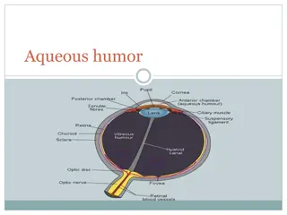
Structure of the Eye: Cornea and Lens
Cornea
•
There are two lenses in your eye, the cornea and the eyelens.
•
The cornea, the front surface of the eye, does most of the
focusing in your eye
•
The eyelens provides adjustable fine-tuning of the focus
Eyelens
Structure of the Eye: Cornea and Lens
air: n = 1
cornea: n ≈ 1.4
humors: n ≈ 1.3
eyelens: n ≈ 1.4
•
This is because the cornea-air surface has a large change in the
index of refraction, so light bends a lot
•
The power of the cornea lens is ~43 diopters (focal length 2.3 cm)
•
The eyelens is surrounded by the humors, which have a very
similar index of refraction as the lens itself.
The Eyelens: Accommodation
•
The eyelens changes its focal length by
changing its shape. Ligaments pull on the
lens to change the amount of “bulge”
Eyelens: Accommodation
Muscles contract,
ligaments relax, more
bulge, more bending
power, shorter focal
length
Muscles relax, ligaments
contract, less bulge, less
bending power, longer
focal length
Ligaments
Eyelens
How Your Eyelens Focuses
•
Your eyelens has
a
small depth of field
–
You can't see something close and far with both objects in
focus at the same time
•
Hold out your thumb about a foot away from your eye
–
Then, alternately focus on thumb and me (right above your
thumb)
•
Note that you cannot see
both
me
and
your thumb
sharply (in focus) at the same time
–
You focus on one or the other by changing the bulge of your
eyelens
Κοντινά αντικείμενα
σύγκλιση ματιών
Χρειάζεται διαστολή κόρης για να εστιαστεί η ακτίνα περισσότερο στη
φοβία, αλλιώς θολή εικόνα
Αλλαγή φακού: συστολή ακτινωτού σώματος
πιο στρογγυλός φακός
Χαλαρό ακτινωτό σώμα επίπεδος φακός καλή όραση για μακρινά
αντικείμενα
Μυωπία: για μακρινά αντικείμενα ο φακός δεν είναι επίπεδος σύγκλιση
εικόνας πριν τον αμφιβληστροειδή (ενώ στο φυσιολογικό μάτι στον
αμφιβληστροειδή)
Υπερμετρωπία: εστίαση φωτινών ακτίνων μετά τον αμφιβληστροειδή
https://www.youtube.com/watch?v=nbwPPcwknPU
(3:44-5:48)
https://www.youtube.com/watch?v=Av1ZiN9P01s
http://castle.eiu.edu/~ddavis/chapter_19/ch19_2.htm
less bulgy, longer
f
thumb is out of focus
less bulgy, longer
f
professor is in focus
Γιατί ανεστραμμένο είδωλο στον αμφιβληστροειδή;
-
Διάθλαση φωτός από τον φακό
αντιστροφή εικόνας στον εγκέφαλο
Ένωση εικόνων από τα δύο μάτια
cheshire cat
illusion:
ανταγωνιστικές εικόνες στα 2 μάτια, μία
στατική, μία με κίνηση, επικρατεί η κίνηση και
«χάνουμε» το στατικό πρόσωπο που βλέπει το
άλλο μάτι.
http://www.exploratorium.edu/snacks/cheshire-cat
Rods & cones: 4 key differences
between scotopic and photopic vision
•
Contrast sensitivity
•
Distribution of rods and cones
•
Spectral sensitivity of rods and cones
•
Sensitivity to light of rods and cones.
•
1. Rods are more sensitive than cones (x50)
•
2. There are more rods than cones (x10)
•
3. Ganglion cells have larger RFs for rods than
cones (i.e. more post-receptoral summation)
1. Contrast sensitivity functions at
three different light levels
Spatial Frequency (cycles/mm on retina)
Spatial frequency (cycles/degree)
Sensitivity (1/threshold contrast)
2. Distribution of rods and cones
visual eccentricity (deg)
spatial density
(cells/square mm)
macula lutea
cones
rods
retinal eccentricity (mm)
3. Spectral sensitivity curves for rod
and cone vision
Relative sensitivity
Wavelength (nm)
Purkinje effect
•
A shift in the colour appearance at dusk.
•
Reds
look
darker
,
blues
look
brighter
Dark adaptation curves
Low
Log. light sensitivity
High
Time in dark (min)
Cones
Rods
Light and Dark Adaptation
•
Light adaptation: Dark → light. Faster.
•
Dark adaptation: Light →dark. Slow.
Υποδεκτικά πεδία στον αμφιβληστροειδή
On-off, off-on
υποδεκτικά πεδία αμφιβληστροειδή
και για δίπολα και για γαγγλιακά κύτταρα: κέντρο
ανταγωνισμός με περιφέρεια.
Animation:
http://www.sumanasinc.com/webcontent/animations
/content/receptivefields.html
The structure and functions of the eye, including the iris, cornea, lens, and accommodation mechanisms are explained. Explore how your eye adjusts to light, focuses images, and perceives depth. Discover the similarities between the eye and a camera's mechanisms.

Download Presentation

Please find below an Image/Link to download the presentation.
The content on the website is provided AS IS for your information and personal use only. It may not be sold, licensed, or shared on other websites without obtaining consent from the author.If you encounter any issues during the download, it is possible that the publisher has removed the file from their server.
You are allowed to download the files provided on this website for personal or commercial use, subject to the condition that they are used lawfully. All files are the property of their respective owners.
The content on the website is provided AS IS for your information and personal use only. It may not be sold, licensed, or shared on other websites without obtaining consent from the author.
E N D
Presentation Transcript
Structure of the Eye: Iris The iris is similar to the diaphragm in a camera Your iris widens in dim light and narrows in bright light The f-number of your eye varies from f/2 (large opening) to f/8 (small opening) Compare this to the range of an average camera lens, which may have f-numbers from f/2.8 to f/22.
Structure of the Eye: Iris The range of intensities that your eye can respond to is a factor of 1013 The main function of the iris is not to control the intensity of light coming into your eye Main functions of iris Reduce aberrations, sharpen image Increase depth of field
Structure of the Eye: Cornea and Lens Cornea Eyelens There are two lenses in your eye, the cornea and the eyelens. The cornea, the front surface of the eye, does most of the focusing in your eye The eyelens provides adjustable fine-tuning of the focus
Structure of the Eye: Cornea and Lens cornea: n 1.4 eyelens: n 1.4 air: n = 1 humors: n 1.3 This is because the cornea-air surface has a large change in the index of refraction, so light bends a lot The power of the cornea lens is ~43 diopters (focal length 2.3 cm) The eyelens is surrounded by the humors, which have a very similar index of refraction as the lens itself.
The Eyelens: Accommodation The eyelens changes its focal length by changing its shape. Ligaments pull on the lens to change the amount of bulge
Eyelens: Accommodation Muscles contract, ligaments relax, more bulge, more bending power, shorter focal length Ligaments Eyelens Muscles relax, ligaments contract, less bulge, less bending power, longer focal length
How Your Eyelens Focuses Your eyelens has a small depth of field You can't see something close and far with both objects in focus at the same time Hold out your thumb about a foot away from your eye Then, alternately focus on thumb and me (right above your thumb) Note that you cannot see both me and your thumb sharply (in focus) at the same time You focus on one or the other by changing the bulge of your eyelens
, : : ( ) : https://www.youtube.com/watch?v=nbwPPcwknPU (3:44-5:48) https://www.youtube.com/watch?v=Av1ZiN9P01s http://castle.eiu.edu/~ddavis/chapter_19/ch19_2.htm
thumb is out of focus less bulgy, longer f less bulgy, longer f professor is in focus thumb is in focus more bulgy, shorter f professor is out of focus
; - cheshire cat illusion: 2 , , , . http://www.exploratorium.edu/snacks/cheshire-cat
Rods & cones: 4 key differences between scotopic and photopic vision Contrast sensitivity Distribution of rods and cones Spectral sensitivity of rods and cones Sensitivity to light of rods and cones.
1. Rods are more sensitive than cones (x50) 2. There are more rods than cones (x10) 3. Ganglion cells have larger RFs for rods than cones (i.e. more post-receptoral summation)
1. Contrast sensitivity functions at three different light levels Spatial frequency (cycles/degree) Sensitivity (1/threshold contrast) Spatial Frequency (cycles/mm on retina)
2. Distribution of rods and cones visual eccentricity (deg) spatial density (cells/square mm) macula lutea cones rods retinal eccentricity (mm)
3. Spectral sensitivity curves for rod and cone vision Relative sensitivity Wavelength (nm)
Purkinje effect A shift in the colour appearance at dusk. Reds look darker, blues look brighter
Dark adaptation curves Low Log. light sensitivity Rods Cones High Time in dark (min)
Light and Dark Adaptation Light adaptation: Dark light. Faster. Dark adaptation: Light dark. Slow.
On-off, off-on : . Animation: http://www.sumanasinc.com/webcontent/animations /content/receptivefields.html
")


")