Small Bowel Anatomy and Barium Meal Technique
ANATOMY
ANATOMY
OF
OF
SMALLBOWEL
SMALLBOWEL
&
&
TECHNIQUE OF
TECHNIQUE OF
BARIUM MEAL
BARIUM MEAL
FOLLOW THROUGH
FOLLOW THROUGH
SMALL BOWEL=>
SMALL BOWEL=>
Convoluted tube ext. from pylorus to I.C.
valve.
Avg length : 6 to 7 m.
Div into Duodenum, Jejunum, Ileum.
DUODENUM=>
DUODENUM=>
-‘C’ shaped tube measuring 25 cm long.
-Runs from pylorus to proxi. Jejunum.
-Curves around head of pancreas &
arches over aorta & I.V.C.
-First inch (2.5 cm) of duo. is
intraperitoneal, remainder is
retroperitoneal, being covered only ante.
by peritoneum.
-Duo. lacks mesentery.
Div. into 4 parts:-
1
st
part :-
continuous with pylorus.
- 5 cm long, first 2 cm is duo. cap (duo. bulb)
& is conical in shape.
- lies in transpyloric plane at L
1
level.
2
nd
part :-
8 cm long.
- runs inferiorly over hilus of rt. kidney & to
rt. of L
2
& L
3
vert.
- Posteromedially at jn. of upper ⅔
rd
& lower
⅓
rd
of second part is duo. papilla or
ampulla of vater-opening of common bile
duct & pancreatic ducts into gut.
3
rd
part :-
8 cm long.
- crosses from rt. to lt. curving forward over
rt. psoas muscle, IVC & aorta following
lower margin of head of pancreas.
4
th
part :-
4 cm long.
- runs upward & to lt. and lies on lt. psoas
muscle.
- Ends by turning forward at duodeno-
jejunal jn. at level of L
2
vert., where it is
held in position by Ligament of Treitz.
- Lig. of Treitz is a peritoneal fold which
ascends to rt. crus of diaphragm.
VASCULAR SUPPY OF DUO. =>
VASCULAR SUPPY OF DUO. =>
Duo. Cap.- small branches from hepatic &
gastroduodenal arteries.
Rest of duo.- sup. & inf. panereatico-duo.
arteries.
Venous bl. drains into prepyloric vn. &
then into portal vn.
JEJUNUM & ILEUM =>
JEJUNUM & ILEUM =>
-
Together are appro. 6.5 m in length.
-
Jej. begins at Lig. of Treitz (Duo.-jej.
Flexure) in lt. upper abdomen.
-
attached to post. abd. wall by its own fan
shaped mesentery which confers
considerable mobility & measures approx.
15 cm
.
STRUCTURE OF SMALL
STRUCTURE OF SMALL
INTESTINE=>
INTESTINE=>
In duo. mucous membrane of 1
st
part is smooth,
but that of rest of small bowel is broken up
into plicae circularis or valvulae conniventes
which are circular folds which encircle 2/3
rd
of
inner mucosal wall.
They become less numerous & less abundant in
ileum, until at termi. ileum, folds are almost
entirely absent.
RAD. DIFF. BET
RAD. DIFF. BET
N
N
JEJ. & ILEUM =>
JEJ. & ILEUM =>
JEJUNUM ILEUM
Proxi. 2/5
th
of sm. int. Distal 3/5
th
.
Position:-
Upper lt. & peri-umbilical Lower rt. hypogastric
region. & pelvic region.
Max.Diameter
:-
4cm. 3 cm.
Plicae circularis
:-
numerous & deeper set in jej. than in
ileum.
Feathery pattern
On BMFT :-
Present Less feathery or
absent.
Vascu. Supply :-
-
by branches of SMA – Jejunal, Ileal &
Ileocolic arteries.
- vns. drainage follows art. supply to drain
into sup. mes. vn.
Radiological investigations of Small
Radiological investigations of Small
bowel :
bowel :
1)
Plain abdominal radiography – Intestinal
obstruction & intestinal perforation.
2)
Contrast (Barium) studies.
3)
Angiography: Inv
n
of bleeding, detecting vascu.
Malformation & inv
n
of mesentric ischaemia.
4)
Radionuclide studies: To detect occult bleeding ,
Meckel’s diverticulum with gastric mucosa &
assessment of I.B.D.
5)
C.T. & M.R.I.: Limited role.
6)
U.S.G.:
Limited application d/t bowel gas.
: To detect intra-abd. Collections &
in children, diagnosis of
Pyloric stenosis , Appendicitis ,
Intussusception.
The major methods used for the
barium examination of the small
bowel are :-
1
) Small bowel follow through examination.
2) Dedicated small bowel follow through
examination.
3) Enteroclysis (or) small bowel enema.
4) Peroral pneumocolon.
5) Retrograde small bowel examination
.
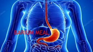
BARIUM MEAL FOLLOW THROUGH
BARIUM MEAL FOLLOW THROUGH
-Radiographic examination of GIT –
Oesophagus, Stomach, Duodenum, Small
bowel & Ileo-caecal junction by oral
administration of contrast media.
-So called because, is performed following
a Ba meal examination of oesophagus,
stomach and duodenum.
-Done to provide information about small
bowel calibre, its disposition, wall
thickness & distribution of volvulae
conniventes.
INDICATIONS=>
INDICATIONS=>
1) Patients who have low
suspicion
of small
bowel disease – abdominal pain and
diarrhoea.
2) Patients with
suspected
complete (or) near
complete small bowel obstruction.
3) Patients who are
suspected
of suffering from
Crohn’s disease.
4) Elderly pts. with jejunal diverticulosis &
present with malabsorption.
5) Patients who refuse placement of
nasogastric tube / failed intubation
.
CONTRA INDICATIONS=>
CONTRA INDICATIONS=>
1)
Colonic Obstruction
2)
Suspected perforation.
3)
Paralytic Ileus
.
CONTRAST =>
CONTRAST =>
Medium density Ba suspension (50-60% w/v)
containing a suspending agent ( to maintain its
stability & prevent flocculation ) is used.
When small bowel obstruction is suspected & colonic
obstruction is ruled out, Ba can be used to find
cause & site of lesion.
PREPARATION=>
PREPARATION=>
-
Colon should be cleaned by suitable
purgative.
-
Low fibre diet & high fluid intake – 48
hrs. prior to investigation.
-
NBM for 12 hrs. prior to investigation.
Small Bowel Follow Through =>
Small Bowel Follow Through =>
Initially 150ml of high density Ba and
effervescent agents are used to evaluate
oesophagus, stomach & duodenum by
double contrast examination.
Later 200 ml of Ba (20 – 25 %) followed
by 250 ml of Ba ( 40 – 45 %) is given.
Series of overhead radiographs obtained
at ½ hourly intervals till terminal ileum
is reached.
DEDICATED SMALL BOWEL
DEDICATED SMALL BOWEL
FOLLOW THROUGH =>
FOLLOW THROUGH =>
Single contrast technique.
Double contrast technique
.
SINGLE CONTRAST TECHNIQUE =>
SINGLE CONTRAST TECHNIQUE =>
Ba (600-900 ml) 50-60% is given &
patient is asked to drink as rapidly as
possible.
Patient put in right side dependent
position to aid gastric emptying.
Subsequent films taken at 15-30 min.
intervals till ileo-caecal junction is
opacified.
To demonstrate ileocaecal junction,
supine right side up is best position
since ileum enters caecum in postero-
medial part.
Always empty bladder prior to these spot
films.
4 spots of I.C. jn should be taken with varying
degrees of compression.
Compression applied on bowel loops to avoid
overlap & to efface the mucosa so that small
lesions may not be missed & mobility of the
loops can be well assessed.
Abnormalities must be shown in at least 2
spot films taken at different times to
demonstrate persistence of lesion.
*
Overlap of contrast filled bowel loops in
pelvis is often a problem & it can be
overcome by :-
- Table head down.
- 30
0
caudal angled view of pelvis.
- Emptying U. B. prior to filming the ileal loops.
*
Gastric & Bowel peristalsis may be
increased by:-
DRUGS:- Metoclopramide (M/cly used)
- Neostigmine
- Glucagon
- Cholecystokinin.
APPEARANCE IN SOME
CONDITIONS:-
Polyposis
–
Films with collapsed bowel loops
shows polyps.
Diverticulosis
– Delayed films may show
persistence of Ba in diverticulae.
Large Ulcers
–
Large collection of Ba seen in
delayed film after bowel loops have emptied
Ba
.
DOUBLE CONTRAST TECHNIQUE=>
DOUBLE CONTRAST TECHNIQUE=>
-
Similar to single contrast technique.
-
Gas producing agent (effervescent
powder) given when head of Ba column
reaches caecum.
-
Patient placed on left side slightly head
down to allow gas to leave stomach &
enter small bowel.
ADVANTAGES=>
Better distension.
Separation of loops.
Improved mucosal detail.
DISADVANTAGES=>
-Difficultly in interpretation by gas
bubbles
.
ADVANTAGES OF BMFT=>
ADVANTAGES OF BMFT=>
-
Easily performed.
-
No discomfort / intubation to patient unlike in
enteroclysis.
-
It is physiological process. Hence transit time
can be assessed.
DISADVANTAGES OF BMFT=>
DISADVANTAGES OF BMFT=>
-
Overlapping of Ba filled bowel loops in pelvis.
-
Poor distension of bowel loops.
-
Operator dependant.
-
Time consuming.
PER ORAL PNEUMOCOLON :-
-Used to evaluate distal ileum.
-When Ba has reached rt. & proximal
transverse colon ,air is insufflated into
rectum and refluxed into distal ileum.
-Colon preparation is imp.
-CROHN’S DISEASE & Ca CAECUM
visualized well.
REFLUX EXAMINATION
(RETROGRADE SMALL BOWEL
EXAM.:-
-Ba & Air refluxed through IC valve.
-Good view of terminal ileum.
-Now a days replaced by Enteroclysis
.
THANK YOU!
Exploring the anatomy of the small bowel, including the duodenum, jejunum, and ileum, as well as the technique of a barium meal follow-through. The small bowel is a convoluted tube extending from the pylorus to the ileocecal valve, averaging 6-7 meters in length. The duodenum, characterized by its C-shaped tube, has four distinct parts and receives vascular supply from various arteries. The jejunum and ileum together measure approximately 6.5 meters in length and have their own mesentery for mobility.

Download Presentation

Please find below an Image/Link to download the presentation.
The content on the website is provided AS IS for your information and personal use only. It may not be sold, licensed, or shared on other websites without obtaining consent from the author.If you encounter any issues during the download, it is possible that the publisher has removed the file from their server.
You are allowed to download the files provided on this website for personal or commercial use, subject to the condition that they are used lawfully. All files are the property of their respective owners.
The content on the website is provided AS IS for your information and personal use only. It may not be sold, licensed, or shared on other websites without obtaining consent from the author.
E N D
Presentation Transcript
ANATOMY OF SMALLBOWEL & TECHNIQUE OF BARIUM MEAL FOLLOW THROUGH
SMALL BOWEL=> Convoluted tube ext. from pylorus to I.C. valve. Avg length : 6 to 7 m. Div into Duodenum, Jejunum, Ileum.
DUODENUM=> - C shaped tube measuring 25 cm long. -Runs from pylorus to proxi. Jejunum. -Curves around head of pancreas & arches over aorta & I.V.C. -First inch (2.5 cm) of duo. is intraperitoneal, remainder is retroperitoneal, being covered only ante. by peritoneum. -Duo. lacks mesentery.
Div. into 4 parts:- 1st part :- continuous with pylorus. - 5 cm long, first 2 cm is duo. cap (duo. bulb) & is conical in shape. - lies in transpyloric plane at L1 level. 2nd part :- 8 cm long. - runs inferiorly over hilus of rt. kidney & to rt. of L2 & L3 vert. - Posteromedially at jn. of upper rd & lower rd of second part is duo. papilla or ampulla of vater-opening of common bile duct & pancreatic ducts into gut.
3rd part :- 8 cm long. - crosses from rt. to lt. curving forward over rt. psoas muscle, IVC & aorta following lower margin of head of pancreas. 4th part :- 4 cm long. - runs upward & to lt. and lies on lt. psoas muscle. - Ends by turning forward at duodeno- jejunal jn. at level of L2 vert., where it is held in position by Ligament of Treitz. - Lig. of Treitz is a peritoneal fold which ascends to rt. crus of diaphragm.
VASCULAR SUPPY OF DUO. => Duo. Cap.- small branches from hepatic & gastroduodenal arteries. Rest of duo.- sup. & inf. panereatico-duo. arteries. Venous bl. drains into prepyloric vn. & then into portal vn.
JEJUNUM & ILEUM => -Together are appro. 6.5 m in length. -Jej. begins at Lig. of Treitz (Duo.-jej. Flexure) in lt. upper abdomen. -attached to post. abd. wall by its own fan shaped mesentery which confers considerable mobility & measures approx. 15 cm.
STRUCTURE OF SMALL INTESTINE=> In duo. mucous membrane of 1st part is smooth, but that of rest of small bowel is broken up into plicae circularis or valvulae conniventes which are circular folds which encircle 2/3rd of inner mucosal wall. They become less numerous & less abundant in ileum, until at termi. ileum, folds are almost entirely absent.
RAD. DIFF. BETN JEJ. & ILEUM => JEJUNUM ILEUM Proxi. 2/5th of sm. int. Distal 3/5th . Position:-Upper lt. & peri-umbilical Lower rt. hypogastric region. & pelvic region. Max.Diameter:- 4cm. 3 cm. Plicae circularis :- numerous & deeper set in jej. than in ileum. Feathery pattern On BMFT :-Present Less feathery or absent.
Vascu. Supply :- - by branches of SMA Jejunal, Ileal & Ileocolic arteries. - vns. drainage follows art. supply to drain into sup. mes. vn.
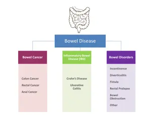
Radiological investigations of Small bowel : 1) Plain abdominal radiography Intestinal obstruction & intestinal perforation. 2) Contrast (Barium) studies. 3) Angiography: Invn of bleeding, detecting vascu. Malformation & invn of mesentric ischaemia. 4) Radionuclide studies: To detect occult bleeding , Meckel s diverticulum with gastric mucosa & assessment of I.B.D. 5) C.T. & M.R.I.: Limited role. 6) U.S.G.: Limited application d/t bowel gas. : To detect intra-abd. Collections & in children, diagnosis of Pyloric stenosis , Appendicitis , Intussusception.
The major methods used for the barium examination of the small bowel are :- 1) Small bowel follow through examination. 2) Dedicated small bowel follow through examination. 3) Enteroclysis (or) small bowel enema. 4) Peroral pneumocolon. 5) Retrograde small bowel examination.
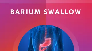
BARIUM MEAL FOLLOW THROUGH -Radiographic examination of GIT Oesophagus, Stomach, Duodenum, Small bowel & Ileo-caecal junction by oral administration of contrast media. -So called because, is performed following a Ba meal examination of oesophagus, stomach and duodenum. -Done to provide information about small bowel calibre, its disposition, wall thickness & distribution of volvulae conniventes.
INDICATIONS=> 1) Patients who have low suspicion of small bowel disease abdominal pain and diarrhoea. 2) Patients with suspected complete (or) near complete small bowel obstruction. 3) Patients who are suspected of suffering from Crohn s disease. 4) Elderly pts. with jejunal diverticulosis & present with malabsorption. 5) Patients who refuse placement of nasogastric tube / failed intubation.
CONTRA INDICATIONS=> 1) Colonic Obstruction 2) Suspected perforation. 3) Paralytic Ileus. CONTRAST => Medium density Ba suspension (50-60% w/v) containing a suspending agent ( to maintain its stability & prevent flocculation ) is used. When small bowel obstruction is suspected & colonic obstruction is ruled out, Ba can be used to find cause & site of lesion.
PREPARATION=> - Colon should be cleaned by suitable purgative. - Low fibre diet & high fluid intake 48 hrs. prior to investigation. - NBM for 12 hrs. prior to investigation.
Small Bowel Follow Through => Initially 150ml of high density Ba and effervescent agents are used to evaluate oesophagus, stomach & duodenum by double contrast examination. Later 200 ml of Ba (20 25 %) followed by 250 ml of Ba ( 40 45 %) is given. Series of overhead radiographs obtained at hourly intervals till terminal ileum is reached.
DEDICATED SMALL BOWEL FOLLOW THROUGH => Single contrast technique. Double contrast technique. SINGLE CONTRAST TECHNIQUE => Ba (600-900 ml) 50-60% is given & patient is asked to drink as rapidly as possible. Patient put in right side dependent position to aid gastric emptying.
Subsequent films taken at 15-30 min. intervals till ileo-caecal junction is opacified. To demonstrate ileocaecal junction, supine right side up is best position since ileum enters caecum in postero- medial part.
ORDER POSITIONING PURPOSE FIRST RIGHT SIDE DOWN DEPENDANT TO AID GASTRIC EMPTYING SECOND PRONE TO SEPARATE BOWEL LOOPS THIRD RIGHT SIDE UP (SUPINE) TO VISUALIZE I.C. Jn.
Always empty bladder prior to these spot films. 4 spots of I.C. jn should be taken with varying degrees of compression. Compression applied on bowel loops to avoid overlap & to efface the mucosa so that small lesions may not be missed & mobility of the loops can be well assessed. Abnormalities must be shown in at least 2 spot films taken at different times to demonstrate persistence of lesion.
* Overlap of contrast filled bowel loops in pelvis is often a problem & it can be overcome by :- - Table head down. - 300 caudal angled view of pelvis. - Emptying U. B. prior to filming the ileal loops. * Gastric & Bowel peristalsis may be increased by:- DRUGS:- Metoclopramide (M/cly used) - Neostigmine - Glucagon - Cholecystokinin.
APPEARANCE IN SOME CONDITIONS:- Polyposis Films with collapsed bowel loops shows polyps. Diverticulosis Delayed films may show persistence of Ba in diverticulae. Large Ulcers Large collection of Ba seen in delayed film after bowel loops have emptied Ba.
DOUBLE CONTRAST TECHNIQUE=> - Similar to single contrast technique. - Gas producing agent (effervescent powder) given when head of Ba column reaches caecum. - Patient placed on left side slightly head down to allow gas to leave stomach & enter small bowel.
ADVANTAGES=> Better distension. Separation of loops. Improved mucosal detail. DISADVANTAGES=> -Difficultly in interpretation by gas bubbles.
ADVANTAGES OF BMFT=> - Easily performed. - No discomfort / intubation to patient unlike in enteroclysis. - It is physiological process. Hence transit time can be assessed. DISADVANTAGES OF BMFT=> - Overlapping of Ba filled bowel loops in pelvis. - Poor distension of bowel loops. - Operator dependant. - Time consuming.
PER ORAL PNEUMOCOLON :- -Used to evaluate distal ileum. -When Ba has reached rt. & proximal transverse colon ,air is insufflated into rectum and refluxed into distal ileum. -Colon preparation is imp. -CROHN S DISEASE & Ca CAECUM visualized well.
REFLUX EXAMINATION (RETROGRADE SMALL BOWEL EXAM.:- -Ba & Air refluxed through IC valve. -Good view of terminal ileum. -Now a days replaced by Enteroclysis.