Rhinosinusitis: Symptoms, Causes, and Classification
Rhinosinusitis, formerly known as sinusitis, is an inflammation affecting the paranasal sinuses, nasal mucosa, and nasal cavity. This condition is categorized based on the duration of symptoms as acute, subacute, or chronic. Various factors contribute to the development of rhinosinusitis, including infections, nasal deformities, adenoid enlargement, environmental factors, and more. Acute rhinosinusitis can be either bacterial or viral, with distinct clinical manifestations such as pressure over infected sinuses, nasal discharge, headache, and fever.

Download Presentation

Please find below an Image/Link to download the presentation.
The content on the website is provided AS IS for your information and personal use only. It may not be sold, licensed, or shared on other websites without obtaining consent from the author.If you encounter any issues during the download, it is possible that the publisher has removed the file from their server.
You are allowed to download the files provided on this website for personal or commercial use, subject to the condition that they are used lawfully. All files are the property of their respective owners.
The content on the website is provided AS IS for your information and personal use only. It may not be sold, licensed, or shared on other websites without obtaining consent from the author.
E N D
Presentation Transcript
Learning Objectives Learning Objectives On completion of this chapter, the student will be able to identify Sinusitis Tonsillitis Otitis media
Rhinosinusitis Rhinosinusitis
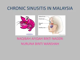
DEFINITION 1- Rhinosinusitis:- formerly called sinusitis, is an inflammation of the paranasal sinuses, nasal mucosa & nasal cavity. The American Academy of Otolaryngology Head and Neck Surgery Foundation recommends the use of the term Rhinosinusitis because sinusitis is almost always accompanied by inflammation of the nasal mucosa. Inflammation of the membranous lining of 1 of the sinuses, leads to sinus cavity obstruction.
Rhinosinusitis is classified by the duration of symptoms: 1- Acute (less than 4 weeks). 2- Subacute (4 to 12 weeks). 3- Chronic (more than 12 weeks).
Causes and Risk Factors 1- Bacterial or viral infection. 2- Nasal deformities (nasal contaminated) conditions polyps or a deviated septum. 3- Enlargement of adenoids. 4- Foreign objects. 5- Tooth infection. 6- Trauma to the nose. 7- Swimming in contaminated conditions. 8- Environmental factors. 9- Chemicals agents. 10- Tumors. 11- Smoking.
Acute Rhinosinusitis Acute rhinosinusitis is classified as acute bacterial rhinosinusitis (ABRS) or acute viral rhinosinusitis (AVRS). Recurrent acute rhinosinusitis is characterized by four or more acute episodes of ABRS per year. 1- Infectious sinusitis is spread through viral, fungal, or bacterial infections. Typical pathogens include Streptococcus pneumonia, Haemophilus influenza, and less commonly Staphylococcus aureus, and Moraxella catarrhalis. 2- Noninfectious sinusitis cannot be spread because they are by causes specific to the person.
Clinical Manifestations 1- Pressure, pain, or tenderness over infected sinus areas (Pain over the cheek, with radiation to teeth). 2- Persistent nasal discharge sometimes yellow or green or nasal congestion. 3- Headache and high fever. 4- Mouthing breathing, snoring. 5- Sore throat, bad breath. 6- Mucosal polyps (hyperplastic sinusitis). 7- Swelling of turbinate's and fatigue.
Diagnostic Tests A careful history and physical examination guide diagnosis generally. Flexible endoscopic culture techniques and sensitivity testing of purulent nasal drainage show the causative bacterial organism. Transillumination of the affected area may reveal a decrease in the transmission of light with rhinosinusitis. Imaging -Sinus X-rays show cloudiness in the affected sinus, air-fluid levels, or thickened mucosal lining. -Ultrasonography and computed tomography scans show recurrent or chronic sinusitis.
Medical Management Treatment of acute rhinosinusitis depends on the cause; a 5 to 7-day course of antibiotics is prescribed for bacterial cases. Antibiotics should be given as soon as the diagnosis of Acute Bacterial Rhinosinusitis (ABRS) is established. Amoxicillin clavulanic acid (Augmentin). Cefuroxime. And moxifloxacin Decongestants (guaifenesin/pseudoephedrine) or nasal saline sprays can increase the patency of the ostiomeatal unit and improve drainage of the sinuses. Topical decongestants should not be used for longer than 3 or 4 days. Pain relief.
Surgical Management If standard medical therapy fails and symptoms persist, Functional Endoscopic Sinus Surgery (FESS) may be indicated to correct structural deformities that obstruct the ostia (openings) of the sinuses. FESS is a minimally invasive surgical procedure that is associated with reduced postoperative discomfort and improvement in the patient s quality of life. In particular, FESS is associated with either complete or moderate relief of symptoms in more than 85% to 91% of patients. Some of the specific procedures performed include excising and cauterizing nasal polyps, correcting a deviated septum, incising and draining the sinuses, aerating the sinuses, and removing tumors. Antimicrobial agents may be given before and after surgery. Computer- assisted or computer-guided surgery is used to increase the precision of the surgical procedure and minimize complications.
Complications of Sinusitis Local complications include osteomyelitis and mucocele (cyst of the paranasal sinuses). Osteomyelitis requires prolonged antibiotic therapy and at times removal of necrotic bone. Intracranial complications, although rare, include cavernous sinus thrombosis, meningitis, brain abscess, ischemic brain infarction, and severe orbital cellulitis.
2- Tonsillitis The tonsils are composed of lymphatic tissue and are situated on each side of the oropharynx. Acute tonsillitis can be confused with pharyngitis. Chronic tonsillitis is less common and may be mistaken for other disorders such as allergy, asthma, and rhinosinusitis.
Adenoiditis The adenoids or pharyngeal tonsils consist of lymphatic tissue near the center of the posterior wall of the nasopharynx. Infection of the adenoids frequently accompanies acute tonsillitis. Frequently occurring bacterial pathogens include group A beta- hemolytic streptococcal (GABHS), the most common organism. The most common viral pathogen is Epstein Barr virus, although cytomegalovirus may also cause tonsillitis and adenoiditis. Often thought of as a childhood disorder, tonsillitis can occur in adults.
TONSILLITIS Inflammation of the tonsils. It may be acute or chronic More common in children than adults. CAUSES Tonsillitis can occur through infected droplets with bacterial or viral such as:- Streptococcus bacteria. Adenovirus.
Predisposing factors Foods with artificial colors and preservatives. Cold foods, cold drinks, ice creams. Changes in weather. Air pollution. Sour fruits.
Clinical Manifestations 1- Red and swollen tonsils. 3- Enlarged lymph nodes in the neck. 2- White spots on the tonsils. 4- Fever. 5-Mild to a severe sore throat. 6- Difficulty swallowing. 7- Mouth-breathing foul-smelling breath. 8- Muscle and joint pain. 9- Chills. 10- Malaise. 11- Headache. 12- Pain, commonly referred to as the ears.
Nursing Process To Pt. With Upper Airway Infection Ineffective airway clearance related to excessive mucus production secondary to retained secretions and inflammation. Planning:- maintenance of a patent airway. Acute pain related to upper airway irritation secondary to an infection. Planning:- Relief of pain. Increase body temperature related to the infectious process. Planning:- The patient will have a temperature lower than (39.4 C).
NURSING INTERVENTION Increasing fluid intake helps thin the mucus. Use of room vaporizers or steam inhalation also loosens secretions and reduces inflammation of the mucous membranes. Keep the patient with sinusitis or rhinitis in the upright position to enhance drainage from the sinuses. Use hot packs to relieve the congestion of sinusitis and promote drainage, and warm-water gargles or irrigations to relieve the pain of a sore throat.
NURSING INTERVENTION Provide a rest period to relieve the generalized discomfort and fever that accompany many upper airway conditions. Instructs the patient in general hygiene techniques to prevent the spread of infection. Use topical or systemic medications to relieve nasal or throat congestion as prescribed. Encourages the patient to take analgesics, such as acetaminophen with codeine, as prescribed, to relieve discomfort.
NURSING INTERVENTION Tonsillectomy (with or without adenoidectomy) continues to be a commonly performed surgical procedure and remains the treatment of choice for patients with chronic tonsillitis. Indications of Tonsillectomy The patient has had repeated episodes of tonsillitis despite antibiotic therapy; hypertrophy of the tonsils and adenoids that could cause obstruction and obstructive sleep apnea; repeated attacks of purulent otitis media; and suspected hearing loss due to serous otitis media that has occurred in association with enlarged tonsils and adenoids. chronic nasal airway obstruction, chronic rhinorrhea, obstruction of the eustachian tube with related ear infections, and abnormal speech. The patient has developed a peritonsillar abscess that occludes the pharynx, making swallowing difficult and endangering the patency of the airway (particularly during sleep).
Otitis Media Acute otitis media (AOM) is an acute infection of the middle ear, lasting less than 6 weeks. Pathogens that cause AOM are usually bacterial or viral and enter the middle ear after Eustachian tube dysfunction caused by obstruction related to upper respiratory infections, inflammation of surrounding structures (e.g., rhino sinusitis, adenoid hypertrophy), or allergic reactions (e.g., allergic rhinitis).
CAUSES Bacterial infection (streptococcus pneumonia, &Haemophilus influenza). Chronic upper respiratory infections. Other diseases such as (down syndrome, cystic fibrosis, and cleft palate). Chronic exposure to secondhand cigarette smoke.
PATHOPHYSIOLOGY Bacteria can enter after eustachian tube dysfunction caused by obstruction from contaminated secretions in the nasopharynx to the middle ear from a tympanic membrane perforation. It can result from inflammation of surrounding structures (eg, rhinosinusitis, adenoid hypertrophy), or allergic reactions (eg, allergic rhinitis). Conductive hearing loss resulting in a purulent exudate which is usually present in the middle ear.
CLINICAL MANIFESTATIONS 1- Otalgia. 2- Discharge from the ear. 3- Fever. 4. Hearing loss. 5- Erythematous of the tympanic membrane and often bulging. 6- Headache. 7- Irritability.
MEDICAL MANAGEMENT Suctioning of the ear under otoscopic guidance. Instillation of antibiotic drops or application of antibiotic powder is used to treat purulent discharge. Systemic antibiotics are prescribed only in cases of acute infection. Surgical management Tympanoplasty. Mastoidectomy.
Surgical Management A myringotomy (i.e., tympanotomy) is an incision in the tympanic membrane. The tympanic membrane is numbed with a local anesthetic agent such as phenol or by iontophoresis (i.e., in which electrical current flows through a lidocaine and epinephrine solution to numb the ear canal and tympanic membrane). The procedure is painless and takes less than 15 minutes. Under microscopic guidance, an incision is made through the tympanic membrane to relieve pressure and drain serous or purulent fluid from the middle ear. Normally, this procedure is unnecessary for treating AOM, but it may be performed if pain persists.
Myringotomy also allows the drainage to be analyzed (by culture and sensitivity testing) so that the infecting organism can be identified and appropriate antibiotic therapy prescribed. The incision heals within 24 to 72 hours. If AOM recurs and there is no contraindication, a ventilating, or pressure-equalizing, the tube may be inserted. The ventilating tube, which temporarily takes the place of the Eustachian tube in equalizing pressure, is retained for 6 to 18 months. The ventilating tube is then extruded with normal skin migration of the tympanic membrane, with the hole healing in nearly every case. Ventilating tubes are used to treat recurrent episodes of AOM.
Ossiculoplasty Ossiculoplasty is the surgical reconstruction of the middle ear bones to restore hearing. Prostheses made of materials such as Teflon, stainless steel, and hydroxyapatite are used to reconnect the ossicles, thereby re-establishing the sound conduction mechanism. However, the greater the damage, the lower the success rate for restoring normal hearing. Mastoidectomy The objectives of mastoid surgery are to remove the cholesteatoma, gain access to diseased structures, and create a dry (noninfected) and healthy ear. If possible, the ossicles are reconstructed during the initial surgical procedure. Occasionally, extensive disease or damage dictates that this be performed as part of a two-stage operation.
A mastoidectomy is usually performed through a postauricular incision. Infection is eliminated by removing the mastoid air cells. A second mastoidectomy may be necessary to check for recurrent or residual cholesteatoma. The hearing mechanism may be reconstructed at this time. The success rate for correcting this conductive hearing loss is approximately 75%. Surgery is usually performed in an outpatient setting. The patient has a mastoid pressure dressing, which can be removed 24 to 48 hours after surgery. Although infrequently injured, the facial nerve, which runs through the middle ear and mastoid, is at some risk for injury during mastoid surgery. As the patient awakens from anesthesia, any evidence of facial paresis should be reported to the primary provider.
Nursing Interventions Providing preoperative patient education. Reducing anxiety. Maintaining a patent airway. Promoting alternative communication methods. Promoting adequate nutrition and hydration. Promoting positive body image and self-esteem. Promoting self-care management. Monitoring and managing potential complications.
Serous Otitis Media Middle ear effusion, or serous otitis media, involves the presence of fluid, without evidence of active infection, in the middle ear. In theory, this fluid results from a negative pressure in the middle ear caused by Eustachian tube obstruction. When this condition occurs in adults, an underlying cause for the Eustachian tube dysfunction must be sought. Middle ear effusion is frequently seen in patients after radiation therapy or barotrauma and in patients with Eustachian tube dysfunction from a concurrent upper respiratory infection or allergy. Barotrauma results from sudden pressure changes in the middle ear caused by changes in barometric pressure, as in scuba diving or airplane descent. A carcinoma (e.g., nasopharyngeal cancer) obstructing the Eustachian tube should be ruled out in adults with persistent unilateral serous otitis media.
Clinical Manifestations Patients may complain of hearing loss, fullness in the ear or a sensation of congestion, or popping and crackling noises that occur as the Eustachian tube attempts to open. The tympanic membrane appears dull on otoscopy, and air bubbles may be visualized in the middle ear. Usually, the audiogram shows conductive hearing loss.
Management Serous otitis media need not be treated medically unless infection (i.e., AOM) occurs. If the hearing loss associated with middle ear effusion is significant, a myringotomy can be performed, and a tube may be placed to keep the middle ear ventilated. Corticosteroids in small doses may decrease the edema of the Eustachian tube in cases of barotrauma. Decongestants have not proved to be effective. A Valsalva maneuver, which forcibly opens the Eustachian tube by increasing nasopharyngeal pressure, may be cautiously performed; this maneuver may cause worsening pain or perforation of the tympanic membrane.
Chronic Otitis Media Chronic otitis media is recurrent AOM that causes irreversible tissue pathology. Chronic infections of the middle ear damage the tympanic membrane, destroy the ossicles, and involve the mastoid but are rare in developed countries. Clinical Manifestation Symptoms may be minimal, with varying degrees of hearing loss and persistent or intermittent, foul-smelling otorrhea. Pain is not usually experienced, except in cases of acute mastoiditis, when the postauricular area is tender and may be erythematous and oedematous. The otoscopic examination may show a perforation, and cholesteatoma can be identified as a white mass behind the tympanic membrane or coming through to the external canal from a perforation.
Summary Rhinosinusitis:- formerly called sinusitis, is an inflammation of the paranasal sinuses, nasal mucosa & nasal cavity. Tonsillitis is inflammation of the tonsils. Acute otitis media is an acute middle ear infection lasting less than 6 weeks.
Reference Textbook of medical-surgical nursing Description: 14th edition. | Philadelphia : Wolters Kluwer, [2018], ISBN 9781496355157 (2-volume American edition).
Questions 1- A patient visiting the clinic is diagnosed with acute sinusitis. To promote sinus drainage, the nurse should instruct the patient to perform which of the following? A) Apply a cold pack to the affected area. B) Apply a mustard poultice to the forehead. C) Perform postural drainage. D) Increase fluid intake. 2- Classifications of sinusitis include the following, except: A- Acute. B- Subacute. C- Chronic. D- Recurrent. 3-Subacute rhinosinusitis last for: A- Less than 4 weeks. B- 4 to 12 weeks. C- More than 12 weeks. D- 16-20 weeks.
Questions 4- The perioperative nurse has admitted a patient who has just undergone a tonsillectomy. The nurses postoperative assessment should prioritize which of the following potential complications of this surgery? A) Difficulty ambulating B) Hemorrhage C) Infrequent swallowing D) Bradycardia 5-Acute otitis media is an acute infection of the middle ear, lasting less than . A- 6 weeks B- 5 weeks C- 4 weeks D- 3 weeks 6-Supportive drugs for viral sinusitis include, except: A- Antipyretic. B- Normal Saline. C- Vitamin C. D- Amoxicillin.
Questions 7- Tonsils are on the back of the throat. Where are the adenoids located? A- Under the tongue B. In the ears C. Behind the nose and roof of the mouth D. None of the above 8. What do your tonsils and adenoids do? A- They don't do anything B. They help your nose with the sense of smell C. They are part of your immune system D. None of the above 9. Which of these is a common problem with tonsils? A- They make an infection worse B. They become swollen C. They can become infected themselves D. B and C
Questions 10- The nurse is providing patient teaching to a patient diagnosed with acute rhinosinusitis. For what possible complication should the nurse teach the patient to seek immediate follow-up? A) Periorbital edema. B) Headache unrelieved by OTC medications. C) Clear drainage from the nose. D) Blood-tinged mucus when blowing the nose.