Recombinant DNA Technology and Plasmid Vectors

Old and Primrose
Chapter 4
Plasmids of Bacterial Origins
Insertional inactivation
is a technique used in recombinant DNA
technology. In this procedure, a fragment of foreign DNA is inserted into a restriction
site inside a gene, hence causing the gene to turn non-functional or in an inactivated
state.
b) Recombinant vector having insert: Loss of Marker gene function
Plasmid vectors
Plasmids are autonomously replicating circular, double
stranded DNA molecules found in bacteria.
They have their own origin of replication (
ori region), and
can replicate independently of the host chromosome.
Plasmid vectors are often used for cloning DNA segments of
small size (upto 10 kilobases).
Insert capacity: 0.1 to 10 Kb
Old and primrose, chapter 4
The plasmid pBR322 is one of the most commonly used
E. coli
cloning vectors. pBR322 is
4361 bp in length and contains: (1) the replicon
rep
responsible for the replication of plasmid
(source – plasmid pMB1); (2)
rop
gene coding for the Rop protein, which promotes conversion
of the unstable RNA I – RNA II complex to a stable complex and serves to decrease copy
number (source – plasmid pMB1); (3)
bla
gene, coding for beta-lactamase that confers
resistance to ampicillin (source – transposon Tn3/RSF2124); (4)
tet
gene, encoding tetracycline
resistance protein (source – plasmid pSC101).
pBR322
The first plasmid vector that has been constructed
artificially is pBR322.
It is named after the scientists
Bolivar and Rodriguez
who
constructed it in 1977.
It is 4361bp in size and most widely used cloning vector.
It has an origin of replication derived from a colicin-
resistance plasmid (
ColE1
).
Medium copy Number (around 20 copies per cell)
Plasmid pBR322 carries two selectable markers viz. genes
for resistance to ampicillin (Ap
r)
and tetracycline (Tc
r
).
Several (over 40 enzymes) unique RE sites are present in pBR322
plasmid vector.
A total of 11 unique restriction sites (Example: Eco
RV,
Bam
HI,
Sph
I,
Sal
I, and
Nru
I are present within the gene coding for
tetracycline resistance, two sites (
Hin
dIII and
Cla
I) within the
promoter of the tetracycline resistance gene and the six sites
(Example:
Pst
I,
Pvu
I and
Sca
I) within the
β
lactamase gene that
provide resistance to ampicillin.
When a foreign DNA segment is inserted in any of these genes,
the antibiotic resistance by that particular gene is lost. This is
called insertional inactivation.
For instance, insertion of a restriction fragment in the
SalI site of
the Tc
r
gene inactivates that gene.
One can still select for Ap
r
colonies, and then screen to see which
ones have lost Tc
r
.
Recombinant selection with pBR322 [
Cloning in pBR322]
Cloning in pBR322
Screening of Clones with insert
Strategy for selecting host cells that have been transformed with
pBR322:
(1)
The transformation mixture, which contains three cell types, viz., nontransformed
cells, cells with the intact original plasmid, and cells with DNA cloned into the BamHI
site of pBR322, is plated on complete medium with ampicillin.
(2)
The mixture is diluted beforehand to ensure separate colonies are formed on the
agar. The nontransformed cells (Amp
s
) are killed.
(3)
The cells with the intact plasmid and cloned DNA– plasmid constructs are Amp
r
and
therefore form colonies. Samples of the surviving colonies on the ampicillin plate are
transferred to a plate with complete medium and tetracycline, keeping the same
position of each colony on the second plate, i.e., replica plating. Only cells with
intact plasmids (Tet
r
) will form colonies in the presence of tetracycline.
(4)
The colonies that did not grow on the tetracycline plate (dashed circles) but grew on
the ampicillin plate carry pBR322 with DNA that was cloned into the BamHI site. The
colonies with cloned DNA inserts are picked from the original plate, pooled, and
grown.
The use of plasmid pBR322 as a vector:
isolation of DNA fragments which carry promoters
Cloning into the
HindIII site of pBR322 generally
results in loss
of tetracycline resistance.
However, in some recombinants, Tc
R
is retained or even
increased. This is because the
HindIII site lies within
the promoter rather than the coding sequence.
Thus whether or not insertional inactivation occurs
depends on whether the cloned DNA carries a
promoter-like sequence able to initiate transcription
of the
Tc
R
gene.
α
-complementation and blue-white screening
Ref: Chapter 3, Old and primrose
α
-complementation
The
lac Z
fragment, whose synthesis can be induced by IPTG, is capable of intra-allelic
complementation with a defective form of β-galactosidase enzyme encoded by host
chromosome (mutation lacZDM15 in E. coli JM109, DH5α and XL1-Blue strains).
In the
presence of IPTG in growth medium, bacteria synthesise both fragments of the
enzyme. Both the fragments can together hydrolyse X-gal (5-bromo-4-chloro-3-indolyl-
beta-D-galactopyranoside) and form blue colonies when grown on media.
Insertion of foreign DNA into the MCS located within the
lac Z
gene causes
insertional
inactivation
of this gene at the
N-terminal
fragment of
beta-galactosidase
and
abolishes intra-allelic complementation. Thus bacteria carrying recombinant plasmids
in the MCS cannot hydrolyse X-gal, giving rise to white colonies, which can be
distinguished on culture media from non-recombinant cells, which are blue.
The rescue of function of the mutant β-galactosidase
by the α-peptide is called α-complementation.
The host
E. coli
strain carries the
lacZ
deletion mutant (
lacZΔM15
) which contains
the ω-peptide, while the plasmids used carry the
lacZα
sequence which encodes
the first 59 residues of β-galactosidase, the α-peptide. Neither is functional by
itself. However, when the two peptides are expressed together, as when a plasmid
containing the
lacZα
sequence is transformed into a
lacZΔM15
cells, they form a
functional
β-galactosidase
enzyme.
The blue/white screening method works by disrupting this α-complementation
process. The plasmid carries within the
lacZα
sequence an internal
multiple cloning
site
(MCS). This MCS within the
lacZα
sequence can be cut by restriction enzymes
so that the foreign DNA may be inserted within the
lacZ
α gene, thereby disrupting
the gene that produces α-peptide. Consequently, in cells containing the plasmid
with an insert, no functional
β-galactosidase
may be formed.
The blue–white screen is a
screening technique
that allows for the rapid and
convenient detection of recombinant bacteria in
vector
-based
molecular
cloning
experiments. This method of screening is usually performed using a
suitable
bacterial strain
, but other organisms such as yeast may also be
used.
DNA
of transformation is ligated into a
vector
. The vector is
then
inserted
into a
competent host cell
viable for transformation, which are then
grown in the presence of
X-gal
. Cells transformed with vectors
containing
recombinant DNA
will produce white colonies; cells transformed with
non-recombinant plasmids (i.e. only the vector) grow into blue colonies.
An LB agar plate showing the
result of a blue white screen
pUC
vector
series
A series of small plasmids (about 2.7 kb) have been developed (by
Messings and co-workers in 1983) at the University of California and
hence the name pUC e.g. pUC7, 8,9,12,13, 18 and 19 etc.
These are high copy number plasmids that carry an ampicillin resistance
gene and an origin of replication, both from pBR322.
High Copy Number:
Because of the lack of
rop
gene and single point
mutation in the
ori
of pMB1.
They also have a multiple cloning site (MCS) –
a sequence of
DNA that carries unique sites for many REs.
The MCS is inserted in to a portion of lacZ gene (lacZ’; alpha peptide)
that codes for the (part of) enzyme
β-
galactosidase.
When such plasmids are introduced into
E. coli
, the colonies are blue on
plates containing X-gal (5-bromo-4-chloro-3-indolyl-
β-
d-
galactopyranoside, the substrate for
β-
galactosidase) and IPTG (isopropyl
thiogalactoside, an inducer).
The insertion of the MCS into the lacZ′
fragment does not affect the ability of the
α-peptide to mediate complementation,
but cloning DNA fragments into the MCS
does.
Recombinants and non-recombinants can therefore be
distinguished simply by plating the transformed cells onto agar
containing ampicillin and X-gal.
All colonies that grow on this medium are made up of
transformed cells because only transformants are ampicillin
resistant.
Blue colonies contain cells with functional β-galactosidase
enzymes and hence with undisrupted
lacZ
′ genes these
colonies are therefore non-recombinants.
The white colonies comprise cells without β-galactosidase
activity and hence with disrupted
lacZ
′ genes; these are
recombinants.
Thus cells containing recombinant plasmids form white (not
blue) colonies.
Recombinant selection with pUC8
1) ori
2) A dominant selectable
Marker
3) Cleavage sites for
cloning
4) (high copy no.)
The plasmid cloning
vector pUC19. This
plasmid has an origin
of replication (
ori
), an
amp
R selectable
marker, and a
polylinker located
within part of the
-
galactosidase gene
lacZ
+.
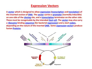
Explore the world of recombinant DNA technology through the utilization of plasmids as vectors for gene cloning. Learn about techniques like insertional inactivation and the characteristics of common plasmid vectors such as pBR322. Discover the intricacies of genetic manipulation in bacterial origins and the applications of these technologies in modern biotechnology.

Download Presentation

Please find below an Image/Link to download the presentation.
The content on the website is provided AS IS for your information and personal use only. It may not be sold, licensed, or shared on other websites without obtaining consent from the author.If you encounter any issues during the download, it is possible that the publisher has removed the file from their server.
You are allowed to download the files provided on this website for personal or commercial use, subject to the condition that they are used lawfully. All files are the property of their respective owners.
The content on the website is provided AS IS for your information and personal use only. It may not be sold, licensed, or shared on other websites without obtaining consent from the author.
E N D
Presentation Transcript
Old and Primrose Chapter 4
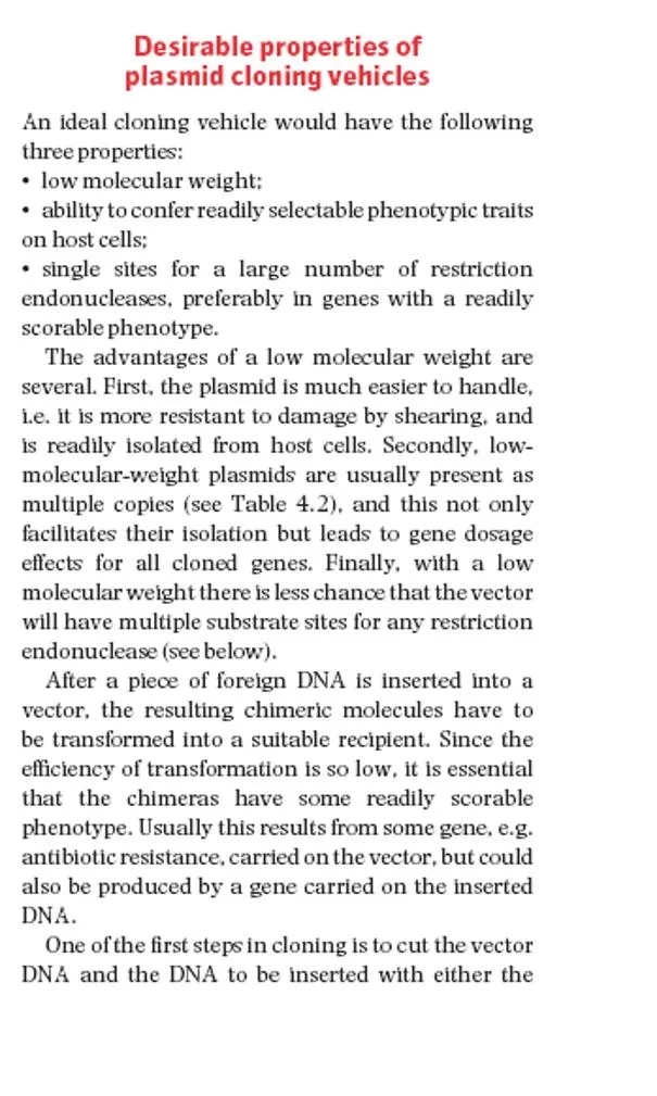
Insertional inactivation is a technique used in recombinant DNA technology. In this procedure, a fragment of foreign DNA is inserted into a restriction site inside a gene, hence causing the gene to turn non-functional or in an inactivated state. b) Recombinant vector having insert: Loss of Marker gene function
Plasmid vectors Plasmids are autonomously replicating circular, double stranded DNA molecules found in bacteria. They have their own origin of replication (ori region), and can replicate independently of the host chromosome. Plasmid vectors are often used for cloning DNA segments of small size (upto 10 kilobases). Insert capacity: 0.1 to 10 Kb
The plasmid pBR322 is one of the most commonly used E. coli cloning vectors. pBR322 is 4361 bp in length and contains: (1) the replicon rep responsible for the replication of plasmid (source plasmid pMB1); (2) rop gene coding for the Rop protein, which promotes conversion of the unstable RNA I RNA II complex to a stable complex and serves to decrease copy number (source plasmid pMB1); (3) bla gene, coding for beta-lactamase that confers resistance to ampicillin (source transposon Tn3/RSF2124); (4) tet gene, encoding tetracycline resistance protein (source plasmid pSC101).
pBR322 The first plasmid vector that has been constructed artificially is pBR322. It is named after the scientists Bolivar and Rodriguez who constructed it in 1977. It is 4361bp in size and most widely used cloning vector. It has an origin of replication derived from a colicin- resistance plasmid (ColE1). Medium copy Number (around 20 copies per cell) Plasmid pBR322 carries two selectable markers viz. genes for resistance to ampicillin (Apr) and tetracycline (Tcr ).
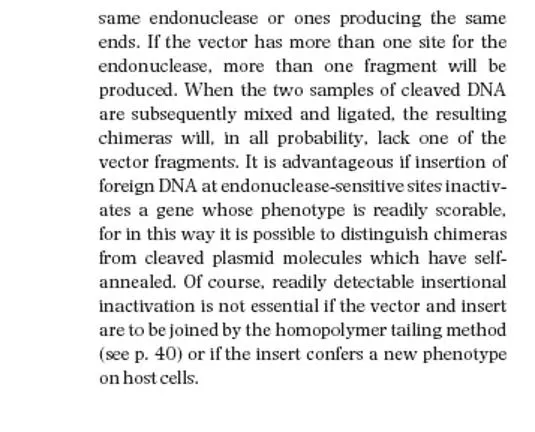
Several (over 40 enzymes) unique RE sites are present in pBR322 plasmid vector. A total of 11 unique restriction sites (Example: EcoRV, BamHI, SphI, SalI, and NruI are present within the gene coding for tetracycline resistance, two sites (HindIII and ClaI) within the promoter of the tetracycline resistance gene and the six sites (Example: PstI, PvuI and ScaI) within the lactamase gene that provide resistance to ampicillin. When a foreign DNA segment is inserted in any of these genes, the antibiotic resistance by that particular gene is lost. This is called insertional inactivation. For instance, insertion of a restriction fragment in the SalI site of the Tcr gene inactivates that gene. One can still select for Apr colonies, and then screen to see which ones have lost Tcr .
Screening of Clones with insert Strategy for selecting host cells that have been transformed with pBR322: (1) The transformation mixture, which contains three cell types, viz., nontransformed cells, cells with the intact original plasmid, and cells with DNA cloned into the BamHI site of pBR322, is plated on complete medium with ampicillin. (2) The mixture is diluted beforehand to ensure separate colonies are formed on the agar. The nontransformed cells (Amps) are killed. (3) The cells with the intact plasmid and cloned DNA plasmid constructs are Ampr and therefore form colonies. Samples of the surviving colonies on the ampicillin plate are transferred to a plate with complete medium and tetracycline, keeping the same position of each colony on the second plate, i.e., replica plating. Only cells with intact plasmids (Tetr) will form colonies in the presence of tetracycline. (4) The colonies that did not grow on the tetracycline plate (dashed circles) but grew on the ampicillin plate carry pBR322 with DNA that was cloned into the BamHI site. The colonies with cloned DNA inserts are picked from the original plate, pooled, and grown.
The use of plasmid pBR322 as a vector: isolation of DNA fragments which carry promoters Cloning into the HindIII site of pBR322 generally results in loss of tetracycline resistance. However, in some recombinants, TcR is retained or even increased. This is because the HindIII site lies within the promoter rather than the coding sequence. Thus whether or not insertional inactivation occurs depends on whether the cloned DNA carries a promoter-like sequence able to initiate transcription of the TcR gene.
The lac Z fragment, whose synthesis can be induced by IPTG, is capable of intra-allelic complementation with a defective form of -galactosidase enzyme encoded by host chromosome (mutation lacZDM15 in E. coli JM109, DH5 and XL1-Blue strains).In the presence of IPTG in growth medium, bacteria synthesise both fragments of the enzyme. Both the fragments can together hydrolyse X-gal (5-bromo-4-chloro-3-indolyl- beta-D-galactopyranoside) and form blue colonies when grown on media. Insertion of foreign DNA into the MCS located within the lac Z gene causes insertional inactivation of this gene at the N-terminal fragment of beta-galactosidase and abolishes intra-allelic complementation. Thus bacteria carrying recombinant plasmids in the MCS cannot hydrolyse X-gal, giving rise to white colonies, which can be distinguished on culture media from non-recombinant cells, which are blue. -complementation
The host E. coli strain carries the lacZ deletion mutant (lacZM15) which contains the -peptide, while the plasmids used carry the lacZ sequence which encodes the first 59 residues of -galactosidase, the -peptide. Neither is functional by itself. However, when the two peptides are expressed together, as when a plasmid containing the lacZ sequence is transformed into a lacZ M15 cells, they form a functional -galactosidase enzyme. The blue/white screening method works by disrupting this -complementation process. The plasmid carries within the lacZ sequence an internal multiple cloning site (MCS). This MCS within the lacZ sequence can be cut by restriction enzymes so that the foreign DNA may be inserted within the lacZ gene, thereby disrupting the gene that produces -peptide. Consequently, in cells containing the plasmid with an insert, no functional -galactosidase may be formed. The rescue of function of the mutant -galactosidase by the -peptide is called -complementation.
The bluewhite screen is a screening technique that allows for the rapid and convenient detection of recombinant bacteria in vector-based molecular cloning experiments. This method of screening is usually performed using a suitable bacterial strain, but other organisms such as yeast may also be used. DNA of transformation is ligated into a vector. The vector is then inserted into a competent host cell viable for transformation, which are then grown in the presence of X-gal. containing recombinant DNA will produce white colonies; cells transformed with non-recombinant plasmids (i.e. only the vector) grow into blue colonies. Cells transformed with vectors An LB agar plate showing the result of a blue white screen
pUCvectorseries A series of small plasmids (about 2.7 kb) have been developed (by Messings and co-workers in 1983) at the University of California and hence the name pUC e.g. pUC7, 8,9,12,13, 18 and 19 etc. These are high copy number plasmids that carry an ampicillin resistance gene and an origin of replication, both from pBR322. High Copy Number: Because of the lack of rop gene and single point mutation in the ori of pMB1. They also have a multiple cloning site (MCS) a sequence of DNA that carries unique sites for many REs. The MCS is inserted in to a portion of lacZ gene (lacZ ; alpha peptide) that codes for the (part of) enzyme -galactosidase. When such plasmids are introduced into E. coli, the colonies are blue on plates containing X-gal galactopyranoside, the substrate for - galactosidase) and IPTG (isopropyl thiogalactoside, an inducer). (5-bromo-4-chloro-3-indolyl- -d-
The insertion of the MCS into the lacZ fragment does not affect the ability of the -peptide to mediate complementation, but cloning DNA fragments into the MCS does.
Recombinants and non-recombinants can therefore be distinguished simply by plating the transformed cells onto agar containing ampicillin and X-gal. All colonies that grow on this medium are made up of transformed cells because only transformants are ampicillin resistant. Blue colonies contain cells with functional -galactosidase enzymes and hence with undisrupted lacZ genes these colonies are therefore non-recombinants. The white colonies comprise cells without -galactosidase activity and hence with disrupted lacZ genes; these are recombinants. Thus cells containing recombinant plasmids form white (not blue) colonies.
1) ori 2) A dominant selectable Marker 3) Cleavage sites for cloning 4) (high copy no.) The plasmid cloning vector pUC19. This plasmid has an origin of replication (ori), an ampR selectable marker, and a polylinker located within part of the - galactosidase gene lacZ+.


 unique RE sites are present in")


 ori")