Pseudomonas and Proteus Bacteria
Pseudomonas is a group of gram-negative, motile, and aerobic bacteria found in various environments, with Pseudomonas aeruginosa being a significant pathogen in humans. It can produce distinctive pigments and is resistant to many antimicrobial agents. Lab diagnosis involves culture testing on specific media. On the other hand, Proteus bacteria are motile, swarm on solid media, and are urease positive. Understanding the characteristics and identification of these bacteria is crucial for proper diagnosis and treatment.

Download Presentation

Please find below an Image/Link to download the presentation.
The content on the website is provided AS IS for your information and personal use only. It may not be sold, licensed, or shared on other websites without obtaining consent from the author.If you encounter any issues during the download, it is possible that the publisher has removed the file from their server.
You are allowed to download the files provided on this website for personal or commercial use, subject to the condition that they are used lawfully. All files are the property of their respective owners.
The content on the website is provided AS IS for your information and personal use only. It may not be sold, licensed, or shared on other websites without obtaining consent from the author.
E N D
Presentation Transcript
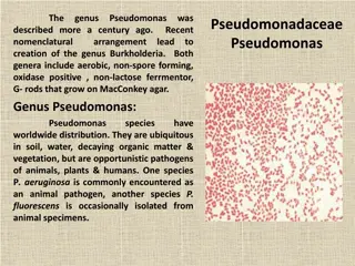
Pseudomonas The pseudomonads are gram-negative, motile, aerobic rods some of which produce water-soluble pigments. The pseudomonads occur widely in soil, water, plants, and animals. P aeruginosa is frequently present in small numbers in the normal intestinal flora and on the skin of humans and is the major pathogen of the group.
Pseudomonas aeruginosa P aeruginosa is widely distributed in nature and is commonly present in moist environments in hospitals. It can colonize normal humans, in whom it is a saprophyte. It causes disease in humans with abnormal host defenses.
Morphology & Identification A. P aeruginosa is motile and rod shaped, measuring about 0.6 2 m. It is gram negative and occurs as single bacteria, in pairs, and occasionally in short chains. B. Culture: *It is an obligate aerobe *sometimes producing a sweet or grapelike or corn taco like odor. *Some strains hemolyze blood. *P aeruginosa forms smooth round colonies with a fluorescent greenish color: bluish pigment pyocyanin, which diffuses into the agar. pyoverdin, which gives a greenish color to the agar. Some strains produce the dark red pigment pyorubin or the black pigment pyomelanin.
P aeruginosa grows well at 3742C; its growth at 42C helps differentiate it from other Pseudomonas species in the fluorescent group. *It is oxidase positive. * It does not ferment carbohydrates, but many strains oxidize glucose. Identification: is usually based on *colonial morphology, *oxidase positivity, *the presence of characteristic pigments, *and growth at 42 C. P aeruginosa and other pseudomonads are resistant to many antimicrobial agents and therefore become dominant and important when more susceptible bacteria of the normal microbiota are suppressed.
Lab diagnosis A. Specimens: Specimens from skin lesions, pus, urine, blood, spinal fluid, sputum, and other material should be obtained as indicated by the type of infection. B. Smears Gram-negative rods are often seen in smears. No specific morphologic characteristics differentiate pseudomonads in specimens from enteric or other gram-negative rods. C. Culture Specimens are plated on blood agar and the differential media commonly used to grow the enteric gram-negative rods. Pseudomonads grow readily on most of these media, but they may grow more slowly than the enterics. P aeruginosa does not ferment lactose and is easily differentiated from the lactose-fermenting bacteria. Culture is the specific test for diagnosis of P aeruginosa infection.
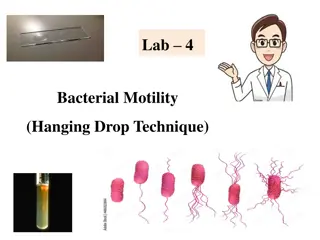
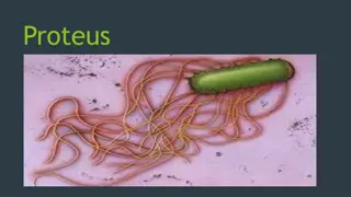
Proteus This bacterium are motile, grow on potassium cyanide medium (KCN), and ferment xylose. Proteus species move very actively by means of peritrichous flagella, resulting in swarming on solid media unless the swarming is inhibited by chemicals, such as phenylethyl alcohol or CLED (cystine-lactose-electrolytedeficient) medium. Proteus species are urease positive.
Proteus mirabilis and P. vulgaris are widely recognized human pathogens. Isolated from urine, wounds, and ear and bacteremic infections Both produce swarming colonies on non-selective media. Both are strongly urease positive Both are phenylalanine deaminase positive
Proteus species produce urease, resulting in rapid hydrolysis of urea with liberation of ammonia. Thus, in urinary tract infections with Proteus species, the urine becomes alkaline, promoting stone formation and making acidification virtually impossible. The rapid motility of Proteus may contribute to its invasion of the urinary tract. Strains of Proteus vary greatly in antibiotic susceptibility. P mirabilis is often inhibited by penicillins; the most active antibiotics for other members of the group are aminoglycosides and cephalosporins.
Yersinia Short, pleomorphic gram-negative rods that can exhibit bipolar staining: catalase positive oxidase negative, and microaerophilic or facultatively anaerobic The genus Yersinia includes: Yersinia pestis, the cause of plague Yersinia pseudotuberculosis and Yersinia enterocolitica, important causes of human diarrheal diseases
Plague Plague is an infection of wild rodents transmitted from one rodent to another and occasionally from rodents to humans by the bites of fleas. Serious infection often results, which in previous centuries produced pandemics of black death with millions of fatalities. The ability of this organism to be transmitted by aerosol and the severity and high mortality associated with pneumonic plague make Y pestis a potential biological weapon.
Yersinia pestis Y pestis is a gram-negative rod that exhibits striking bipolar staining with special stains such as Wright, Giemsa, Wayson, or methylene blue. It is nonmotile. It grows as a facultative anaerobe on many bacteriologic media. blood agar at 37 C, colonies may be very small at 24 hours. A virulent inoculum, derived from infected tissue, produces gray and viscous colonies, but after passage in the laboratory, the colonies become irregular and rough. The organism has little biochemical activity, and this is somewhat variable.
Diseases Pneumonic plague: The incubation period is shorter about (2 to 3 days). Patients experience fever and malaise, and pulmonary signs develop within 1 day. Mortality rate in untreated patients exceeds 90%.
Diseases Y. pestis causes two clinical manifestations: Bubonic plague: is characterized by an incubation period of no more than 7 days after a person has been bitten by an infected flea. patients have a high fever and a painful bubo (inflammatory swelling of the lymph nodes) in groin or axilla Bacteremia develops rapidly if patients are not treated, and as many as 75% die
Pathogenesis When a flea feeds on a rodent infected with Y pestis , the ingested organisms multiply in the gut of the flea. Subsequently, the blocked and hungry flea bites ferociously, and the aspirated blood, contaminated with Y pestis from the flea, is regurgitated into the bite wound. The inoculated organisms may be phagocytosed by polymorphonuclear cells and macrophages. The Y pestis organisms are killed by the polymorphonuclear cells but multiply in the macrophages; because the bacteria are multiplying at 37 C, they produce the antiphagocytic protein and subsequently are able to resist phagocytosis.
Lab diagnosis A. Specimens; Blood is taken for culture and aspirates of enlarged lymph nodes for smear and culture. Acute and convalescent sera may be examined for antibody levels. In pneumonia, sputum is cultured; in possible meningitis, cerebrospinal fluid is taken for smear and culture. B. Smears; Y pestis are small gram-negative bacilli that appear as single cells or as pairs or short chains in clinical material. Wright, Giemsa, or Wayson stains may be more useful when staining material from a suspected buboe or a positive blood culture result because of the striking bipolar appearance (safety pin shape) of the organism using these stains that is not evident on a direct Gram stain. More specific direct staining methods (possibly available through reference laboratories) include the use of fluorescent antibody stains targeting the capsular F1 antigen.
c. Culture; All materials are cultured on blood agar, chocolate, and MacConkey agar plates and in brain heart infusion broth. Growth on solid media may be slow, requiring more than 48 hours, but blood culture results are often positive in 24 hours. Cultures can be tentatively identified by biochemical reactions. Y pestis produces nonlactose-fermenting colonies on MacConkey agar, and it grows better at 25 C than at 37 C. The organism is catalase positive; indole, oxidase, urease negative; and nonmotile. The last two reactions are useful in differentiating Y pestis from other pathogenic yersiniae. An organism with these characteristics should be referred to a public health laboratory for more confirmatory testing. D. Serology In patients who have not been previously vaccinated, a convalescent serum antibody titer of 1:16 or greater is presumptive evidence of Y pestis infection. A titer rise in two sequential specimens confirms the serologic diagnosis