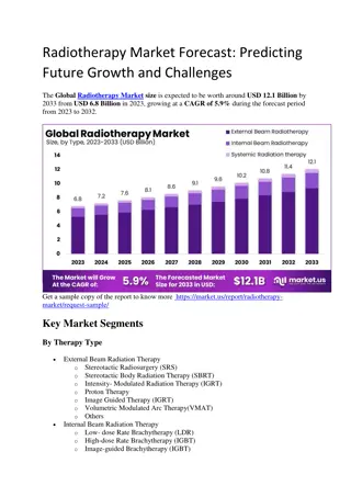
Overview of General Principles of Radiotherapy


G
e
n
e
r
a
l
P
r
i
n
c
i
p
l
e
s
o
f
R
a
d
i
o
t
h
e
r
a
p
y
Anita O’Donovan
Discipline of Radiation Therapy, School of Medicine, Trinity College Dublin

Email:


Disclosure
No conflicts of interest to declare


Objectives
•
What is radiotherapy?
•
How does it work?
•
Patient pathway through radiotherapy
•
Advances in radiation therapy
Objectives
•
What is radiotherapy?
•
How does it work?
•
Patient pathway through radiotherapy
•
Advances in radiation therapy
What is Radiotherapy?
Radiation therapy uses high-energy X-rays to destroy or
damage cancer cells
Goal:
to target radiation to cancer cells while keeping
the dose to normal cells as low as possible
Radical Vs Palliative Vs Prophylactic Vs Abscopal
Different Modalities
•
External
Beam Radiotherapy
•
Internal
– Brachytherapy
(Interstitial/Intracavitary)
Dose is measured in Gray (Gy)
Prescribed by Radiation Oncologist as Total
Dose/Fraction Dose e.g. 50Gy/25#
(conventional fractionation)
50% of cancer patients require RT at some
stage of their cancer journey
Objectives
•
What is radiotherapy?
•
How does it work?
•
Patient pathway through radiotherapy
•
Advances in radiation therapy
Cancer cell (rapidly
proliferating)
die as a
result
Normal cells (more slowly
dividing) can repair
damage caused
Effect is at
the cellular
level
Damages
the cell’s
DNA
Radiation therapy
delivery
Radiation is precisely
targeted to cancer
How Does it Work?
How Does it Work?
•
DNA damage results from DNA strand breaks
•
Two types
of DNA strand breaks:
Maximising the Therapeutic Ratio
•
Tumour control probability is
proportional to the dose of radiation that
is delivered
•
The limiting factor in escalating the
radiation dose is the collateral damage
caused to nearby organs
•
The main goal of radiotherapy research is
to improve this TCP–NTCP balance
Maximum
probability of
complication free
tumour control
Mediated by:
•
Comorbidities
•
Medications
•
Frailty syndrome
•
Cognition
•
Others…..
Studies ongoing:
We need to know more
about age-specific changes
in radiosensitivity and
molecular mechanisms at
play
Balancing Risk in Older Patients
Toxicity
•
Acute/Early
:
up to 90 days following the start
of radiotherapy
•
Late
:
beyond 90 days after the onset of
radiotherapy
•
Minimised by 2 main mechanisms:
–
Geometric avoidance (how our beams hit the
target)
–
Biological sparing (fractionation)
Accurate dose delivery
matters!
Toxicity
Classification of Organs at Risk
a. Series:
Whole organ is a
continuous unit; damage at
any one point will damage
entire organ (
e.g. spinal cord,
digestive system)
b. Parallel:
Organ made up of
a number of Functional
Subunits (FSUs), and if one
part damaged the rest of the
organ compensates (
e.g. lung,
bladder)
Valdagni, R., & Rancati, T. (2013). Reducing rectal injury during external beam radiotherapy
for prostate cancer.
Nature Reviews Urology
,
10
(6), 345-357.
Toxicity
O'Donovan, A., Leech, M., & Gillham, C. (2017). Assessment and management of radiotherapy
induced toxicity in older patients.
Journal of geriatric oncology
,
8
(6), 421-427.
Objectives
•
What is radiotherapy?
•
How does it work?
•
Patient pathway through radiotherapy
•
Advances in radiation therapy
Patient Pathway
Pre-Treatment Phase
Positioning and Immobilisation
Ed.ac.uk
Pre-Treatment Phase
Localisation
•
Patient scanned in
treatment position
•
Reference scan generated
for daily treatment
•
Reference marks placed
on patient’s skin/mask for
daily localisation of the
target
Planning Phase
Treatment Phase
Daily treatment
Treatment Phase
Linear Accelerator
Fractionation Schedules
•
Conventional
: 1.8-2Gy/day/5 days per week
•
Single fraction:
usually palliative setting where late
effects are not a concern e.g. 8Gy for bone metastases
•
Hypofractionation:
↑dose/fraction;↓overall dose &
OTT
•
Hyperfractionation:
↓ dose/fraction; ↑overall dose;
OTT
unchanged
•
Accelerated fractionation:
conventional dose/fraction
& overall dose; OTT
significantly reduced
•
Split course:
Conventional fractionation schedule with
treatment gap of 1-2 weeks to overcome toxicity;
↑OTT
*OTT=Overall Treatment Time
Objectives
•
What is radiotherapy?
•
How does it work?
•
Patient pathway through radiotherapy
•
Advances in radiation therapy
2D-3D
IMRT
•
Greater conformity
•
Steeper dose gradients
•
Greater sparing of Organs at
Risk (OARs)
IGRT
•
Any imaging that leads to an action that can improve or verify the
accuracy of treatment
•
2D
(
kV or MV 2D planar for verification of setup based on bony
landmarks)
•
3D
(kV or MV 3D
CBCT
for verification of setup based on volumetric
information
)
Thariat, J., et al. "Using Proton Beam Therapy
in the Elderly Population: A Snapshot of
Current Perception and Practice."
International Journal of Radiation
Oncology*Biology*Physics
.
Proton Therapy
http://provisionproton.com/static/uploads/VV-button-PvC.jpg
Stereotactic
•
Stereotactic: refers to precise positioning in
three-dimensional space
i.e. focal irradiation
•
Much smaller treatment volumes than
conventional radiotherapy=less toxicity
•
SRS (single fraction) Vs SRT (Generally 2-6
fractions)
•
3 Main Types:
Gamma knife (cranial only)
Cyberknife (cranial and extra-cranial)
Linear accelerator (cranial and extra-cranial)
Stereotactic
Cyberknife
MR Linac
The Future of Radiotherapy
www.elekta.com
Objectives
•
What is radiotherapy?
•
How does it work?
•
Patient pathway through radiotherapy
•
Advances in radiation therapy
Thank You!
anita.odonovan@tcd.ie
@anitaodonovan1
Email:
Radiotherapy is a crucial treatment modality that uses high-energy X-rays to target and destroy cancer cells while minimizing damage to normal cells. This summary covers the fundamentals of radiotherapy, including its objectives, patient pathway, different modalities, and how it works at the cellular level to combat cancer effectively.

Download Presentation

Please find below an Image/Link to download the presentation.
The content on the website is provided AS IS for your information and personal use only. It may not be sold, licensed, or shared on other websites without obtaining consent from the author.If you encounter any issues during the download, it is possible that the publisher has removed the file from their server.
You are allowed to download the files provided on this website for personal or commercial use, subject to the condition that they are used lawfully. All files are the property of their respective owners.
The content on the website is provided AS IS for your information and personal use only. It may not be sold, licensed, or shared on other websites without obtaining consent from the author.
E N D
Presentation Transcript
General Principles of Radiotherapy Anita O Donovan Discipline of Radiation Therapy, School of Medicine, Trinity College Dublin anita.odonovan@tcd.ie @anitaodonovan1 Email:
Disclosure No conflicts of interest to declare
Objectives What is radiotherapy? How does it work? Patient pathway through radiotherapy Advances in radiation therapy
Objectives What is radiotherapy? How does it work? Patient pathway through radiotherapy Advances in radiation therapy
What is Radiotherapy? Radiation therapy uses high-energy X-rays to destroy or damage cancer cells Goal: to target radiation to cancer cells while keeping the dose to normal cells as low as possible Radical Vs Palliative Vs Prophylactic Vs Abscopal
Different Modalities External Beam Radiotherapy Internal Brachytherapy (Interstitial/Intracavitary) Dose is measured in Gray (Gy) Prescribed by Radiation Oncologist as Total Dose/Fraction Dose e.g. 50Gy/25# (conventional fractionation) 50% of cancer patients require RT at some stage of their cancer journey
Objectives What is radiotherapy? How does it work? Patient pathway through radiotherapy Advances in radiation therapy
How Does it Work? Radiation therapy delivery Effect is at the cellular level Radiation is precisely targeted to cancer Cancer cell (rapidly proliferating) die as a result Damages the cell s DNA Normal cells (more slowly dividing) can repair damage caused
How Does it Work? DNA damage results from DNA strand breaks Two types of DNA strand breaks: Single Strand Breaks Double Strand Breaks Normal tissue response Tumour response
Maximising the Therapeutic Ratio Maximum probability of complication free tumour control Tumour control probability is proportional to the dose of radiation that is delivered The limiting factor in escalating the radiation dose is the collateral damage caused to nearby organs The main goal of radiotherapy research is to improve this TCP NTCP balance
Balancing Risk in Older Patients Studies ongoing: We need to know more about age-specific changes in radiosensitivity and molecular mechanisms at play Mediated by: Comorbidities Medications Frailty syndrome Cognition Others ..
Toxicity Acute/Early: up to 90 days following the start of radiotherapy Late: beyond 90 days after the onset of radiotherapy Minimised by 2 main mechanisms: Geometric avoidance (how our beams hit the target) Biological sparing (fractionation) Accurate dose delivery matters!
Toxicity Classification of Organs at Risk a. Series: Whole organ is a continuous unit; damage at any one point will damage entire organ (e.g. spinal cord, digestive system) b. Parallel: Organ made up of a number of Functional Subunits (FSUs), and if one part damaged the rest of the organ compensates (e.g. lung, bladder) Valdagni, R., & Rancati, T. (2013). Reducing rectal injury during external beam radiotherapy for prostate cancer. Nature Reviews Urology, 10(6), 345-357.
Toxicity Acute Effects During radiotherapy and up to 90 days thereafter Late Effects Later than 90 days after the commencement of radiotherapy Time of onset Occurrence Rapidly proliferating tissues e.g. skin, gastrointestinal tract, haematopoietic system Overall treatment time More slowly proliferating tissues e.g. kidney, heart and central nervous system Dose per fraction (daily dose) Relationship to radiotherapy parameters Reversibility Site Specific Examples Usually reversible Generalised: Fatigue Skin: radiation dermatitis Head and Neck: mucositis, xerostomia, dysgeusia CNS: Alopecia, lethargy, somnolence, headache Thoracic: Radiation pneumonitis, oesophagitis dyspnoea Gastrointestinal: Nausea, vomiting, proctitis, diarrhoea Genitourinary: Urinary frequency, dysuria, nocturia Not reversible Generalised: Fatigue Skin: telangiectasia (rare) Head and Neck: osteoradionecrosis, trismus, hypothyroidism CNS: Cognitive impairment, cataracts, radiation necrosis Thoracic: Pulmonary fibrosis, bronchial stenosis (SABR), cardiac toxicity, rib fracture Gastrointestinal: proctitis, small bowel obstruction, fistula Genitourinary: urethral stricture O'Donovan, A., Leech, M., & Gillham, C. (2017). Assessment and management of radiotherapy induced toxicity in older patients. Journal of geriatric oncology, 8(6), 421-427.
Objectives What is radiotherapy? How does it work? Patient pathway through radiotherapy Advances in radiation therapy
Pre-Treatment Phase Positioning and Immobilisation http://t2.gstatic.com/images?q=tbn:ANd9GcRMOSTqEX2XqmE-sUIv3uca9r9TBGFyi4xTlKtbbS8s_C0gCjgK http://www.dartmouth.edu/~humananatomy/figures/chapter_33/33-1_files/image002.jpg http://akiavintage.com/wp-content/uploads/2011/12/Stage-3-Colon-Cancer-radiotherapy-image7.jpg Ed.ac.uk
Pre-Treatment Phase Localisation http://t0.gstatic.com/images?q=tbn:ANd9GcShR7qvyJKkcVJSSp6tb79GuUutRa8vhpTCa4Kg-ttnRCSliVMecQ Patient scanned in treatment position Reference scan generated for daily treatment Reference marks placed on patient s skin/mask for daily localisation of the target
Treatment Phase Daily treatment http://akiavintage.com/wp-content/uploads/2011/12/Stage-3-Colon-Cancer-radiotherapy-image7.jpg
Treatment Phase Linear Accelerator
Fractionation Schedules Conventional: 1.8-2Gy/day/5 days per week Single fraction: usually palliative setting where late effects are not a concern e.g. 8Gy for bone metastases Hypofractionation: dose/fraction; overall dose & OTT Hyperfractionation: dose/fraction; overall dose; OTT unchanged Accelerated fractionation: conventional dose/fraction & overall dose; OTT significantly reduced Split course: Conventional fractionation schedule with treatment gap of 1-2 weeks to overcome toxicity; OTT *OTT=Overall Treatment Time
Objectives What is radiotherapy? How does it work? Patient pathway through radiotherapy Advances in radiation therapy
2D-3D Conformality 3DCRT
IMRT Greater conformity Steeper dose gradients Greater sparing of Organs at Risk (OARs) VMAT
IGRT Any imaging that leads to an action that can improve or verify the accuracy of treatment 2D (kV or MV 2D planar for verification of setup based on bony landmarks) 3D (kV or MV 3D CBCT for verification of setup based on volumetric information)
Proton Therapy Thariat, J., et al. "Using Proton Beam Therapy in the Elderly Population: A Snapshot of Current Perception and Practice." International Journal of Radiation Oncology*Biology*Physics. http://provisionproton.com/static/uploads/VV-button-PvC.jpg
Stereotactic Stereotactic: refers to precise positioning in three-dimensional space i.e. focal irradiation Much smaller treatment volumes than conventional radiotherapy=less toxicity SRS (single fraction) Vs SRT (Generally 2-6 fractions) 3 Main Types: Gamma knife (cranial only) Cyberknife (cranial and extra-cranial) Linear accelerator (cranial and extra-cranial)
Stereotactic Cyberknife
MR Linac The Future of Radiotherapy www.elekta.com
Objectives What is radiotherapy? How does it work? Patient pathway through radiotherapy Advances in radiation therapy
Thank You! anita.odonovan@tcd.ie @anitaodonovan1 Email: