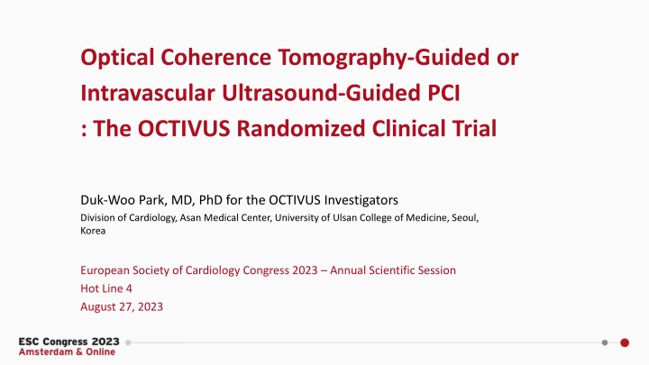
OCTIVUS Randomized Clinical Trial: OCT-Guided vs IVUS-Guided PCI
The OCTIVUS Randomized Clinical Trial compared the clinical efficacy and safety of Optical Coherence Tomography (OCT)-guided and Intravascular Ultrasound (IVUS)-guided strategies in patients undergoing PCI for significant CAD. The study aimed to determine if OCT-guided PCI is noninferior to IVUS-guided PCI in terms of target-vessel failure at 1 year. The trial design included 2,000 patients with obstructive CAD, with primary endpoints being cardiac death, target-vessel myocardial infarction, or ischemia-driven target vessel revascularization at 1 year.
Download Presentation

Please find below an Image/Link to download the presentation.
The content on the website is provided AS IS for your information and personal use only. It may not be sold, licensed, or shared on other websites without obtaining consent from the author.If you encounter any issues during the download, it is possible that the publisher has removed the file from their server.
You are allowed to download the files provided on this website for personal or commercial use, subject to the condition that they are used lawfully. All files are the property of their respective owners.
The content on the website is provided AS IS for your information and personal use only. It may not be sold, licensed, or shared on other websites without obtaining consent from the author.
E N D
Presentation Transcript
Optical Coherence Tomography-Guided or Intravascular Ultrasound-Guided PCI : The OCTIVUS Randomized Clinical Trial Duk-Woo Park, MD, PhD for the OCTIVUS Investigators Division of Cardiology, Asan Medical Center, University of Ulsan College of Medicine, Seoul, Korea European Society of Cardiology Congress 2023 Annual Scientific Session Hot Line 4 August 27, 2023
Disclosure The OCTIVUS was an investigator-initiated trial supported by the CardioVascular Research Foundation (Seoul, Korea) and Abbott Vascular (US), and Medtronic (US). The funders did not participate in the trial design, data collection, data analysis, or manuscript preparation.
Background Intracoronary imaging-guided PCI with intravascular ultrasound (IVUS) and optical coherence tomography (OCT) showed superior clinical outcomes compared to angiography-guided PCI.1-4 Each intracoronary imaging modality has different imaging technologies, lesion applications, advantages, or limitations.5 However, data on the comparative effectiveness of OCT or IVUS for PCI guidance for broad-range of patients with respect to relevant clinical outcomes are still limited.6-7 PCI, percutaneous coronary intervention 1Hong SJ, et al. JAMA 2015;314:2155-63. 2Kim BK, et al. Circ Cardiovasc Interv 2015;8. 3Zhang J, et al. J Am Coll Cardiol 2018;72:3126-37. 4Lee JM, et al. New Engl J Med 2023;388:1668-79. 5Schlofmitz E, et al. Circ Cardiovasc Interv 2020;13. 6Ali ZA, et al. Lancet 2016;388. 7Kubo T, et al. Eur Heart J 2017;38.
Background: Current Guidelines Recommendations for intravascular imaging for PCI optimization OCT IVUS IVUS, intravascular ultrasound; OCT, optical coherence tomography Neumann FJ et al. Eur Heart J 2019;40:87-165
Objective To compare the clinical efficacy and safety of OCT-guided and IVUS-guided strategies inpatients who underwent PCI for significant CAD. We hypothesize that OCT-guided PCI is noninferior to IVUS-guided PCI with respect to target-vessel failure at 1 year. Kang DY, Park DW et al. Am Heart J 2020;228:72-80
Pragmatic Trial Design Optical Coherence Tomography guided versus IntraVascular UltraSound guided percutaneous coronary intervention OCTIVUS Trial 2,000 patients with obstructive CAD undergoing PCI in routine PCI clinical practice A permuted block size of 4 or 6, stratified randomization by (1) trial center OCT-guided PCI strategy (N=1,000) IVUS-guided PCI strategy (N=1,000) The composite primary end point (target-vessel failure) was cardiac death, target-vessel myocardial infarction, or ischemia-driven target vessel revascularization at 1-year Kang DY, Park DW et al. Am Heart J 2020;228:72-80
Inclusion and Exclusion Criteria EXCLUSION INCLUSION 1. ST-elevation myocardial infarction. 2. Severe renal dysfunction (eGFR <30 mL/min/1.73 m2), unless patient is on renal replacement therapy. 3. Cardiogenic shock or decompensated heart failure with severe left ventricular dysfunction (left ventricular ejection fraction < 30%). 4. Life expectancy < 1 years for any non-cardiac or cardiac causes. 5. Any lesion characteristics resulting in the expected inability to deliver the intracoronary imaging catheter during PCI (e.g., severe vessel calcification or tortuosity). 1. Men or women at least age 19 years. 2. Patients with obstructive coronary artery disease (native or restenotic) undergoing PCI with contemporary drug-eluting stents or drug- coated balloons (only for in-stent restenotic lesion) under intracoronary imaging guidance. 3. The patient or guardian agreed to the study protocol and the schedule for clinical follow-up, and provided informed written consent, as approved by the appropriate Institutional Review Board/Ethical Committee of the respective clinical site. PCI, percutaneous coronary intervention
Imaging-guided PCI PCI procedure was performed using standard techniques. In each group, either IVUS (Opticross or Opticross HD, Boston Scientific, CA) or OCT (C7-XR and OPTIS , Abbott, CA) was used before, during, and immediately after PCI; a final imaging assessment for PCI optimization was mandated. Stent size, length, and optimization of the stented segment was determined with the use of a predefined common algorithm for IVUS or OCT on the basis of EAPCI expert consensus.1 A distal lumen or external elastic membrane reference-based stent sizing strategy was used and a sufficient stent expansion of more than 80% of the mean reference lumen area with avoiding major stent malapposition or edge dissection was achieved. All intravascular imaging data were measured by the independent imaging core laboratories (the Asan Medical Center, Core-lab). 1Raber L, et al. Eur Heart J 2018;39:3281-3300.
Endpoints Primary endpoint Target-vessel failure (a composite of death from cardiac cause, target vessel-MI, or ischemia-driven target-vessel revascularization) at 1 year after randomization Secondary endpoints Individual components of the primary composite outcome Target-lesion failure* Stent thrombosis Stroke Repeat revascularization Rehospitalization Bleeding events contrast-induced acute kidney injury procedural complications requiring active intervention MI, myocardial infarction *Target-lesion failure was a composite of death from cardiac causes, target-vessel MI, or ischemia-driven target-lesion revascularization.
Statistical Considerations Power Calculation (N = 2,000) Assuming a 1-year event rate of 8.0% in the IVUS-guided PCI group Statistical power of 80% to detect noninferiority of OCT-guided PCI group with 3.1% of noninferiority margin in primary endpoint (which represented 39% of the expected percentage of patients with an event) Calculated with the likelihood-score method by Farrington and Manning at a one-sided type I error of 0.05 Pre-Specified Statistical Analysis Primary intention-to-treat analysis Kaplan-Meier estimates for calculating cumulative event rates Cox proportional hazard models Estimate the relative risks if proportional hazards assumption is not violated Sensitivity analysis Sensitivity analyses in the per-protocol and as-treated populations The interaction term between randomized groups and key subgroups was evaluated for primary outcome.
Participating Investigators and Trial Organization Participating Investigators (9 Sites in South Korea) Do-Yoon Kang, Jung-Min Ahn, Sung-Cheol Yun, Hoyun Kim, Yeonwoo Choi, Jinho Lee, Duk-Woo Park, Seung-Jung Park (Asan Medical Centers); Seung-Ho Hur, Yun-Kyeong Cho, Cheol Hyun Lee (Keimyung University Dongsan Hospital); Soon Jun Hong, Subin Lim (Korea University Anam Hospital); Sang- Wook Kim, Hoyoun Won (Chung-Ang University Hospital); Jun-Hyok Oh, Jeong Cheon Choe (Pusan National University Hospital); Young Joon Hong (Chonnam National University Hospital); Young Won Yoon (Gangnam Severance Hospital); Soo-Joong Kim (Kyunghee University College of Medicine); Jang-Ho Bae (Konyang University Hospital). Executive Committee Duk-Woo Park (Trial PI) Seung-Jung Park (Trial Co-PI) Do-Yoon Kang Jung-Min Ahn Additional Steering Committee Seung-Ho Hur Cheol Hyun Lee Soon Jun Hong Sang-Wook Kim Event Adjudication Committee Hanbit Park Ha Hye Jo Junghoon Lee Joong Min Lee Ju Hyeon Kim Do-Hyeon Kim Data & Safety Monitoring Board Jong-Min Song Mineok Jang So-Yeon Choi Elly Jeong-youn Bae Byeong-Keuk Kim Trial Funding CardioVascular Research Foundation (CVRF) Abbott Vascular Medtronic
Patient Flow and Follow-Up (from April 12, 2018, through January 14, 2022) Screened (3879) Randomized (2008) OCT-guided PCI (1005) IVUS-guided PCI (1003) 1 Failure to pass imaging device 25 Cross-over to IVUS-guided PCI by the operator s discretion 1 Did not use intravascular imaging devices by the operator s discretion 1 Failed PCI 1 Failure to pass imaging device 6 Cross-over to OCT-guided PCI by the operator s discretion 4 Withdrew consent at <12 mo 8 Were lost to follow-up at <12 mo 2 Withdrew consent 6 Were lost to follow-up At 12-month follow-up: 993 (98.8%) Completed follow-up At 12-month follow-up: 995 (99.2%) Completed follow-up 1005 (100%) Were included in the intention-to-treat analysis 1003 (100%) Were included in the intention-to-treat analysis
Key Baseline Characteristics OCT-Guided PCI (N = 1005) 64.3 (10.3) 215 (21.4) 24.9 3.2 325 (32.3) 647 (64.4) 840 (83.6) 217 (21.6) 226 (22.5) 33 (3.3) 66 (6.6) 28 (2.8) 20 (2.0) 58.8 (9.1) IVUS-Guided PCI (N = 1003) 65.1 (10.5) 218 (21.7) 25.0 3.1 345 (34.4) 639 (63.7) 841 (83.9) 189 (18.8) 202 (20.1) 18 (1.8) 73 (7.3) 38 (3.8) 26 (2.6) 58.3 (10.1) Age [yrs], mean (SD) Female sex Body-mass index Diabetes mellitus no. (%) Hypertension no. (%) Dyslipidemia no. (%) Current smoking no. (%) Previous PCI no. (%) Previous CABG no. (%) Previous stroke no. (%) Atrial fibrillation no. (%) End-stage renal disease on dialysis no. (%) Left ventricular ejection fraction [%], mean (SD) Clinical indication for index PCI no. (%) Silent ischemia Chronic coronary syndrome Acute coronary syndrome Unstable angina NSTEMI 106 (10.6) 663 (66.0) 236 (23.5) 137 (13.6) 99 (9.9) 115 (11.5) 654 (65.2) 234 (23.3) 135 (13.5) 99 (9.9)
Anatomical Characteristics OCT-Guided PCI (N = 1005) 608 (60.5) IVUS-Guided PCI (N = 1003) 629 (62.7) Multivessel disease no. (%) Treated complex coronary lesions no. (%) Left main disease Any bifurcation disease no. (%) Ostial lesion no. (%) Chronic total occlusion no. (%) Severely calcified lesion no. (%) In-stent restenotic lesion no. (%) Diffuse long lesion no. (%) Bypass graft disease no. (%) SYNTAX score Mean Category no./total no. (%) Low, 0 to 22 Intermediate, 23 to 32 High, >32 116 (11.5) 516 (51.3) 96 (9.6) 56 (5.6) 76 (7.6) 87 (8.7) 575 (57.2) 3 (0.3) 148 (14.8) 540 (53.8) 99 (9.9) 52 (5.2) 76 (7.6) 77 (7.7) 594 (59.2) 0 (0.0) 15.1 8.9 15.8 9.5 813 (80.9) 141 (14.0) 51 (5.1) 773 (77.1) 173 (17.3) 57 (5.7) Those with encircling calcium seen on angiography Lesion length 28 mm or stent length 32 mm of treated segment SYNTAX, Synergy between Percutaneous Coronary Intervention with Taxus and Cardiac Surgery.
Procedural Characteristics OCT-Guided PCI (N = 1005) IVUS-Guided PCI (N = 1003) P Value PCI approach 0.99 Radial access 639 (63.6) 638 (63.6) Femoral access 366 (36.4) 365 (36.4) PCI modality Use of drug-eluting stents 970 (96.5) 973 (97.1) 0.45 Used of drug-coated balloons (only for ISR lesion) 35 (3.5) 29 (2.9) 1.3 0.6 1.4 0.6 Total no. of lesions treated per patient 0.36 1.6 1.0 1.6 1.0 Mean number of stents per patient 0.38 47.2 32.4 47.8 32.2 Total stent length per patient mm 0.69 Post-dilatation with larger or high-pressure balloon no. (%) 931 (92.6) 917 (91.5) 0.35 238.3 112.4 199.8 109.7 Total amount of contrast media used mL <0.001 46.1 23.6 48.9 25.1 Total PCI time min <0.001 ISR, In-stent restenosis.
Procedural Outcomes OCT-Guided PCI (N = 1005) IVUS-Guided PCI (N = 1003) P Value Procedural success no. (%) Angiography-based 993 (98.8) 990 (98.7) 0.84 Imaging-based 476 / 986 (48.3) 546 / 995 (54.9) 0.003 Procedural complications requiring active intervention no. (%) Any 22 (2.2) 37 (3.7) 0.047 IVUS or OCT procedure related complications 0 (0) 0 (0) Angiographic device success is defined as successful PCI at the intended target lesion with final in-stent residual stenosis of less than 30% by quantitative coronary angiography. By patient-level analyses: imaging-based device success is defined as successful PCI at the intended target lesion, which fulfills all optimal criteria for stent implantation by IVUS or OCT. Among patients with multivessel interventions, all treated lesions should be met for optimization criteria. Procedural complications requiring active intervention, which were related to PCI or use of intravascular imaging (i.e., procedural safety outcomes).
Core Lab-Imaging Analysis : Lesion-Level Analysis OCT-Guided PCI (N = 1005 Patients) (N = 1279 Lesions) IVUS-Guided PCI (N = 1003 Patients) (N = 1271 Lesions) P Value Core Lab Imaging analysis final post-PCI 5.60 2.01 6.70 2.37 Minimum stent area mm2 <0.001 85.36 17.49 91.37 22.31 Minimum stent expansion % <0.001 134.81 47.75 126.04 39.12 Minimum stent area by distal reference lumen area % <0.001 Optimization Imaging-Guided PCI Criteria All stent-optimization criteria met no./total no. (%) 613 / 1148 (53.4%) 743 / 1236 (60.1%) 0.001 Optimal stent expansion 712 / 1148 (62.0%) 860 / 1236 (69.6%) <0.001 Plaque burden at stent landing zone < 50% 708 / 800 (88.5%) 1016 / 1186 (85.7%) 0.07 No major malapposition 1059 / 1147 (92.3%) 1209 / 1235 (97.9%) <0.001 No large dissection 1114 / 1141 (97.6%) 1222 / 1231 (99.3%) 0.001 A relative stent expansion of >90% no./total no.(%) 394 / 1025 (38.4%) 575 / 1179 (48.8%) <0.001 *Plus minus values are means SD. Optimal stent expansion was defined as a relative stent expansion of >80% (an in-stent minimum stent area divided by average reference lumen area). In lesions with non-evaluable reference lumen area, optimal stent expansion was defined as an absolute in-stent minimum stent area of >5.5 mm2 by IVUS and >4.5 mm2 by OCT. Extensive stent malapposition was defined as an acute stent malapposition of 0.4 mm with longitudinal extension >1 mm of the stent over its entire length against the vessel wall. Large dissection was defined as a dissection that occurred 5mm from the edge of the stent, extended to extensive lateral >60 , longitudinal extension >2mm, and flap extending to media or adventitia.
Primary Endpoint of TVF: Cardiac Death, TV-MI, or TVR 5% Hazard ratio, 0.80 (95% CI, 0.47 1.36) P for noninferiority <0.001 4% 3.1% Patients (%) IVUS-guided 3% 2% 2.5% OCT-guided 1% 0% 0 3 6 9 12 Months since Randomization No. at Risk OCT-guided PCI IVUS-guided PCI 1005 1003 990 985 984 981 979 969 912 893 CI, confidence interval; TV-MI, target-vessel myocardial infarction; TVR, target-vessel revascularization
Types of CV Outcomes OCT-Guided PCI (N = 1005) IVUS-Guided PCI (N = 1003) Risk Difference (95% CI) HR (95% CI) Outcome* Primary composite outcome 25 (2.5%) 31 (3.1%) 0.6 ( 2.0 to 0.8) 0.80 (0.47 to 1.36) Secondary outcomes Target-lesion failure 22 (2.2%) 29 (2.9%) 0.7 ( 2.1 to 0.7) 0.76 (0.43 to 1.31) Death 10 (1.0%) 14 (1.4%) 0.4 ( 1.4 to 0.6) 0.71 (0.32 to 1.60) From cardiac cause 3 (0.3%) 6 (0.6%) 0.3 ( 0.9 to 0.3) 0.71 (0.32 to 1.60) From noncardiac cause 7 (0.7%) 8 (0.8%) 0.1 ( 0.9 to 0.6) 0.87 (0.32 to 2.40) Target-vessel myocardial infarction 9 (0.9%) 14 (1.4%) 0.5 ( 1.4 to 0.4) 0.64 (0.28 to 1.48) Periprocedural 7 (0.7%) 11 (1.1%) 0.4 ( 1.2 to 0.4) 0.64 (0.25 to 1.64) Spontaneous 2 (0.2%) 3 (0.3%) 0.1 ( 0.5 to 0.3) 0.67 (0.11 to 3.98) Repeat revascularization 16 (1.6%) 19 (1.9%) 0.3 ( 1.5 to 0.8) 0.84 (0.43 to 1.63) Target-lesion revascularization 11 (1.1%) 14 (1.4%) 0.3 ( 1.3 to 0.7) 0.78 (0.36 to 1.72) Target-vessel revascularization 14 (1.4%) 16 (1.6%) 0.2 ( 1.3 to 0.9) 0.87 (0.43 to 1.79) Contrast-induced nephropathy no. (%)** 14 (1.4%) 15 (1.5%) 0.1 ( 1.1 to 0.9) 0.93 (0.45 to 1.91) *The percentages were estimated by the Kaplan Meier estimates. Hazard ratios are for the OCT-guided PCI group, as compared with the IVUS-guided PCI group by use of Cox proportional hazard models. The primary composite outcome was death from cardiac cause, target-vessel myocardial infarction, or target vessel revascularization. Target-lesion failure was a composite of death from cardiac causes, target-vessel MI, or ischemia-driven target-lesion revascularization. ** Contrast-induced nephropathy was defined as either a greater than 25% increase of serum creatinine or an absolute increase in serum creatinine of 0.5 mg/dL from baseline within 72 h after the index PCI procedure. Event rates (%) of contrast-induced nephropathy are presented as calculated percentages. CI, confidence interval; HR, hazard ratio; PCI, percutaneous coronary intervention
Sensitivity Analysis Primary Endpoint in the Per-Protocol Population Primary Endpoint in the As-Treated Population
Prespecified Key Subgroups Analysis: by Clinical Factors Estimated 1-Yr Event Rate (%) Percent of Patients P-for- Interaction Hazard Ratios (95% CI) Subgroup OCT-guided IVUS-guided Age 0.419 48.2 2.3 2.1 < 65 1.07 (0.45 to 2.51) 2.8 51.8 4.0 65 0.68 (0.35 to 1.34) Sex 0.212 21.6 2.3 5.1 Female 0.46 (0.16 to 1.31) 78.4 2.6 2.6 Male 0.99 (0.53 to 1.85) Diabetes mellitus 0.222 33.4 4.4 3.8 Yes 1.14 (0.54 to 2.43) 66.6 1.6 2.8 No 0.59 (0.28 to 1.25) Acute coronary syndrome 0.710 23.4 3.0 4.3 Yes 0.69 (0.26 to 1.81) 76.6 2.4 2.7 No 0.86 (0.46 to 1.61) Left ventricular ejection fraction 0.847 11.2 6.3 8.1 50% 0.78 (0.26 to 2.38) 88.8 2.0 2.9 > 50% 0.69 (0.35 to 1.34) 0.1 1 10 IVUS-guided PCI better OCT-guided PCI better
Estimated 1-Yr Event Rate (%) Prespecified Key Subgroups Analysis: by Anatomical Factors Percent of Patients P-for- Interaction Hazard Ratios (95% CI) Subgroup OCT-guided IVUS-guided Left main disease 0.868 Yes 0.78 (0.28 to 2.16) 13.2 5.3 6.8 No 0.87 (0.47 to 1.61) 86.9 2.2 2.5 Bifurcation disease 0.901 Yes 0.83 (0.43 to 1.61) 52.6 3.1 3.7 No 0.78 (0.32 to 1.87) 47.4 1.9 2.4 Diffuse long coronary artery lesion 0.077 Yes 1.15 (0.60 to 2.22) 58.2 3.3 2.9 No 0.41 (0.16 to 1.05) 41.8 1.4 3.4 Severely calcified lesion 0.149 Yes 1.36 (0.60 to 3.07) 7.6 7.9 8.1 No 0.61 (0.31 to 1.23) 92.4 2.1 2.7 Multivessel disease 0.547 Yes 0.75 (0.42 to 1.36) 61.6 3.2 4.2 No 1.13 (0.35 to 3.71) 38.4 1.5 1.4 SYNTAX score 0.096 Low 0.52 (0.26 to 1.04) 79.0 1.5 2.9 Intermediate 1.63 (0.57 to 4.70) 15.6 5.7 3.5 High 1.93 (0.46 to 8.06) 5.4 9.9 5.3 0.1 1 10 IVUS-guided PCI better OCT-guided PCI better
CV Outcomes during the Entire Follow-up Period (Median 2.0 years; range 1.0-4.8 years) OCT-Guided PCI (N = 1005) IVUS-Guided PCI (N = 1003) HR (95% CI) Outcome* Primary composite outcome 58 (5.8%) 61 (6.1%) 0.91 (0.63 to 1.30) Secondary outcomes Target-lesion failure 50 (5.0%) 56 (5.6%) 0.85 (0.58 to 1.25) Death 28 (2.8%) 27 (2.7%) 0.99 (0.58 to 1.69) From cardiac cause 11 (1.1%) 11 (1.1%) 0.93 (0.39 to 2.18) From noncardiac cause 17 (1.7%) 16 (1.6%) 1.03 (0.52 to 2.05) Target-vessel myocardial infarction 9 (0.9%) 20 (2.0%) 0.45 (0.21 to 0.99) Periprocedural 7 (0.7%) 11 (1.1%) 0.64 (0.25 to 1.64) Spontaneous 2 (0.2%) 9 (0.9%) 0.22 (0.05 to 1.04) Repeat revascularization 52 (5.2%) 50 (5.0%) 1.01 (0.68 to 1.49) Target-lesion revascularization 31 (3.1%) 33 (3.3%) 0.90 (0.55 to 1.48) Target-vessel revascularization 39 (3.9%) 38 (3.8%) 0.98 (0.63 to 1.54) *The listed percentages were estimated as the ratio of the numerator and denominator. Hazard ratios are for the OCT-guided PCI group, as compared with the IVUS-guided PCI group by use of Cox proportional hazard models. The primary composite outcome was death from cardiac cause, target-vessel myocardial infarction, or target vessel revascularization. Target-lesion failure was a composite of death from cardiac causes, target-vessel MI, or ischemia-driven target-lesion revascularization.
Study Limitations It was not possible to mask the imaging modalities from the patients and investigators (the possibility of ascertainment or selection bias). The observed number of primary-outcome events was lower than expected (several explanations would be possible). There would be the possibility of discrepancy on a site-determined and core-laboratory measured imaging interpretation. The generalizability and reproducibility of our trial findings may be potentially limited due to the geographic variability in the use of intravascular imaging in the daily PCI practice. Our trial did not include an angiography-guided arm.
Summary for the OCTIVUS Key Findings In this large-scale, pragmatic RCT comparing OCT and IVUS for PCI guidance in patients with diverse anatomical or clinical characteristics, we found that OCT-guided PCI was noninferior to IVUS-guided PCI procedures with respect to a primary composite of death from cardiac causes, target-vessel MI, or ischemia-driven TVR at 1 year. The incidence of primary outcomes was lower than expected, possibly due to improvements in the methods/techniques to perform PCI and general improvements in cardiovascular care during the past few years. The amount of contrast dye used during the procedures was higher in the OCT group than in the IVUS group, but it was not related to an increase of contrast-induced nephropathy.
Conclusions In this OCTIVUS trial involving patients who are undergoing PCI for diverse coronary-artery lesions, 1. OCT-guided PCI was noninferior to IVUS-guided PCI with respect to a composite of death from cardiac causes, target- vessel myocardial infarction, or ischemia-driven target-vessel revascularization at 1 year. 2. However, the selected study population and lower than expected event rates should be considered in interpreting the trial.
CIRCULATION. 2023; [PUBLISHED ONLINE AHEAD OF PRINT]. DOI: 10.1161/CIRCULATIONAHA.123.066429 OPTICAL COHERENCE TOMOGRAPHY-GUIDED OR INTRAVASCULAR ULTRASOUND-GUIDED PERCUTANEOUS CORONARY INTERVENTION: THE OCTIVUS RANDOMIZED CLINICAL TRIAL DO-YOON KANG, MD; JUNG-MIN AHN, MD; SUNG-CHEOL YUN, PHD; SEUNG-HO HUR, MD; YUN-KYEONG CHO, MD; CHEOL HYUN LEE, MD; SOON JUN HONG, MD; SUBIN LIM, MD; SANG-WOOK KIM, MD; HOYOUN WON, MD; JUN-HYOK OH, MD; JEONG CHEON CHOE, MD; YOUNG JOON HONG, MD; YONG-HOON YOON, MD; HOYUN KIM, MD; YEONWOO CHOI, MD; JINHO LEE, MD; YOUNG WON YOON, MD; SOO-JOONG KIM, MD; JANG-HO BAE, MD; DUK-WOO PARK, MD; AND SEUNG-JUNG PARK, MD FOR THE OCTIVUS INVESTIGATORS CIRCULATION HTTPS://WWW.AHAJOURNALS.ORG/DOI/10.1161/CIRCULATIONAHA.123.066429 27
