Management of Acute Respiratory Distress Syndrome: Case Study and Diagnostics
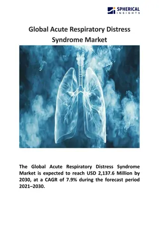
A 54-year-old woman post-laparotomy presents with respiratory distress and bilateral infiltrates on CXR. The case raises suspicion for Acute Respiratory Distress Syndrome (ARDS). Diagnostic criteria and potential investigations, such as CT pulmonary angiogram, thoracic USS, echocardiogram, bronchoscopy, or ECG, are discussed. The patient's deteriorating condition despite CPAP leads to the decision for intubation, highlighting the challenges in managing ARDS.
Uploaded on Nov 16, 2024 | 1 Views
Download Presentation

Please find below an Image/Link to download the presentation.
The content on the website is provided AS IS for your information and personal use only. It may not be sold, licensed, or shared on other websites without obtaining consent from the author.If you encounter any issues during the download, it is possible that the publisher has removed the file from their server.
You are allowed to download the files provided on this website for personal or commercial use, subject to the condition that they are used lawfully. All files are the property of their respective owners.
The content on the website is provided AS IS for your information and personal use only. It may not be sold, licensed, or shared on other websites without obtaining consent from the author.
E N D
Presentation Transcript
SpR 2; Intensive Care in the SCE Annette Fisher ST3 Respiratory Medicine Sunderland Royal Hospital
Question 1. A 54 year old woman is referred to critical care 48 hrs post laparotomy for a perforated duodenal ulcer with four-quadrant peritonitis. She has deteriorated over the past 12 hrs with respiratory distress. On examination she appears confused and is speaking in broken sentences. RR is 30 and SpO2 on 92 % via 15 L/min non-rebreathe mask. Auscultation reveals fine crackles bilaterally. HR is 110 bpm and BP is 100/60. Her JVP is not elevated and she has normal heart sounds. There is no peripheral oedema and her calves are soft and non-tender. Fluid balance is 800 ml positive. A portable CXR demonstrates extensive bilateral infiltrates. A blood gas is performed with the following result; pH 7.32 pCO2 3.8 kPa pO2 8.1 kPa HCO3 18.7 mmol/L What would be the most useful next investigation in assessing the suspected cause for her deterioration? a) CT pulmonary angiogram b) Thoracic USS c) Echocardiogram d) Bronchoscopy with bronchoalveolar lavage e) ECG
Acute Respiratory Distress Syndrome1, 2 Severe end of spectrum of acute lung injury due to many different insults; Sepsis/pneumonia Gastric aspiration Trauma/burns Panceatitis TRALI Post stem cell transplant Overdose Near drowning
ARDS - Pathophysiology Acute inflammation affecting alveolar-capillary membrane, either by locally produced pro-inflammatory mediators or remotely produced and arriving via pulmonary artery. Increase in permeability of the membrane allowing fluid and protein leakage into alveolar space manifesting as high permeability pulmonary oedema. Resulting acute inflammatory exudate collapse and consolidation of distal airpaces with progressive loss of gas exchange surface area. No compensatory hypoxic pulmonary vasoconstriction resulting in gross V/Q mismatch.
ARDS diagnostic criteria (2012 Berlin Definition)2 Timing; Onset within 1 week of known clinical insult Radiology; Bilateral opacities consistent with pulmonary oedema on CXR or CT scan Origin of oedema; Not fully explained by left sided heart failure or fluid overload. Echo indicated to assess LV function. Respiratory failure; Oxygenation impairment defined by PaO2/FiO2 ratio at least <40 kPA (<300 mmHg) despite PEEP > 5 mmH2O Mild PaO2/FiO2 >27 kPa (> 200 mmHg) Moderate PaO2/FiO2 > 13 kPa (> 100 mmHg) Severe PaO2/FiO2 <13 kPa (<100 mmHg)
Question 2. The patient is transferred to ICU. A bedside echocardiogram is performed which confirms preserved LV function and no signs of hypo/hypervolaemia. She is initially trialled on CPAP with PEEP of 10 mmHg and FiO2 of 60 % but continues to deteriorate with progressive hypoxia over the next 4 hours. The decision is made to to proceed with intubation and invasive mechanical ventilation. What would be the most appropriate initial ventilator settings? a) SIMV mode, FiO2 80 %, PEEP 16, tidal volume 6 ml/kg IBW, RR 16 b) PS/CPAP mode, FiO2 25 %, PEEP 5, Tidal volume 6 ml/kg IBW, RR 16 c) SIMV mode, FiO2 80 %, PEEP 16, tidal volume 12 ml/kg IBW, RR 16 d) PS/CPAP mode, FiO2 50 %, PEEP 16, Tidal volume 6 ml/kg IBW, RR 16 e) SIMV mode, FiO2 80 %, PEEP 5, tidal volume 6 ml/kg IBW, RR 26
Question 3. Over the next 6 days, the patient is managed with lung protective ventilation, high PEEP, prone positioning and a 48 hr infusion of neuromuscular blocking agent. In spite of this she fails to improve, as evidenced by worsening hypoxia. She has also developed an acute kidney injury necessitating renal replacement therapy, and her vasopressor requirements are increasing. The ICU team decide to refer her for consideration of Extra-Corporeal Membrane Oxygenation. What is most likely to lead to a decline in the referral? a) Age >50 years b) Recent extensive intra-abdominal surgery c) Refractory multi-organ failure d) Invasive ventilation for >5 days e) CPAP treatment preceding invasive ventilation
ECMO referral3,4 Indications Age >16 yrs Potentially reversible severe acute respiratory failure Severe hypoxaemia (Eg PaO2/FiO2 < 13.3 kPa) Uncompensated hypercapnia with pH <7.2 No limitation to ongoing life- sustaining treatment Inability to achieve lung protective tidal volumes and pressures Murray lung injury score >3 Contraindications Age >65 Intracranial bleed (current/recent) Other contraindications to heparinisation Multi-organ failure High pressure (PIP > 30 mmH20 and/or high FiO2 (>80%)) ventilation for more than 7 days (CPAP counts) Prolonged cardiac arrest
Question 4. The patient is declined for ECMO on the basis of established multi-organ failure. Her condition does, however, stabilise over the next few days, with reducing ventilatory support and the recovery of some renal function. The ICU team decide to proceed with full active management. During a night shift as the ICU resident you are called by nursing staff to review her urgently. Her oxygen saturations have dropped rapidly they are currently 85 % on 100 % FiO2 with PEEP of 16. HR is 130 bpm, sinus tachycardia, BP is 90/50. On examination there is decreased right sided chest expansion with minimal breath sounds. The oral ETT is 24 cm at the lips, which is unchanged from previously. Peak inspiratory pressures have risen to >40 mmH20. What is the most appropriate treatment to improve the patient s condition? a) Therapeutic bronchoscopy b) Needle decompression followed by chest drain insertion c) Broad spectrum antibiotics d) Thrombolysis c) Pull back ETT 2 cm
Complications of ARDS1 Barotrauma; Pneumothorax, surgical emphysema, pneumomediastium Nosocomial infections/ventilator associated pneumonia Myopathy VTE GI haemorrhage Inadequate nutrition Complications of proning; Loss of venous access ETT displacement Nerve compression Pressure sores Retinal damage Vomiting Transient arrythmia/hypotension/desaturation
Question 5. Following decompression of the tension pneumothorax, the patient continues to make gradual recovery. A percutaneous tracheostomy is inserted to aid with prolonged ventilator wean. The patient is eventually stepped down from ICU to the respiratory ward with the tracheostomy still in situ. Which of the following options would suggest tracheostomy decannulation is inappropriate? a) No upper airway obstructions b) Adequate independent secretion clearance c) Tolerates cuff deflation for 24 hrs d) Delirium with fluctuating GCS e) Ongoing oxygen requirement
Tracheostomy1, 5 Indications Airway protection following major head and neck surgery Prolonged ventilator wean Neurospinal indications; cervical cord pathology, reduced GCS following neurological injury Advantages Improved patient communication Improved tube tolerance Reduced sedation requirement Disadvantages Increased tendency to aspirate Reduced ability to talk Increased infection risk Tracheal stenosis
Tracheostomy decannulation Should be carried out as soon as; Adequate secretion clearance Low probability of thick mucous plugs blocking large airways No upper airway obstruction No significant aspiration No need to continue ventilation or reduce dead space for gas exchange Patient tolerates cuff deflation for 24 hrs/speaking valve for 12 hrs Haemodynamic stability No new lung infiltrates on CXR MDT agreement for decannulation
References 1. Oxford Handbook of Respiratory Medicine 4thEdition 2. Faculty of Intensive Care Medicine/Intensive Care Society (2018) Guidelines on the Management of Acute Respiratory Distress Syndrome Version 1 3. Royal Bromptom and Harefield Hospitals ECMO Referrals and Transfer Pathways https://www.rbht.nhs.uk/our-services/clinical_support/critical- care-and-anaesthesia/ecmo-and-severe-respiratory-failure/ecmo-referrals- and-transfer-pathway 4.NHS England ECMO National Referral Pathway https://www.signpost.healthcare/ecmo-referral-pathway 5. Stelfox HT, Crimi C, Berra L, Noto A, Schmidt U, Bigatello LM, Hess D (2008) Determinants of tracheostomy decannulation: an international survey Critical care 12:R26.
Thank you for listening, any questions?
