Malaria Parasites and Their Life Cycle


Malaria parasite
Plasmodium vivax
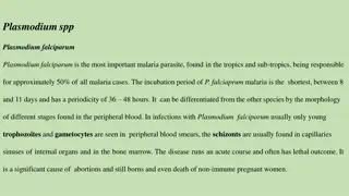
Plasmodium falciparum
Plasmodium malariae
Plasmodium ovale

Malaria parasites as a group
(general information)
* This parasite has the alternation of generations
phenomena. That means the life cycle includes
an asexual phase (
schizogony
) alternating with
sexual one (
gametogony
) followed by
(
sporogony
).
* The malarial parasites require two hosts:
1. Vertebrate host, in which the asexual phase
develops and gametocytes are produced.
2. Vector host (invertebrate host), in which the
gametocytes become mature gametes.
Malaria parasites as a group
(general information)
* Following maturation, the microgamete
unites with the macrogamete to form the
zygote, which then become an oocyst
which will produce the sporozoites later.
* When the numerous sporozoites are
introduced into the vertebrate host, they
will develop into the asexual stage.
Malaria parasites as a group
(general information)
* There are two separate transfer stages, the
gametocyte and the sporozoites.
* The asexual phase or cycle called (
schizogony
)
and is found only in the vertebrate host, while
maturation and union of sex cells followed by
production of sporozoites take place only in
blood sucking invertebrates, mostly arthropods
and predominantly
mosquitoes
.
The factors influencing the effectiveness of a
particular species of
Anopheles
in transmission of
malaria are:
1. Susceptibility to infection by the parasite.
2. Survival long enough for development and
transmission of the sporozoites.
3. A preference for human blood.
4. Presence in sufficient numbers.
Life cycle
1. Schizogony (in the human):
A. Pre-erythrocytic development (Asexual
Development out of the R.B.Cs.):
*
Inoculation occurs when an infected female
Anopheles
mosquito injects saliva containing sporozoites into
cutaneous blood vessels in a preparation to take a
blood meal. The insect injects spindle shape bodies
(the
sporozoites
) which circulate in the blood stream,
but within a half an hour they will disappear.
Life cycle
*
The first colonization takes place in the
parenchymal cells of the liver where the
sporozoites will pass through two schizogony,
the first called (primary exoerythrocytic
schizogony) the second one called (secondary
exoerythrocytic schizogony) except in
Plasmodium falciparum
where the parasite
passing single schizogony in the liver.
Life cycle
* The first generation called (cryptozoites) and
sometimes called (merozoites), while the second
generation individuals called (metacryptozoites), after
(7-10) days in the liver the parasite enters the R.B.Cs.
* The first evidence of infection can be seen (48hours) to
(7days) later in the parenchymal cells of the liver,
where young schizonts in active nuclear division
observes, these are termed primary exoerythrocytic
(EE) schizonts or pre-erythrocytic schizonts.
Life cycle
* The average incubation period, is (12) days
for
P. falciparum
,
(13) to (17) days for
P.
vivax
and
P. ovale
and (28 to 30) days for
P. malariae
.
* The incubation period: is the time between
the inoculation of sporozoites and the first
appearance of clinical sings, of which fever
is the most common.
Life cycle
B. Erythrocytic development:
(Asexual Development in R.B.Cs.):
*
When the merozoites which have developed in the pre-
erythrocytic foci enters the red blood cells, they transform
into trophozoites, which grow and develop into schizonts,
each producing the number of merozoites characteristic of
the species of
Plasmodium
.
When fully matured, the
merozoites break out of the parasitized R. B.C. and soon
actively enter other R.B.Cs. to repeat the asexual cycle.
Life cycle
* The merozoites that enters the R.B.Cs. appears in the
beginning in a ring phase called (ring stage) because of the
presence of a vacuole or space in the middle of the parasite
surrounded by a ring of cytoplasm and a minute nucleus in
one side.
* when it is grow because of its feeding on the R.B.Cs. Content,
it is become rounded then irregular so called (amoeboid shape
trophozoite) which is produce the schizont stage then.
* in the case of
P. falciparum
the R.B.C. become more viscous,
so it aggregates in the internal organs and not appear in
circulation. (Important)
Life cycle
* The infected R.B.Cs. with
P.
vivax
, after the
appearance of the ring stage it characterized by the
appearance of minute dots stains with red color or
(reddish-orange)
in Giemsa or right stained thin blood
films called
(Schüffner’s dots)
or granules, also the
R.B.C. appears swelling and paler; however, this dots
appears also in the case of
P. ovale
, but in this case
the infected R.B.C. becomes oval in shape (the cause
of the name). In the case of
P. falciparum
appears
reddish
dots called (
Maurer’s dots
) this dots less in
number and larger than the first type. In
P. malariae
appears other dots called (
Ziemann’s dots
).
Life cycle
* After the destruction of the R.B.Cs. the dots, many
material & wastes which produced by the parasites
will be freeing to the blood stream, when it reaches
to the spleen & other organs, sometimes under the
skin, the infected person feels with
chills or
shivering then fever then sweating sequently
.
* The merozoites spend a regular period from the
entrance to the growing & forming new merozoites,
these periods are:
Life cycle
(48)hr. in
P. vivax
&
P. ovale
(72)hr. in
P. malariae
(36-48)hr. in
P. falciparum
.
* After many generations of schizont stage, some of the
merozoites grow slowly and form more pigments and each
one grow to gametocyte.
* There are two type of gametocyte microgametocyte (♂) &
macrogametocyte (♀). These gametocytes will continue in
the circulation for many weeks, but it isn’t growing in the
human body.
* It is important to note that these gametocytes are
crescent in case of
P. falciparum
while they are
rounded in the other types.
Life cycle
2. Gametogony (in the mosquito):
*
Once the ripe gametocytes are ingested by female
Anopheles
mosquitoes with the blood meal and reach the
midgut, they transform into mature gametes.
* One macrogametocyte develops a single macrogamete
(unfertilized oocyst or ovum), and one microgametocyte
produces several flagellated microgametes.
* A microgamete then enters a macrogamete, resulting in a
zygote which become a motile Ookinete, migrates through
the stomach wall and becomes an oocyst just under the
outer membrane of the stomach.
Life cycle
*
The oocyst grows rapidly and develops internal nuclear
centers, each center then produces a large number of
delicate, spindle-shaped sporozoites. By time the
sporozoites become mature, the wall of the greatly enlarged
oocyst bursts, releasing the sporozoites in to the hemocoele
of the mosquito.
Life cycle
* The sporozoites then migrate to the salivary glands,
and from there they pass down through the salivary
ducts into the median tube (hypopharyngeal tube) of
the mosquito proboscis. When the mosquito take its
next blood meal, the sporozoites are injected into the
cutaneous blood vessels of the victim and initiate a
new infection.
Symptomatology
* The incubation period extends from many weeks to
months until the symptoms appear, which is sequent
paroxysm in regular periods of shivering or chill then
fever then sweating.
* Other important symptoms characterized by
splenomegaly and hepatomegaly, and increase in the
bone marrow activity.
* There is also secondary sings like constipation, diarrhea
and anemia (pernicious anemia).
Symptomatology
* Sometimes relapses occurs, after the
disappearance of the symptoms, when the
immune system become non-efficient;
whenever, the symptoms of the infection
appears without new exposure for mosquito
bite,
the cause of that is some hidden stages
in the liver cells
.
Diagnosis
By making (blood films):
1. Thick blood films: are frequently necessary to detect the
parasites.
This type allows rapid examination of a large
volume of blood in a small area on the slide
. Staining of thick
blood films is carried out
according to Giemsa technique. These
films provide a concentration of the parasites.
2. Thin blood films: it is also essential that thin films be prepared
because the malarial species can be more readily identified on
these, especially less experienced examiners. It is stain by
Giemsa stain or Right stain.
Treatment
* All malaria infection except resistant
P. falciparum
:
Chloroquine diphosphate (orally).
* Treatment of attack: If oral dose can’t be given,
Quinine dihydrochloride (IV),
Chloroquine hydrochloride (IM).
Table show “Comparison between the 4 species of
Plasmodium
”
Malaria parasites, including Plasmodium vivax, falciparum, malariae, and ovale, exhibit an alternation of generations phenomenon with a life cycle involving asexual and sexual phases. These parasites require both vertebrate and invertebrate hosts for development, with maturation stages involving gametocytes and sporozoites. The transmission of malaria is influenced by specific species of Anopheles mosquitoes and factors like susceptibility, survival, and blood preference. The life cycle includes schizogony in humans, characterized by pre-erythrocytic development initiated by infected female Anopheles mosquitoes injecting sporozoites into the bloodstream.

Download Presentation

Please find below an Image/Link to download the presentation.
The content on the website is provided AS IS for your information and personal use only. It may not be sold, licensed, or shared on other websites without obtaining consent from the author.If you encounter any issues during the download, it is possible that the publisher has removed the file from their server.
You are allowed to download the files provided on this website for personal or commercial use, subject to the condition that they are used lawfully. All files are the property of their respective owners.
The content on the website is provided AS IS for your information and personal use only. It may not be sold, licensed, or shared on other websites without obtaining consent from the author.
E N D
Presentation Transcript
Malaria parasite Malaria parasite Plasmodium vivax Plasmodium falciparum Plasmodium malariae Plasmodium ovale
Malaria parasites as a group (general information) * This parasite has the alternation of generations phenomena. That means the life cycle includes an asexual phase (schizogony) alternating with sexual one (gametogony) (sporogony). * The malarial parasites require two hosts: 1. Vertebrate host, in which the asexual phase develops and gametocytes are produced. 2. Vector host (invertebrate host), in which the gametocytes become mature gametes. followed by
Malaria parasites as a group (general information) * Following maturation, the microgamete unites with the macrogamete to form the zygote, which then become an oocyst which will produce the sporozoites later. * When the numerous sporozoites are introduced into the vertebrate host, they will develop into the asexual stage.
Malaria parasites as a group (general information) * There are two separate transfer stages, the gametocyte and the sporozoites. * The asexual phase or cycle called (schizogony) and is found only in the vertebrate host, while maturation and union of sex cells followed by production of sporozoites take place only in blood sucking invertebrates, mostly arthropods and predominantly mosquitoes.
The particular species of Anopheles in transmission of malaria are: factors influencing the effectiveness of a 1. Susceptibility to infection by the parasite. 2. Survival long enough for development and transmission of the sporozoites. 3.Apreference for human blood. 4. Presence in sufficient numbers.
Life cycle Life cycle 1. Schizogony (in the human): A. Pre-erythrocytic development (Asexual Development out of the R.B.Cs.): * Inoculation occurs when an infected female Anopheles mosquito injects saliva containing sporozoites into cutaneous blood vessels in a preparation to take a blood meal. The insect injects spindle shape bodies (the sporozoites) which circulate in the blood stream, but within a half an hour they will disappear.
Life cycle Life cycle * The first colonization takes place in the parenchymal cells of the liver where the sporozoites will pass through two schizogony, the first called (primary schizogony) the second one called (secondary exoerythrocytic schizogony) Plasmodium falciparum where the parasite passing single schizogony in the liver. exoerythrocytic except in
Life cycle Life cycle * The sometimes called (merozoites), while the second generation individuals called (metacryptozoites), after (7-10) days in the liver the parasite enters the R.B.Cs. first generation called (cryptozoites) and * The first evidence of infection can be seen (48hours) to (7days) later in the parenchymal cells of the liver, where young schizonts in active nuclear division observes, these are termed primary exoerythrocytic (EE) schizonts or pre-erythrocytic schizonts.
Life cycle Life cycle * The average incubation period, is (12) days for P. falciparum, (13) to (17) days for vivax and P. ovale and (28 to 30) days for P. malariae. P. * The incubation period: is the time between the inoculation of sporozoites and the first appearance of clinical sings, of which fever is the most common.
Life cycle Life cycle B. Erythrocytic development: (Asexual Development in R.B.Cs.): * When the merozoites which have developed in the pre- erythrocytic foci enters the red blood cells, they transform into trophozoites, which grow and develop into schizonts, each producing the number of merozoites characteristic of the species of Plasmodium. When fully matured, the merozoites break out of the parasitized R. B.C. and soon actively enter other R.B.Cs. to repeat the asexual cycle.
Life cycle Life cycle * The merozoites that enters the R.B.Cs. appears in the beginning in a ring phase called (ring stage) because of the presence of a vacuole or space in the middle of the parasite surrounded by a ring of cytoplasm and a minute nucleus in one side. * when it is grow because of its feeding on the R.B.Cs. Content, it is become rounded then irregular so called (amoeboid shape trophozoite) which is produce the schizont stage then. * in the case of P. falciparum the R.B.C. become more viscous, so it aggregates in the internal organs and not appear in circulation. (Important)
Life cycle Life cycle * The infected R.B.Cs. with P. vivax, after the appearance of the ring stage it characterized by the appearance of minute dots stains with red color or (reddish-orange) in Giemsa or right stained thin blood films called (Sch ffner s dots) or granules, also the R.B.C. appears swelling and paler; however, this dots appears also in the case of P. ovale, but in this case the infected R.B.C. becomes oval in shape (the cause of the name). In the case of P. falciparum appears reddish dots called (Maurer s dots) this dots less in number and larger than the first type. In P. malariae appears other dots called (Ziemann s dots).
Life cycle Life cycle * After the destruction of the R.B.Cs. the dots, many material & wastes which produced by the parasites will be freeing to the blood stream, when it reaches to the spleen & other organs, sometimes under the skin, the infected person feels with chills or shivering then fever then sweating sequently. * The merozoites spend a regular period from the entrance to the growing & forming new merozoites, these periods are:
Life cycle Life cycle (48)hr. in P. vivax & P. ovale (72)hr. in P. malariae (36-48)hr. in P. falciparum. * After many generations of schizont stage, some of the merozoites grow slowly and form more pigments and each one grow to gametocyte. * There are two type of gametocyte microgametocyte ( ) & macrogametocyte ( ). These gametocytes will continue in the circulation for many weeks, but it isn t growing in the human body.
* It is important to note that these gametocytes are crescent in case of P. falciparum while they are rounded in the other types.
Life cycle Life cycle 2. Gametogony (in the mosquito): * Once the ripe gametocytes are ingested by female Anopheles mosquitoes with the blood meal and reach the midgut, they transform into mature gametes. * One macrogametocyte develops a single macrogamete (unfertilized oocyst or ovum), and one microgametocyte produces several flagellated microgametes. * A microgamete then enters a macrogamete, resulting in a zygote which become a motile Ookinete, migrates through the stomach wall and becomes an oocyst just under the outer membrane of the stomach.
Life cycle Life cycle * The oocyst grows rapidly and develops internal nuclear centers, each center then produces a large number of delicate, spindle-shaped sporozoites become mature, the wall of the greatly enlarged oocyst bursts, releasing the sporozoites in to the hemocoele of the mosquito. sporozoites. By time the
Life cycle Life cycle * The sporozoites then migrate to the salivary glands, and from there they pass down through the salivary ducts into the median tube (hypopharyngeal tube) of the mosquito proboscis. When the mosquito take its next blood meal, the sporozoites are injected into the cutaneous blood vessels of the victim and initiate a new infection.
Symptomatology * The incubation period extends from many weeks to months until the symptoms appear, which is sequent paroxysm in regular periods of shivering or chill then fever then sweating. * Other splenomegaly and hepatomegaly, and increase in the bone marrow activity. important symptoms characterized by * There is also secondary sings like constipation, diarrhea and anemia (pernicious anemia).
Symptomatology * Sometimes disappearance of the symptoms, when the immune system become whenever, the symptoms of the infection appears without new exposure for mosquito bite, the cause of that is some hidden stages in the liver cells. relapses occurs, after the non-efficient;
Diagnosis By making (blood films): 1. Thick blood films: are frequently necessary to detect the parasites. This type allows rapid examination of a large volume of blood in a small area on the slide. Staining of thick blood films is carried out according to Giemsa technique. These films provide a concentration of the parasites. 2. Thin blood films: it is also essential that thin films be prepared because the malarial species can be more readily identified on these, especially less experienced examiners. It is stain by Giemsa stain or Right stain.
Treatment * All malaria infection except resistant P. falciparum: Chloroquine diphosphate (orally). * Treatment of attack: If oral dose can t be given, Quinine dihydrochloride (IV), Chloroquine hydrochloride (IM).
Table show Comparison between the 4 species of Plasmodium P. vivax P. ovale P. malariae P. falciparum Distinctive character The infective R.B.Cs. size The infective R.B.Cs. color The infective R.B.Cs. shape The pigment dots The lasting of schizogony in blood Names Enlargement Slight enlargement Pale No enlargement No enlargement Become pale Normal Normal Circular as it is Ovale, may be fimbriated Sch ffner s dots 48 hrs. Ovale Circular, but may be crenated Maurer s dots 36-48 hrs. Ziemann s dots 72 hrs. Sch ffner s dots 48 hrs. Benign malaria Simple malaria Tertian malaria Ovale tertian malaria Quartan malaria Quartan ague Malignant malaria Aestivo-autumnal malaria 50% Percentage of infection/total Spreading 43% Rare 7% Tropical & temperate climates Rarely spreaded Tropical & temperate climates Tropical & sub- tropical, but are not spreaded northward as it is in P. vivax