Kidney Structure, Function and Nephron Physiology
Kidney – Structure, Function
and Urine formation
CLASS: I M.Sc.,
UNIT: 5
PREPARED BY: A. BENNO SUSAI
P
H
Y
S
I
O
L
O
G
I
C
A
N
A
T
O
M
Y
O
F
T
H
E
K
I
D
N
E
Y
S
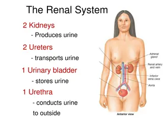
The two kidneys lie on the posterior wall of
the abdomen, outside the peritoneal cavity.
Each kidney of the adult human weighs about
150
grams
and is about the size of a clenched
fist.
The medial side of each kidney contains
an indented region called the
hilum
through
which pass the renal artery
and vein,
lymphatics, nerve supply, and
ureter
, which
carries the final urine from the kidney to the
bladder, where it is stored until emptied.
The kidney is surrounded by a tough,
fibrous
capsule
that protects its
delicate inner
structures.
Kidney
If the kidney is bisected from top to
bottom, the two major regions that can be
visualized are the outer
cortex and the inner
region referred to as the
medulla
.
The medulla is divided into multiple cone-
shaped masses of tissue called
renal
pyramids
.
The base of each pyramid originates at
the border between the cortex and medulla
and terminates in the
papilla
,
which projects
into the space of the
renal pelvis, a
funnel-
shaped continuation of the upper end of the
ureter.
The outer border of the pelvis is divided
into open-ended pouches called
major
calyces
that extend
downward and divide
into
minor calyces
, which collect
urine
from the tubules of each
papilla
.
The walls of the
calyces, pelvis,
and ureter contain contractile elements
that propel the urine toward the
bladder,
where urine
is stored until it is emptied by
micturition
.
T
H
E
N
E
P
H
R
O
N
I
S
T
H
E
F
U
N
C
T
I
O
N
A
L
U
N
I
T
O
F
T
H
E
K
I
D
N
E
Y
Each kidney in the human contains about
1 million
nephrons, each capable of forming urine.
The kidney
cannot regenerate new nephrons. Therefore, with renal
injury, disease, or normal aging, there is a gradual
decrease in nephron number.
After age 40, the number of functioning
nephrons usually decreases about 10 per cent every 10
years; thus, at age 80, many people have 40 per cent
fewer functioning nephrons than they did at age 40.
This loss is not life threatening because adaptive
changes in the remaining nephrons allow them to
excrete the proper amounts of water, electrolytes, and
waste products.
Nephron
Each
Nephron
contains
(1) a tuft of glomerular capillaries called the
glomerulus
, through which large
amounts
of fluid are filtered from the blood, and
(2) a long
tubule
in which the filtered fluid is
converted into
urine on its way to the
pelvis of the kidney
The glomerulus contains a network of
branching and anastomosing glomerular
capillaries that, compared with other
capillaries, have high hydrostatic pressure
(
about 60 mm Hg
).
The glomerular capillaries are covered
by epithelial cells, and the total glomerulus
is encased in
Bowman’s capsule.
Fluid
filtered from the glomerular
capillaries flows into Bowman’s capsule
and then into the
proximal tubule
,
which
lies in the cortex of the kidney.
From the proximal tubule, fluid flows into the
loop
of Henle
, which dips into the renal medulla.
Each loop
consists of a
descending and an
ascending limb. The
walls of the descending limb
and the lower end of the ascending limb are very
thin and therefore are called the
thin segment of the
loop of Henle. After the ascending
limb of the loop
has returned partway back to
The cortex, its wall becomes much thicker, and it
is referred to as the
thick segment of the ascending
limb.
At the end of the thick ascending limb is a
short segment, which is actually a plaque in its
wall, known as the
macula densa
.
The macula
densa plays an important role in
controlling nephron function. Beyond the macula
densa, fluid enters the
distal tubule
, which, like
the proximal tubule, lies in the
renal cortex.
This is followed by the
connecting tubule
and the
cortical collecting tubule
, which lead to the
Cortical collecting duct.
The initial parts of 8 to 10 cortical collecting
ducts join to form a single larger collecting duct
that runs downward into the medulla and
becomes the
medullary collecting duct.
The collecting ducts merge to form
progressively larger ducts that eventually empty
into the renal pelvis through the tips of the
renal
papillae
.
In each kidney, there are about
250 of the
very large collecting ducts, each of which
collects urine from about 4000 nephrons.
D
i
f
f
e
r
e
n
c
e
s
i
n
N
e
p
h
r
o
n
S
t
r
u
c
t
u
r
e
:
C
o
r
t
i
c
a
l
a
n
d
J
u
x
t
a
m
e
d
u
l
l
a
r
y
N
e
p
h
r
o
n
s
.
Those nephrons that have glomeruli
located in the outer cortex are called
cortical
nephrons
; they have
short loops of Henle
that
penetrate only a short distance into the
medulla. About 20 to 30 per cent of the
nephrons have glomeruli that lie deep in the
renal cortex near the medulla and are called
juxtamedullary nephrons
.
These nephrons
have
long loops of Henle
that dip deeply into
the medulla, in some cases all the way to the
tips of the renal papillae.
U
r
i
n
e
F
o
r
m
a
t
i
o
n
(1) Glomerular filtration,
(2) Reabsorption of substances from the renal
tubules into the blood, and
(3) Secretion of substances from the blood into the
renal tubules.
Urinary excretion rate =
Filtration rate
-
Reabsorption rate
+
Secretion
rate
Urine formation begins when a large amount
of fluid that is virtually free of protein is filtered
from the glomerular capillaries into Bowman’s
capsule. Most substances in the plasma, except
for proteins, are freely filtered, so that their
concentration in the glomerular filtrate in
Bowman’s capsule is almost the same as in the
plasma. As filtered fluid leaves Bowman’s
capsule and passes through the tubules, it is
modified by reabsorption of water and specific
solutes back into the blood or by secretion of
other substances from the peritubular capillaries
into the tubules.
G
L
O
M
E
R
U
L
A
R
F
I
L
T
R
A
T
I
O
N
—
T
H
E
F
I
R
S
T
S
T
E
P
I
N
U
R
I
N
E
F
O
R
M
A
T
I
O
N
Urine formation begins with filtration of large
amounts of fluid through the glomerular capillaries
into Bowman’s capsule. Like most capillaries, the
glom-
erular capillaries are relatively impermeable to
proteins,
so that the filtered fluid (called the glomerular filtrate)
is essentially protein-free and devoid of cellular
elements,
including red blood cells.
Almost one half of the plasma calcium and
most of the plasma fatty acids are bound
to proteins, and these bound portions are
not filtered through the glomerular
capillaries
REFERENCE
•
Arthur C. Guyton, 2005, Text Book of Medical Physiology, WB
Saunders’s, USA.
•
C. C Chatterjee, 1985, Human Physiology Vol I & Vol II.
11thEdn, Kalyani Mukerjee
Publications, Kolkata, India.
•
Kathleen, J.W. Wilson and Anne Waugh. 1998. Ross and
Wilson Anatomy and Physiology in health and illness. (8th
Edition). Churchchill Livingstone, New York.
•
Gerald J. Tortora and Sandra Reynolds. 2003. Principles of
Anatomy and Physiology. (10th Edition). John Wiley and Sons.
Inc. Pub. New York.
•
Abraham White., Philip Handler and Emil L. Smith. 1983.
Principles of Biochemistry.
(6th Edition). Tata Mc Graw – Hill Publishing Company, New
Delhi.
The kidneys are vital organs responsible for urine formation. Understanding their structure, function, and the role of nephrons is crucial for health and disease management. Each kidney consists of millions of nephrons, the functional units that filter blood and regulate electrolyte balance. As we age, there is a natural decline in nephron numbers, affecting renal function. Proper knowledge of kidney anatomy and physiology is essential for maintaining overall health.

Download Presentation

Please find below an Image/Link to download the presentation.
The content on the website is provided AS IS for your information and personal use only. It may not be sold, licensed, or shared on other websites without obtaining consent from the author.If you encounter any issues during the download, it is possible that the publisher has removed the file from their server.
You are allowed to download the files provided on this website for personal or commercial use, subject to the condition that they are used lawfully. All files are the property of their respective owners.
The content on the website is provided AS IS for your information and personal use only. It may not be sold, licensed, or shared on other websites without obtaining consent from the author.
E N D
Presentation Transcript
Kidney Structure, Function and Urine formation CLASS: I M.Sc., UNIT: 5 PREPARED BY: A. BENNO SUSAI
PHYSIOLOGIC ANATOMY OF THE KIDNEYS the abdomen, outside the peritoneal cavity. Each kidney of the adult human weighs about 150 grams and is about the size of a clenched fist. The medial side of each kidney contains an indented region called the hilum through which pass the renal artery and vein, lymphatics, nerve supply, and ureter, which carries the final urine from the kidney to the bladder, where it is stored until emptied. The kidney is surrounded by a tough, fibrous capsule that protects its delicate inner structures. The two kidneys lie on the posterior wall of
bottom, the two major regions that can be visualized are the outer cortex and the inner region referred to as the medulla. The medulla is divided into multiple cone- shaped masses of tissue called renal pyramids. The base of each pyramid originates at the border between the cortex and medulla and terminates in the papilla, which projects into the space of the renal pelvis, a funnel- shaped continuation of the upper end of the ureter. If the kidney is bisected from top to
into open-ended pouches called major calyces that extend downward and divide into minor calyces, which collect urine from the tubules of each papilla. The walls of the calyces, pelvis, and ureter contain contractile elements that propel the urine toward the bladder, where urine is stored until it is emptied by micturition. The outer border of the pelvis is divided
THE NEPHRON IS THE FUNCTIONAL UNIT OF THE KIDNEY Each kidney in the human contains about 1 million nephrons, each capable of forming urine. The kidney cannot regenerate new nephrons. Therefore, with renal injury, disease, or normal aging, there is a gradual decrease in nephron number. After age 40, the number of functioning nephrons usually decreases about 10 per cent every 10 years; thus, at age 80, many people have 40 per cent fewer functioning nephrons than they did at age 40. This loss is not life threatening because adaptive changes in the remaining nephrons allow them to excrete the proper amounts of water, electrolytes, and waste products.
Nephron Each Nephron contains (1) a tuft of glomerular capillaries called the glomerulus, through which large amounts of fluid are filtered from the blood, and (2) a long tubule in which the filtered fluid is converted into urine on its way to the pelvis of the kidney
branching and anastomosing glomerular capillaries that, compared with other capillaries, have high hydrostatic pressure (about 60 mm Hg). The glomerular capillaries are covered by epithelial cells, and the total glomerulus is encased in Bowman s capsule. Fluid filtered from the glomerular capillaries flows into Bowman s capsule and then into the proximal tubule, which lies in the cortex of the kidney. The glomerulus contains a network of
From the proximal tubule, fluid flows into the loop of Henle, which dips into the renal medulla. Each loop consists of a descending and an ascending limb. The walls of the descending limb and the lower end of the ascending limb are very thin and therefore are called the thin segment of the loop of Henle. After the ascending limb of the loop has returned partway back to The cortex, its wall becomes much thicker, and it is referred to as the thick segment of the ascending limb.
short segment, which is actually a plaque in its wall, known as the macula densa. The macula densa plays an important role in controlling nephron function. Beyond the macula densa, fluid enters the distal tubule, which, like the proximal tubule, lies in the renal cortex. This is followed by the connecting tubule and the cortical collecting tubule, which lead to the Cortical collecting duct. At the end of the thick ascending limb is a
The initial parts of 8 to 10 cortical collecting ducts join to form a single larger collecting duct that runs downward into the medulla and becomes the medullary collecting duct. The collecting ducts merge to form progressively larger ducts that eventually empty into the renal pelvis through the tips of the renal papillae. In each kidney, there are about 250 of the very large collecting ducts, each of which collects urine from about 4000 nephrons.
Differences in Nephron Structure: Cortical and Juxtamedullary Nephrons. Those nephrons that have glomeruli located in the outer cortex are called cortical nephrons; they have short loops of Henle that penetrate only a short distance into the medulla. About 20 to 30 per cent of the nephrons have glomeruli that lie deep in the renal cortex near the medulla and are called juxtamedullary nephrons. These nephrons have long loops of Henle that dip deeply into the medulla, in some cases all the way to the tips of the renal papillae.
Urine Formation (1) Glomerular filtration, (2) Reabsorption of substances from the renal tubules into the blood, and (3) Secretion of substances from the blood into the renal tubules. Urinary excretion rate =Filtration rate - Reabsorption rate + Secretion rate
of fluid that is virtually free of protein is filtered from the glomerular capillaries into Bowman s capsule. Most substances in the plasma, except for proteins, are freely filtered, so that their concentration in the glomerular filtrate in Bowman s capsule is almost the same as in the plasma. As filtered fluid leaves Bowman s capsule and passes through the tubules, it is modified by reabsorption of water and specific solutes back into the blood or by secretion of other substances from the peritubular capillaries into the tubules. Urine formation begins when a large amount
GLOMERULAR FILTRATIONTHE FIRST STEP IN URINE FORMATION Urine formation begins with filtration of large amounts of fluid through the glomerular capillaries into Bowman s capsule. Like most capillaries, the glom- erular capillaries are relatively impermeable to proteins, so that the filtered fluid (called the glomerular filtrate) is essentially protein-free and devoid of cellular elements, including red blood cells.
Almost one half of the plasma calcium and most of the plasma fatty acids are bound to proteins, and these bound portions are not filtered through the glomerular capillaries
REFERENCE Arthur C. Guyton, 2005, Text Book of Medical Physiology, WB Saunders s, USA. C. C Chatterjee, 1985, Human Physiology Vol I & Vol II. 11thEdn, Kalyani Mukerjee Publications, Kolkata, India. Kathleen, J.W. Wilson and Anne Waugh. 1998. Ross and Wilson Anatomy and Physiology in health and illness. (8th Edition). Churchchill Livingstone, New York. Gerald J. Tortora and Sandra Reynolds. 2003. Principles of Anatomy and Physiology. (10th Edition). John Wiley and Sons. Inc. Pub. New York. Abraham White., Philip Handler and Emil L. Smith. 1983. Principles of Biochemistry. (6th Edition). Tata Mc Graw Hill Publishing Company, New Delhi.