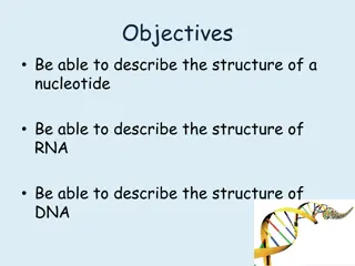
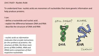
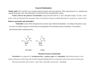
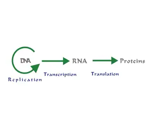
Exploring the Structural Model of DNA and RNA

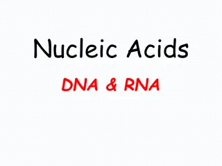
Nucleic Acids
DNA & RNA


The Double Helix (DNA)
Structural model:

•
Model proposed by Watson & Crick, 1953
•
Two sugar-phosphate strands, next to each
other, but running in opposite directions.
•
Specific Hydrogen bonds
occur among bases
from one chain to the other:
A---T
,
C---G
Due to this specificity, a certain base on one
strand indicates a certain base in the other
.
•
The 2 strands intertwine, forming a double-
helix that winds around a central axis
Untwisted it
looks like this:
•
The
sides
of the ladder are:
P =
phosphate
S
=
sugar
molecule
•
The
steps
of the ladder are C, G, T, A =
nitrogenous bases
(Nitrogenous means containing the
element
nitrogen
.
)
A =
Adenine
T =
Thymine
A always pairs with T in DNA
C =
Cytosine
G =
Guanine
C always pairs with G in DNA
(
A
pples are
T
asty)
(
C
ookies are
G
ood)
Secondary Structure: DNA Double Helix
•
In DNA there are two strands of nucleotides that wind together
in a
double helix
- the strands run in opposite directions
- the bases are are arranged in step-like pairs
- the
base pairs
are held together by hydrogen bonding
•
The pairing of the bases from the two strands is very specific
•
The
complimentary base pairs
are
A-T
and
G-C
- two hydrogen bonds form between A and T
- three hydrogen bonds form between G and C
•
Each pair consists of a purine and a pyrimidine, so they are the
same width, keeping the two strands at equal distances from
each other
Model of
DNA
:
•
The model was developed
by
Watson
and
Crick
in
1953.
•
They received a
nobel prize
in 1962 for their work.
•
The model looks like a
twisted ladder –
double
helix
.
N
u
c
l
e
i
c
A
c
i
d
S
t
r
u
c
t
u
r
e
“
B
a
s
e
P
a
i
r
i
n
g
”
D
N
A
b
a
s
e
-
p
a
i
r
i
n
g
i
s
a
n
t
i
p
a
r
a
l
l
e
l
i.e. 5’ - 3’ (l-r) on top : 5’ - 3’ (r-l) on
Discovering the structure of DNA
Erwin Chargaff – (1905-2002)
•
Columbia University, NY
•
Investigated the composition of DNA
•
His findings by 1950 strongly
suggested the base-pairings
of A-T & G-C
•
Met with Watson and Crick in
1952 and shared his findings
•
“Chargaff’s rule” A = T & C = G
Discovering the structure of DNA
•
DNA
= Deoxyribose nucleic acid
•
Present in all living cells
•
Contains all the information
•
Nucleotides
:
•
a subunit that consists of:
•
a sugar (deoxyribose)
•
a phosphate
•
and one nitrogen base – 4 different bases
•
Adenine (A) and Thymine (T)
•
Guanine (G) and Cytosine (C)
The strands separate
N
u
c
l
e
i
c
A
c
i
d
S
t
r
u
c
t
u
r
e
“
B
a
s
e
P
a
i
r
i
n
g
”
RNA [normally] exists as a single stranded polymer
DNA exists as a double stranded polymer
DNA double strand is created by hydrogen bonds between nucleotides
Nucleotides always bind to complementary nucleotides
(
2
H
-
b
o
n
d
s
)
(
3
H
-
b
o
n
d
s
)
Practice DNA Base Pairs
Complementarity of DNA strands
•
Two chains differ in sequence
(sequence is read from 5’ to 3’)
•
Two chains are
complementary
•
Two chains run antiparallel
N
u
c
l
e
i
c
A
c
i
d
S
t
r
u
c
t
u
r
e
“
B
a
s
e
P
a
i
r
i
n
g
”
N
u
c
l
e
i
c
A
c
i
d
S
t
r
u
c
t
u
r
e
P
o
l
y
m
e
r
i
z
a
t
i
o
n
5
’
3
’
N
u
c
l
e
i
c
A
c
i
d
S
t
r
u
c
t
u
r
e
P
o
l
y
m
e
r
i
z
a
t
i
o
n
+
Phosphodiesterase
Delve into the intricate world of nucleic acids, DNA, and RNA through the groundbreaking structural model proposed by Watson and Crick in 1953. Unravel the double helix structure, base pairings, and secondary structures of DNA, alongside the pivotal discoveries by Erwin Chargaff in understanding DNA composition. Witness the beauty of DNA's model and learn about antiparallel base pairings that form the foundation of genetic information storage.

Download Presentation

Please find below an Image/Link to download the presentation.
The content on the website is provided AS IS for your information and personal use only. It may not be sold, licensed, or shared on other websites without obtaining consent from the author.If you encounter any issues during the download, it is possible that the publisher has removed the file from their server.
You are allowed to download the files provided on this website for personal or commercial use, subject to the condition that they are used lawfully. All files are the property of their respective owners.
The content on the website is provided AS IS for your information and personal use only. It may not be sold, licensed, or shared on other websites without obtaining consent from the author.
E N D
Presentation Transcript
Nucleic Acids DNA & RNA
The Double Helix (DNA) Structural model: Model proposed by Watson & Crick, 1953 Two sugar-phosphate strands, next to each other, but running in opposite directions. Specific Hydrogen bonds occur among bases from one chain to the other: A---T , C---G Due to this specificity, a certain base on one strand indicates a certain base in the other. The 2 strands intertwine, forming a double- helix that winds around a central axis
The sides of the ladder are: Untwisted it looks like this: P = phosphate S = sugar molecule The steps of the ladder are C, G, T, A = nitrogenous bases (Nitrogenous means containing the element nitrogen.) A = Adenine T = Thymine A always pairs with T in DNA (Apples are Tasty) C = Cytosine G = Guanine C always pairs with G in DNA (Cookies are Good) Nucleotide
Secondary Structure: DNA Double Helix In DNA there are two strands of nucleotides that wind together in a double helix - the strands run in opposite directions - the bases are are arranged in step-like pairs - the base pairs are held together by hydrogen bonding The pairing of the bases from the two strands is very specific The complimentary base pairs are A-T and G-C - two hydrogen bonds form between A and T - three hydrogen bonds form between G and C Each pair consists of a purine and a pyrimidine, so they are the same width, keeping the two strands at equal distances from each other
Model of DNA: The model was developed by Watson and Crick in 1953. They received a nobel prize in 1962 for their work. The model looks like a twisted ladder double helix.
Nucleic Acid Structure Base Pairing DNA base-pairing is antiparallel i.e. 5 - 3 (l-r) on top : 5 - 3 (r-l) on 5 3 A T C G C G T A G C A T 3 5
Discovering the structure of DNA Erwin Chargaff (1905-2002) Columbia University, NY Investigated the composition of DNA His findings by 1950 strongly suggested the base-pairings of A-T & G-C Met with Watson and Crick in 1952 and shared his findings Chargaff s rule A = T & C = G
Discovering the structure of DNA DNA = Deoxyribose nucleic acid Present in all living cells Contains all the information Nucleotides: a subunit that consists of: a sugar (deoxyribose) a phosphate and one nitrogen base 4 different bases Adenine (A) and Thymine (T) Guanine (G) and Cytosine (C)
The strands separate PO4 PO4 PO4 PO4 PO4 PO4 PO4 PO4 PO4 PO4 PO4 PO4 PO4 PO4 PO4 PO4
Nucleic Acid Structure Base Pairing RNA [normally] exists as a single stranded polymer DNA exists as a double stranded polymer DNA double strand is created by hydrogen bonds between nucleotides Nucleotides always bind to complementary nucleotides A T (2 H-bonds) G C (3 H-bonds)
Practice DNA Base Pairs G A T T A C A C T A A T G T
Complementarity of DNA strands Two chains differ in sequence (sequence is read from 5 to 3 ) Two chains are complementary Two chains run antiparallel
Nucleic Acid Structure Base Pairing
Nucleic Acid Structure Polymerization 3 5 Sugar Phosphate backbone A C C T G A Bases 3 5 TAGCAC
Nucleic Acid Structure Polymerization P P P P P P N N C C S S Phosphodiesterase P + N P P C (PPi) S P P P N C S

")