Exploring Living Cells with Light Microscopes in Classrooms
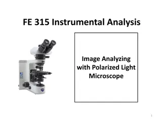
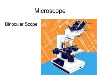
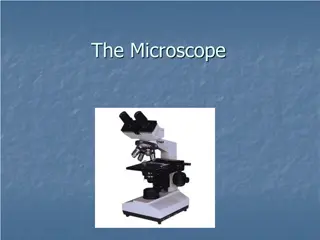
Light microscopes are commonly used in classrooms to examine living cells, even though they do not provide high magnification levels. Magnification, representing the ratio between an object's image and real size, is limited to about 1,000 times with light microscopes.
Download Presentation

Please find below an Image/Link to download the presentation.
The content on the website is provided AS IS for your information and personal use only. It may not be sold, licensed, or shared on other websites without obtaining consent from the author. Download presentation by click this link. If you encounter any issues during the download, it is possible that the publisher has removed the file from their server.
E N D
Presentation Transcript
Light microscopes are commonly used in classrooms. Living cells can be examined. The downfall is that they do not magnify a tremendous amount. Magnificationis the ratio of an object s image to its real size. A light microscope can magnify effectively to about 1,000 times the real size of a specimen. Resolution is a measure of image clarity. It is the minimum distance two points can be separated and still be distinguished as two separate points. 2
In general, electron microscopes are much more powerful than light microscopes. They use electrons to see an image rather than light. The researcher views the image on a screen rather than through an eyepiece. Scanning Electron Microscopes (SEM) Studies the surface of cells, looks at the outside of the cell For the SEM, the sample surface is covered with a thin film of gold. The beam excites electrons on the surface of the sample, and these secondary electrons are collected and focused on a screen, producing a surface image of the specimen. SEMs have great depth of field, resulting in an image that seems three- dimensional. 3
Transmission Electron Microscope (TEM) studies the insides of the cell; the preparation kills the cells and therefore cannot be used on living cells A TEM aims an electron beam through a very thin section of the specimen. To enhance contrast, the thin sections are stained with atoms of heavy metals, which attach to certain cellular structures. 4
This is a process that allows scientists to study different parts of the cell. It separates the different parts of the cell based on density, and then those separated sections can be tested for their functions. 5
Plasma membrane selective barrier (more on this later ) Cytosol clear jelly-like stuff that is made up of mostly water and enzymes and the organelles are suspended in this Cytoplasm In Prokaryotes, the whole interior In Eukaryotes, the region between the nucleus and plasma membrane (cytosol and organelles) Ribosomes site of protein synthesis Chromosomes genetic information In Prokaryotes, this is in the NUCLEOID REGION In Eukaryotes, this is in the NUCLEUS 6
Prokaryotic Cells These cells are a lot simpler and smaller than eukaryotic cells, and they lack organelles. They DO have chromosomes, even though they do NOT have a nucleus. Instead, the chromosomes are confined in the nucleoid region. 7
Eukaryotic Cells these cells are larger and more complex than prokaryotic cells. They have membrane-bound organelles and a nucleus. Plant and animal cells are examples of eukaryotic cells. 8
COPY THIS SLIDE!! The endosymbiotic theory is an evolutionary theory that explains the origin of eukaryotic cells from prokaryotes. It states that several key organelles of eukaryotes originated as a symbiotic relationship between separate single-celled organisms. Observations that support the endosymbiotic theory: Both mitochondria and chloroplasts: Have their own DNA Their chromosomes are circular like prokaryotic ones Have their own ribosomes similar to prokaryotic ones Can self-replicate Can do transcription and translation Their inner membranes are similar to prokaryotic membranes Are approximately the size of bacteria Use many prokaryotic-like enzymes 9
The plasma membrane determines what can come in and go out of the cell. It is made up of a phospholipid bilayer and has proteins throughout it. It helps transport things out of the cell and also brings things into the cell. More on this in Chapter 7... 10
The logistics of carrying out cellular metabolism set limits on cell size. If a cell gets too big, it will not be able to transport out waste quickly enough, and the cell will be poisoned. As a cell increases in size, its volume increases faster than its surface area. So, smaller objects have a higher ratio of Surface Area to Volume than larger cells. (Some cells have features that increase their surface area like folding or microvilli) Eukaryotic cells are typically much larger than prokaryotic cells. Larger organisms do not generally have larger cells than smaller organisms, simply more cells. 11
Nucleus Control center of the cell; contains most of the DNA (genes) of the cell. Nucleolus Part inside the nucleus that makes rRNA (makes ribosomes) Nuclear Envelope Double membrane that surrounds the nucleus; it has pores so that the RNA can get out Nuclear Lamina a network of protein filaments INSIDE the nucleus used to help with the structure Chromatin uncoiled chromosomes for most of the cell cycle the DNA is in this form (they coil up into chromosomes before cell division) 12
Ribosomes are made up of rRNA and do NOT have a membrane surrounding them. They are the site of protein synthesis. They are made up of a small subunit and a larger subunit. There are 3 binding sites. We will look more at this when we do the chapter on protein synthesis. Free Ribosomes are NOT attached to the ER and synthesize proteins that function within the cytosol Bound Ribosomes attached to ER or nuclear envelope; synthesize proteins that are inserted into membranes or that are secreted from the cell Ribosomes can switch between free and bound (they all START as free when they are synthesizing a protein) 13
A system in internal membranes which includes Nuclear Envelope Endoplasmic Reticulum Golgi apparatus Lysosomes Vesicles Vacuoles Plasma Membrane Each of these structures are either directly continuous or connected by vesicles 14
ER the ER is connected to the nuclear envelope and has a very folded structure to increase the surface area; it accounts for more than HALF the membranes in a eukaryotic cell. There are two continuous parts to the ER Smooth and Rough. The ER can also aid in moving substances around the cell like a track for the organelles to move around on. 15
Cells that have high levels of smooth ER can be used for lipid/hormone synthesis or detoxification Smooth ER lacks ribosomes; function = makes lipids (hormones, steroids); detoxifies drugs and alcohol, breaks down carbs; stores calcium ions In the smooth ER of the liver, enzymes help detoxify poisons and drugs such as alcohol and barbiturates. Frequent use of these drugs leads to the proliferation of smooth ER in liver cells, increasing the rate of detoxification. This proliferation of smooth ER increases tolerance to the target and other drugs, so higher doses are required to achieve the same effect. Stores calcium ions which is very important in muscle cells! - - - Rough ER lined with ribosomes; function = makes secretory proteins and makes membranes; LOTS of Rough ER in cells that secrete proteins. As a polypeptide chain grows from a bound ribosome, it is threaded into the ER lumen through a pore formed by a protein complex in the ER membrane. As the new polypeptide enters the ER lumen, it folds into its native shape. Most secretory polypeptides are glycoproteins (proteins and carbs) Secretory proteins are packaged in transport vesicles that can carry proteins from one part of the cell to another. - - - - - 16
The golgi is responsible for packaging, modifying, and secreting substances from the cell. It is can modify substances that have come to it from the ER, OR it can manufacture its own molecules. It has a receiving side (cis) and a shipping side (trans). It generally gets materials from the ER, processes and alters them slightly, puts them in vesicles, and sends them to wherever they are needed inside or outside of the cell. Cells that have a large about of Golgi and Rough ER make lots of protein; they have the ER to make the proteins and the Golgi to pack and secrete them 17
Lysosomes are found only in ANIMAL cellswaste that needs to get broken down in plants gets put into the vacuole and enzymes in there break it down Lysosomes digestive compartments of the cell; sacs of enzymes; can digest and recycle old parts of the cell (AUTOPHAGY!); can digest/destroy dangerous things that are taken into the cell (PHAGOCYTOSIS); some are formed from budding from the golgi; these play an important role in embryo development and also in programmed cell death 18
Animal cells have vacuoles mostly for storage. They are much smaller and more numerous than those in plant cells. Food vacuoles are used when cells take in food by endocytosis. They then combine with lysosomes to digest the food . Contractile vacuoles are found mostly in single celled animals. These structures get rid of excess water so that they cell does not burst. 20
Plant cells have a large central vacuole. Here they store water, waste, and enzymes. It also helps with the structure of a plant cell by increasing or decreasing the turgor pressure depending on how much water is available to the plant cell at that time. Tonoplast membrane surrounding the vacuole in plant cells 21
The endosymbiont theory states that an early ancestor of eukaryotic cells engulfed an oxygen-using non-photosynthetic prokaryotic cell. Over the course of evolution, the host cell and its endosymbiont merged into a single organism, a eukaryotic cell with a mitochondrion. There is considerable evidence to support the endosymbiont theory for the origin of mitochondria and chloroplasts: - They both have double membranes - They both contain ribosomes and their own DNA - They both grow and reproduce on their own Mitochondria site of cellular respiration, use oxygen to generate ATP by extracting energy from sugars, fats, and other fuels Chloroplasts found in plants and algae, site of photosynthesis; they convert solar energy to chemical energy by absorbing sunshine and using it to synthesize new organic compounds such as sugars from CO2 and H2O 22
The mitochondria is considered to be the powerhouse of the cell. This is where cellular respiration takes place. It has a double membrane with the inner membrane highly folded to increase the surface area. Cells that are high in aerobic activity have more mitochondria. Cells that are high in mitochondria are typically involved in activities that utilize a lot of ATP; like cells involved in locomotion/ movement (ex. muscle cells) or if it is paired with cilia/flagella Cristae = folds of the membrane in mitochondria 23
Chloroplasts contains the pigment chlorophyll; This is where photosynthesis takes place in autotrophs. It is an organelle that has a double membrane surrounding clear liquid (stroma Calvin cycle dark reactions occur here) and stacks of thylakoids (where the light reactions happen) called grana. The chloroplast belongs to a family of plant structures called plastids. Amyloplasts are colorless plastids that store starch in roots and tubers. Chromoplasts store pigments for fruits and flowers 24
Peroxisomes have a variety of functions in cells. They produce hydrogen peroxide as a byproduct, but then immediately break it down into water and oxygen. These organelles help detoxify alcohol in the liver. 25
When you think of a cell that lacks membrane-bound organelles you probably think of a prokaryotic cell. However there are eukaryotic cells that lack organelles too! Red Blood Cells lack organelles to have more room to carry oxygen Xylem dead at functional maturity and carry water up through plants 26
The cytoskeleton is the structural support in animal cells. It helps to anchor organelles and also aids in cell movement. There are 3 main components of the cytoskeleton: microtubules, microfilaments, and intermediate filaments. Microtubules are the thickest of the three types of fibers; microfilaments (or actin filaments) are the thinnest; and intermediate filaments are fibers with diameters in a middle range. 27
Microtubules hollow rods that make up part of the cytoskeleton; made of the protein tubulin; helps chromosomes move during cell division and also helps move organelles around Cilia and flagella (cell movement) are made out of microtubules. They use motor proteins to move. 28
Centrosomes are located near the nucleus and this is where the microtubules are made. Centrioles are organelles that are found within centrosomes in ANIMAL cells; they help with cell division. 29
Cilia and flagella are two structures that enable cells to move. They have similar structures, except that flagella are longer. They are made of microtubules that extend from the cell and covered by the PM. Functions of Cilia and Flagella: The sperm of animals, algae, and some plants have flagella. Cilia lining the trachea sweep mucus carrying trapped debris out of the lungs. In the reproductive tract, cilia lining the oviducts help move an egg toward the uterus. 30
Basal Body anchors cilia and flagella in the cell; structure is identical to a centriole The motor molecules (dynein arms) bend the cilia and flagella. Think of a line of 15 people all standing on the ground. Imagine a big log on the floor that they are all straddling. If, at the same time, they all picked it up and shoved it backwards between their legs then dropped it, and then repeated the process again, eventually the log would move down the line, even though the people aren t moving. That is what is happening with the cilia and flagella. Depending on which direction the microtubules are moved, that determines which way the cilia or flagella bends. The general term for their structure is the 9 + 2 arrangement. This translates to 9 pairs of microtubules around the outside with two individual ones in the middle. 31
Microfilaments are the second part of the animal cytoskeleton. They are made up of the protein actin and are solid rods. They are very thin, they are designed to bear tension, and they work with myosin in muscle fibers. They also aid in cytoplasmic streaming. 32
Intermediate filaments are the last part of the animal cytoskeleton. These are more permanent, and don t break down to reassemble as much. They are created from a family of proteins called keratins. Intermediate filaments have a variety of functions in the cell. 34
Cell Walls protect the cell, maintains its shape, prevents excessive water uptake, and supports the plant against gravity. Primary Cell Wall starts as thin and flexible, develops from the cell plate after cell division; once the plant matures, it hardens to be more supportive Secondary Cell Wall this is found under the primary cell wall to strengthen it; it is not in every plant; it is very strong (wood is an example) Middle Lamella this glues plant cells together; it is found between the cell wall of one cell and the cell wall of the next cell 36
The extracellular matrix of animal cells provides support, adhesion, movement, and regulation. Extracellular Matrix (ECM) This is the structure that is found outside the animal cells. Because animals don t have cell walls, they need another structure to help support them. It is composed mostly of glycoproteins such as collagen, proteoglycans, and fibronectins. 37
Plasmodesmata are channels in the cell walls of plants. This allows substances to get from one plant cell to the next. They are similar to gap junctions in animal cells. 38
Tight Junctions fusing of membranes of adjacent cells; prevents leakage of fluid Desmosomes/ Anchoring Junctions/ Adhesion Junctions holds cells together in strong sheets Gap Junctions/ Communicating junctions holes between cells that allow cellular substances to pass through; similar to plasmodesmata in plant cells; very important in development for chemical signaling 40
Phospholipids are amphipathic molecules. This means that they have both hydrophobic (FA tails) and hydrophilic (phosphate heads) regions. The phospholipid molecules make up the structure of the phospholipid bilayer, which is the structure of the plasma membrane in all cells. Plasma membranes are SELECTIVELY PERMEABLE and only allow certain things to pass through 42
Models of membranes were developed long before membranes were first seen with electron microscopes in the 1950s. In 1915, membranes isolated from red blood cells were chemically analyzed and found to be composed of lipids and proteins. In 1925, two Dutch scientists reasoned that cell membranes must be phospholipid BILAYERS. The molecules in the bilayer are arranged such that the hydrophobic fatty acid tails are sheltered from water while the hydrophilic phosphate groups interact with water. 43
Their idea of the structure of the PM was that there was a phospholipid bilayer between two globular protein layers. This was hypothesized in 1935 and became widely accepted in the scientific community. 44
Scientists realized that the Davson-Danielli model was inaccurate for two reasons. First, not all membranes are the same. Second, measurements showed that membrane proteins are not very soluble in water. Since membrane proteins are amphipathic, their hydrophobic regions would be in contact with water. So, in 1972 Singer and Nicolson discovered the correct structure of the PM. They called this the fluid mosaic model. 45
Unsaturated FA chains allow the membrane to be more fluid and flexible because the phospholipid molecules are more spread out due to the kinks in the chains (double bonds = kinks) Saturated FA chains membrane is a little more rigid because the phospholipid molecules are packed tightly together (straight chains = single C-C bonds) Cholesterolis called a fluidity buffer for the membrane because it can make the membrane either more or less fluid depending on the temperature. In warm temperatures, it restrains the movement of phospholipids so decreases the fluidity. In cold temperatures, it increases fluidity by preventing tight packing of the phospholipids. 47
A membrane is a collage of different proteins embedded in the fluid matrix of the lipid bilayer. Proteins determine most of the membrane s specific functions. Two Main Types of Proteins: Integral Proteins Proteins that go all the way through the membrane; also called transmembrane proteins Peripheral Proteins Proteins that are on either on the inside or outside of the PM; they are held in place by either the ECM or cytoskeleton 48
Functions of Membrane Proteins: 1. Transport of specific solutes into or out of cells 2. Enzymatic activity, sometimes catalyzing one of a number of steps of a metabolic pathway 3. Signal transduction, relaying hormonal messages to the cell 4. Cell-cell recognition, allowing other proteins to attach two adjacent cells together 5. Intercellular joining of adjacent cells with gap or tight junctions 6. Attachment to the cytoskeleton and extracellular matrix, maintaining cell shape and stabilizing the location of certain membrane proteins 49
Main Function of membrane carbs is for cell to cell recognition and also to act as markers. Membrane carbohydrates may be covalently bonded to lipids, forming glycolipids, or more commonly to proteins, forming glycoproteins The carbohydrates on the outside of the plasma membrane vary from species to species, from individual to individual, and even from cell type to cell type within an individual. For example, The four human blood groups (A, B, AB, and O) differ in the carbohydrate part of glycoproteins on the surface of red blood cells. 50


