Diagram of Integumentary System Components

I
n
t
e
g
u
m
e
n
t
a
r
y
S
y
s
t
e
m
Skin (cutaneous
membrane)
Skin derivatives
Sweat
glands
Apocrine
Eccrine
Oil
(sebaceous)
glands
Hair
Shaft
Follicle
Nails

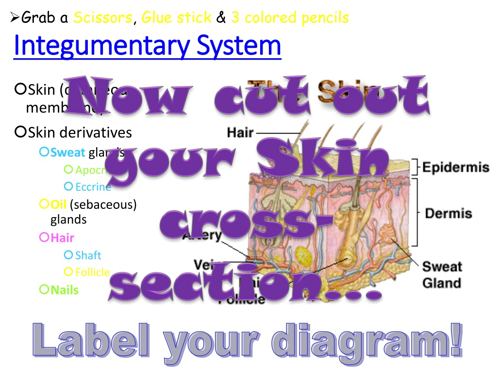
Grab a
Scissors
,
Glue stick
&
3 colored pencils


I
n
t
e
g
u
m
e
n
t
(
S
k
i
n
)
F
u
n
c
t
i
o
n
s
Protects
deeper tissue from:
Bumps
Chemical damage (acid & bases)
Bacterial damage
UV radiation
Thermal damage (heat/cold)
Desiccation
(drying out)
Aids
in:
Body heat loss & retention (controlled by NS)
Excretion of urea & uric acid
(perspiration)
Synthesis of Vitamin D

•
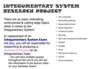
Hair color/dye
•
Burns/skin grafting
•
Sweat dysfunctions
•
Botox injections
•
Freckles & moles
•
Bruising
•
Skin color variations/ birthmarks
•
Tattoos
•
Gauges
•
Scars
•
Permanent make-up
•
Facials
•
Face transplants
There are so many interesting,
controversial & cutting-edge topics
when it comes to the
Integumentary System!
In replacement of an
Integumentary System Exam
(
60 pts
), you will be responsible for
researching & producing a
Research Project
on an
Integumentary topic.
- You will have multiple quizzes
throughout the unit & you will see
the information in the lecture notes
on your Semester Exam!
INTEGUMENTARY SYSTEM
RESEARCH PROJECT
•
Rubric that will be used
(60 pts)
…each criterion is worth
10 pts
.
•
Define
,
discuss
&
explain
the topic & its relevance/
importance to the Integumentary System using
anatomical/Integumentary terminologies
•
Works Cited in MLA Format
with Proper Research Citation
•
Minimum of
5 graphics
(
must pertain to topic
)
•
Minimum of
2 statistical analyses
via graph or raw data
•
Solo OR partners
actively participated
in research &
project
•
A
5 question Quiz
(Multiple Choice, Fill-in &/or
Identification) turned in with Research Project
INTEGUMENTARY SYSTEM
RESEARCH PROJECT
•
FORMAT of Project:
•
Physical model
•
Billboard display
•
Brochure/pamphlet
•
Video
•
Skit/play
•
Any other format EXCEPT a PowerPoint/
Prezi presentation
•
G
E
T
C
R
E
A
T
I
V
E
!!!!
INTEGUMENTARY SYSTEM
RESEARCH PROJECT
•
REMEMBER
: This is your
INTEGUMENTARY SYSTEM EXAM
grade!
•
There will be
NO IN-CLASS WORK TIME
dedicated, so it
will also require
out-of-class work time
!
•
Decided whether you would like to work with a
partner
or
solo
. Stand/sit next to your partner, if you have one.
•
I’m going to make my way around with my “
Can o’ Topics
”.
•
Please pick
1
!
•
Write your
TOPIC
on your
RUBRIC
!
•
Exchange phone numbers &/or e-mails
so you can
contact each other about meeting & dividing up work!
INTEGUMENTARY SYSTEM
RESEARCH PROJECT
I
n
t
e
g
u
m
e
n
t
a
r
y
S
y
s
t
e
m
Skin (cutaneous
membrane)
Skin derivatives
Sweat
glands
Apocrine
Eccrine
Oil
(sebaceous)
glands
Hair
Shaft
Follicle
Nails
Grab a
Scissors
,
Glue stick
&
3 colored pencils
S
k
i
n
S
t
r
u
c
t
u
r
e
•
Epidermis
—outer layer
•
Stratified squamous epithelium
•
Consists of
4-5 distinct layers
•
Often keratinized
(hardened by keratin)
•
Not vascular
•
Regenerated
every 25-45 days
•
Dermis
•
Dense connective tissue
•
Subcutaneous
tissue (
hypodermis
) is
deep
to dermis
•
Not part of skin
•
Anchors skin to underlying organs
•
Composed mostly of
adipose tissue
(fat)
L
a
y
e
r
s
o
f
t
h
e
E
p
i
d
e
r
m
i
s
•
From
deepest
to most
superficial
•
Stratum
basale
•
Deepest layer of epidermis
•
Cells undergoing mitosis
•
Daughter cells pushed upward to
become more superficial
•
Stratum
spinosum
•
Stratum
granulosum
•
Stratum
lucidum
•
Formed from dead cells of deeper
strata
•
Occurs only in thick, hairless skin of palms
of hands & soles of feet
•
Stratum
corneum
•
Dead cells are filled with keratin (
protective
protein prevents water loss from skin
)
Per. 3 - Mon
L
a
y
e
r
s
o
f
t
h
e
E
p
i
d
e
r
m
i
s
•
From most
superficial
to
deepest
•
Stratum
corneum
•
Stratum
lucidum
•
Stratum
granulosum
•
Stratum
spinosum
•
Stratum
basale
Per. 1 - Mon
4 Types of Cells
:
Keratinocytes
Produce keratin, the fibrous
protein that helps give the
epidermis its protective
properties
Melanocytes
Synthesize pigment called
melanin
Melanin picked up by
keratinocytes &
used to shade their nuclei
from UV radiation
Merkel cells
Combo of Merkel cell &
nerve ending (called Merkel
disc) functions as a sensory
receptor for touch
Langerhans’ cells
Macrophages that help to
activate immune system
Layers of the Epidermis
Per. 2, 4 - Mon
Normal Skin Color Determinants
•
Melanin
:
•
Pigment (
melanin
) produced by
melanocytes
•
Melanocytes are mostly in the
stratum
basale
•
Color is
yellow
to
brown
to black
•
Amount of melanin
produced depends upon
genetics & exposure to
sunlight (UV)
Color in the
Epidermis
•
What are the major
characteristics
of the
Epidermis?
•
What are the
4 types of cells
found
within the Epidermis & their functions?
•
What are the
5 layers
(strata) of the
Epidermis from deep to superficial?
Epidermis Review
Where’s this Headed?
•
What is the
picture
below called?
•
Which
of these forms of electromagnetic radiation are most
effected by the Integumentary System?
•
What are some
precautions
we can take to protect our
Int. Sys. from UV radiation?
•
What does “
SPF
”
stands
for & how does it
work
?
•
How do we
know
these
precautions actually work?
•
Do our bodies’ have a
natural defense
against UV?
Dermis
Strong, flexible connective
tissue
Cell types
:
Fibroblasts
Macrophages
Mast cells
White blood cells
Binds
the entire body together
Your HIDE
Innervated
(nerves)
&
vascularized
(blood)
Hair follicles, oil glands & sweat
glands
2 major layers:
Papillary layer
Reticular layer
Papillary Layer
Highly vascularized
Borders the
stratum basale
of
epidermis
Superior portion covered with
dermal papillae
, which house
blood capillaries
Touch & pain receptors
On palms & soles, these papillae
lie atop dermal ridges, which
produce whorled
epidermal
ridges
(
fingerprints
)
Layers of the
Dermis
Reticular Layer
•
Deep to papillary layer
•
~80% of dermis
•
Made up of
dense irregular
connective tissue
(
matrix consist
of high quantities of
collagen fibers
)
•
Helps dermis from being easily penetrated
•
Collagen
binds water, helping to
hydrate
the skin
•
Elastin fibers
give skin
elasticity
Layers of the
Dermis
Extreme stretching
of skin can tear dermis
(
stretch marks
) -
striae
Short-term trauma can cause a
blister
to
form (
fluid sac that forms
between epidermis
& dermis
)
Flexure lines
– dermal
folds
that
occur at
or near joints
where dermis is
tightly attached
to deeper underlying structures
Flexure
lines
Other Stuff about
the Dermis
Hypodermis
Known as
subcutaneous
tissue
or
superficial fascia
Structure
:
Has more adipose than dermis
Functions
:
Energy
reservoir
Thermal
insulation
Hypodermic injections
Into subcutaneous tissue
since
highly vascular
Hypodermis
•
Mid-
section
•
Outer
thighs
•
Buttocks
•
Breasts
Hypodermal (subcutaneous)
fat distribution
Normal Skin Color Determinants
Carotene
:
Yellow
-
orange
pigment found in
certain plant products
Accumulates in
stratum corneum
& in
adipose tissue
of hypodermis
Most obvious in palms & soles where S. corneum is
thickest
Color in the Dermis
& Hypodermis
Normal Skin Color
Determinants
Hemoglobin
:
Red
pigment of red blood cells
(
RBCs
)
Since Caucasian people contain relatively small
amounts of
melanin
, their skin is nearly transparent
which allows hemoglobin’s color to shine through
Color in the Dermis
& Hypodermis
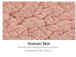
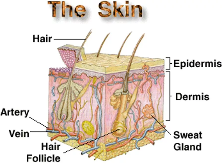
This diagram illustrates the various components of the Integumentary System, including the skin, sweat glands (apocrine and eccrine), oil glands (sebaceous), hair shaft, follicle, and nails. Use scissors, glue stick, and colored pencils to label the diagram effectively.

Download Presentation

Please find below an Image/Link to download the presentation.
The content on the website is provided AS IS for your information and personal use only. It may not be sold, licensed, or shared on other websites without obtaining consent from the author.If you encounter any issues during the download, it is possible that the publisher has removed the file from their server.
You are allowed to download the files provided on this website for personal or commercial use, subject to the condition that they are used lawfully. All files are the property of their respective owners.
The content on the website is provided AS IS for your information and personal use only. It may not be sold, licensed, or shared on other websites without obtaining consent from the author.
E N D
Presentation Transcript
Grab a Scissors, Glue stick & 3 colored pencils Integumentary System Integumentary System Skin (cutaneous membrane) Skin derivatives Sweat glands Apocrine Eccrine Oil (sebaceous) glands Hair Shaft Follicle Nails Label your diagram!
Add to the TOP of your foldable Integument (Skin) Integument (Skin) Functions Functions Protects deeper tissue from: Bumps Chemical damage (acid & bases) Bacterial damage UV radiation Thermal damage (heat/cold) Desiccation (drying out) Aids in: Body heat loss & retention (controlled by NS) Excretion of urea & uric acid (perspiration) Synthesis of Vitamin D
INTEGUMENTARY SYSTEM INTEGUMENTARY SYSTEM RESEARCH PROJECT RESEARCH PROJECT Hair color/dye There are so many interesting, controversial & cutting-edge topics when it comes to the Integumentary System! In replacement of an Integumentary System Exam (60 pts), you will be responsible for researching & producing a Research Project on an Integumentary topic. - You will have multiple quizzes throughout the unit & you will see the information in the lecture notes on your Semester Exam! Burns/skin grafting Sweat dysfunctions Botox injections Freckles & moles Bruising Skin color variations/ birthmarks Tattoos Gauges Scars Permanent make-up Facials Face transplants
INTEGUMENTARY SYSTEM INTEGUMENTARY SYSTEM RESEARCH PROJECT RESEARCH PROJECT Rubric that will be used (60 pts) each criterion is worth 10 pts. Define, discuss & explain the topic & its relevance/ importance to the Integumentary System using anatomical/Integumentary terminologies Works Cited in MLA Format with Proper Research Citation Minimum of 5 graphics (must pertain to topic) Minimum of 2 statistical analyses via graph or raw data Solo OR partners actively participated in research & project A 5 question Quiz (Multiple Choice, Fill-in &/or Identification) turned in with Research Project
INTEGUMENTARY SYSTEM INTEGUMENTARY SYSTEM RESEARCH PROJECT RESEARCH PROJECT FORMAT of Project: Physical model Billboard display Brochure/pamphlet Video Skit/play Any other format EXCEPT a PowerPoint/ Prezi presentation GET CREATIVE!!!!
INTEGUMENTARY SYSTEM INTEGUMENTARY SYSTEM RESEARCH PROJECT RESEARCH PROJECT REMEMBER: This is your INTEGUMENTARY SYSTEM EXAM grade! There will be NO IN-CLASS WORK TIME dedicated, so it will also require out-of-class work time! Decided whether you would like to work with a partner or solo. Stand/sit next to your partner, if you have one. I m going to make my way around with my Can o Topics . Please pick 1! Write your TOPIC on your RUBRIC! Exchange phone numbers &/or e-mails so you can contact each other about meeting & dividing up work!
Grab a Scissors, Glue stick & 3 colored pencils Integumentary System Integumentary System Skin (cutaneous membrane) Skin derivatives Sweat glands Apocrine Eccrine Oil (sebaceous) glands Hair Shaft Follicle Nails Label your diagram!
Skin Structure Skin Structure Epidermis outer layer Stratified squamous epithelium Consists of 4-5 distinct layers Often keratinized (hardened by keratin) Not vascular Regenerated every 25-45 days Dermis Dense connective tissue Subcutaneous tissue (hypodermis) is deep to dermis Not part of skin Anchors skin to underlying organs Composed mostly of adipose tissue (fat) Add these Add these tidbits tidbits to the area to the area each flap! BEHIND BEHIND each flap!
Per. 3 - Mon Layers of the Epidermis Layers of the Epidermis From deepest to most superficial Stratum basale Deepest layer of epidermis Cells undergoing mitosis Daughter cells pushed upward to become more superficial Stratum spinosum Stratum granulosum Stratum lucidum Formed from dead cells of deeper strata Occurs only in thick, hairless skin of palms of hands & soles of feet Stratum corneum Dead cells are filled with keratin (protective protein prevents water loss from skin) Write this in the
Per. 1 - Mon Layers of the Epidermis Layers of the Epidermis From most superficial to deepest Stratum corneum Stratum lucidum Stratum granulosum Stratum spinosum Stratum basale Color code your diagram Can you Can you come up come up with a with a Mnemonic Mnemonic to to remember remember the the layers? layers?
Per. 2, 4 - Mon Layers of the Epidermis 4 Types of Cells: Keratinocytes Produce keratin, the fibrous protein that helps give the epidermis its protective properties Melanocytes Synthesize pigment called melanin Melanin picked up by keratinocytes & used to shade their nuclei from UV radiation Merkel cells Combo of Merkel cell & nerve ending (called Merkel disc) functions as a sensory receptor for touch Langerhans cells Macrophages that help to activate immune system
Color in the Epidermis Normal Skin Color Determinants Melanin: Pigment (melanin) produced by melanocytes Melanocytes are mostly in the stratum basale Color is yellow to brown to black Amount of melanin produced depends upon genetics & exposure to sunlight (UV)
Epidermis Review What are the major characteristics of the Epidermis? What are the 4 types of cells found within the Epidermis & their functions? What are the 5 layers (strata) of the Epidermis from deep to superficial?
Wheres this Headed? What is the picture below called? Which of these forms of electromagnetic radiation are most effected by the Integumentary System? What are some precautions we can take to protect our Int. Sys. from UV radiation? What does SPF stands for & how does it work? How do we know these precautions actually work? Do our bodies have a natural defense against UV?
Add this info to your Foldable! Strong, flexible connective tissue Cell types: Fibroblasts Macrophages Mast cells White blood cells Binds the entire body together Your HIDE Innervated (nerves) & vascularized (blood) Hair follicles, oil glands & sweat glands 2 major layers: Papillary layer Reticular layer Dermis
Keep writing! Layers of the Dermis Papillary Layer Highly vascularized Borders the stratum basale of epidermis Superior portion covered with dermal papillae, which house blood capillaries Touch & pain receptors On palms & soles, these papillae lie atop dermal ridges, which produce whorled epidermal ridges (fingerprints)
Layers of the Dermis One more slide for Dermis! Reticular Layer Deep to papillary layer ~80% of dermis Made up of dense irregular connective tissue (matrix consist of high quantities of collagen fibers) Helps dermis from being easily penetrated Collagen binds water, helping to hydrate the skin Elastin fibers give skin elasticity
Other Stuff about the Dermis Flexure lines Extreme stretching of skin can tear dermis (stretch marks) - striae Short-term trauma can cause a blister to form (fluid sac that forms between epidermis & dermis) Last slide under Dermis! Flexure lines dermal folds that occur at or near joints where dermis is tightly attached to deeper underlying structures
Add this info to your Foldable under Hypodermis! Hypodermis Known as subcutaneous tissue or superficial fascia Structure: Has more adipose than dermis Functions: Energy reservoir Thermal insulation Hypodermic injections Into subcutaneous tissue since highly vascular Hypodermis
Hypodermal (subcutaneous) fat distribution Mid- section Outer thighs Buttocks Breasts Why is there
Add this info to your Foldable Color in the Dermis & Hypodermis Normal Skin Color Determinants Carotene: Yellow-orange pigment found in certain plant products Accumulates in stratum corneum & in adipose tissue of hypodermis Most obvious in palms & soles where S. corneum is thickest enter here
Add this info to your Foldable Color in the Dermis & Hypodermis Normal Skin Color Determinants Hemoglobin: Red pigment of red blood cells (RBCs) Since Caucasian people contain relatively small amounts of melanin, their skin is nearly transparent which allows hemoglobin s color to shine through


")