Diagnosis and Treatment Planning in Implantology
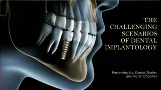
Dental implants play a crucial role in restoring the oral health of edentulous patients. This discipline covers the entire process, from diagnosis to restoration, ensuring optimal function and aesthetics. Understanding the indications and contraindications is essential for successful implant treatment.
Download Presentation

Please find below an Image/Link to download the presentation.
The content on the website is provided AS IS for your information and personal use only. It may not be sold, licensed, or shared on other websites without obtaining consent from the author.If you encounter any issues during the download, it is possible that the publisher has removed the file from their server.
You are allowed to download the files provided on this website for personal or commercial use, subject to the condition that they are used lawfully. All files are the property of their respective owners.
The content on the website is provided AS IS for your information and personal use only. It may not be sold, licensed, or shared on other websites without obtaining consent from the author.
E N D
Presentation Transcript
Diagnosis and Treatment Planning in Implantology Pavan Jadhav
A Dental Implant is a permucosal device that is biocompatible and biofunctional and is placed on or within the bone associated with the oral cavity to provide support for fixed or removable prosthesis.
Oral Implantology is the science and discipline concerned with the diagnosis, design, insertion, restoration and for management of alloplastic or autogenous oral structures to restore the loss of contour, comfort, function, esthetics, speech and/or health of the partially or completely edentulous patient.
Indications Edentulous patient: One of the first indication for dental implant treatment is to treat complete edentulism. Partially Edentulous patient. Single tooth loss: Implant maintains bone volume after tooth extraction. Anchorage for the maxillofacial prosthesis: Patients with maxillofacial deformities uses implant for the maxillofacial prosthesis. For rehabilitation of congenital and developmental defects like cleft palate, ectodermal dysplasia, etc. For orthodontic anchorage.
Contraindications Immunologically compromised patients: Systemic diseases such as developing cancer and AIDS. Cardiac diseases: Implant surgery should be carefully considered in patients with heart valve replacements and should not be performed on patients having suffered from recent infarcts, i.e. within the latest 6 months period. Deficient hemostasis and blood dyscrasias. Anticoagulant medications. Certain psychiatric disorders: Patients with psychological disorders have difficulties in cooperating and maintaining sufficient oral hygiene.
Uncontrolled acute infections, as in the respiratory tract, may negatively influence the surgical procedure or may affect the treatment result and are thus, a contraindication for surgical treatment. Recent history of orofacial irradiation: Irradiation of the jaw may be another potential risk factor for implant treatment, specifically if the jaw has been exposed to irradiation over the level of 50 Gy. Heavy smoking and alcohol abuse. Xerostomia, Macroglossia and unfavorable intermaxillary occlusal relationship.
Classification 1 2 3 Endosteal Implants Subperiosteal Implants Transosteal Implants
Bone Implant Interface The relationship between endosseous implants and bone involves mechanisms like: Fibro-osseous Integration : Tissue to implant contact by interposition of a healthy dense collagenous tissue between the implant and the bone interface". weak union. Osseointegration : A direct structural and functional connection between ordered living bone and the surface of the load carrying implant. Bioactive Integration : Integration which results by a physiochemical interaction between collagen of bone and hydroxyapatite crystals of the implants.
Pretreatment Evaluation
Pretreatment Evaluation A comprehensive evaluation is indicated for any patient who is being considered for dental implant therapy. Conducting an organized, systematic history and examination is essential to obtaining an accurate diagnosis and creating a treatment plan that is appropriate for the patient. The evaluation should assess all aspects of the patient s current health status, including a review the patient s past medical history, medications, and medical treatments. Patients should be questioned about parafunctional habits, such as clenching or grinding teeth, as well as any substance use or abuse, including tobacco, alcohol, and drugs.
The assessment should also include an evaluation of the patients motivations, level of understanding, compliance, and overall behavior. For most patients, this involves simply observing their demeanor and listening to their comments for an impression of their overall sensibility and coherence with other patient norms. An intraoral and radiographic examination must be done to determine whether it is possible to place implant(s) in the desired location(s). Properly mounted diagnostic study models and intraoral clinical photographs are useful parts of the clinical examination and treatment- planning process to aid in the assessment of spatial and occlusal relationships. Once the data collection is completed, the clinician will be able to determine whether implant therapy is possible, practical, and indicated for the patient.
Chief Complaint The patient's chief concern, desires for treatment, and vision of the successful outcome must be taken into consideration. The patient will measure implant success according to his or her personal criteria. The clinician could consider an implant and the retained prosthesis a success using standard criteria of symptom-free implant function, implant stability, and lack of peri- implant infection or bone loss.
At the same time, however, the patient who does not like the aesthetic result or does not think the condition has improved could consider the treatment a failure. Therefore it is critical to inquire, as specifically as possible, about the patient s expectations before initiating implant therapy and to appreciate the patient s desires and values.
Medical History A thorough medical history is required for any patient in need of dental treatment, regardless of whether implants are part of the plan. Patients must be in reasonably good health to undergo surgical therapy for the placement of dental implants. Any disorder that may impair the normal wound-healing process, especially as it relates to bone metabolism, should be carefully considered as a possible risk factor or contraindication to implant therapy Appropriate laboratory tests should be requested to evaluate further any conditions that may affect the patient s ability to undergo the planned surgical and restorative procedures safely and effectively.
Dental History A review of a patient's past dental experiences can be a valuable part of the overall evaluation. The individual's previous experiences with surgery and prosthetics should be discussed. If a patient reports numerous problems and difficulties with past dental care, including a history of dissatisfaction with past treatment, the patient may have similar difficulties with implant therapy. The clinician must also assess the patient's dental knowledge and understanding of the proposed treatment, as well as the patient's attitude and motivation toward implants.
Intraoral Examination The oral examination is performed to assess the current health and condition of existing teeth, as well as to evaluate the condition of the oral hard and soft tissues. The condition of the oral hard and soft tissues should be evaluated. Level of oral hygiene, overall dental and periodontal health, occlusion, jaw relationship, temporomandibular joint condition, interarch spaces and ability to open the mouth wide should be assessed. All oral lesions, especially infections, should be diagnosed and appropriately treated before implant therapy. After a thorough intraoral examination, the clinician can evaluate potential implant sites. All sites should be clinically evaluated to measure the available space in the bone for the placement of implants and in the dental space for prosthetic tooth replacement
Bone Height : Measured from the crest of the alveolar ridge to the opposing border of the bone or an anatomical structure.According to the bone height, implant length is determined. Bone Width : It is the bucco-lingual width of the alveolar bone. Implant diameter is selected based on this parameter. Width and Height of the maxillary alveolar ridge measured at a specific point prior to implant placement
After adequate height is available, the next most significant criterion affecting long-term survival of implants is the width of the available bone. Axial cross-section on the root level showing the space around the implant. Minimum Bone height needed: Implant length (at least 10mm)+ 2mm. This additional 2mm to permit for surgical error / osteoplasty / additional margin of safety over the mandibular nerve The minimum needed bone width is: 2mm more than implant diameter for predictable survival. These dimensions provide 1 mm of bone on each side of the implant at the crest.
The mesial-distal and buccal-lingual dimensions of edentulous spaces can be approximated with a periodontal probe or more precisely measured using diagnostic study models and imaging techniques.
There Should be at least 1 to 1.5 mm of bone around all surfaces of the implant at the crest. The implant should be at least >1.5 mm from an adjacent tooth and 3 mm from an adjacent implant.
Minimum 7mm of space required between implant/restoration interface and opposing occlusal surfaces for restoration of an implant.
Bone density(bone mineral density) is the amount of mineral matter per squre centimeter of bones. It is a Determining factor in treatment planning, implant design, surgical approach, healing time, and initial progressive bone loading. Type of Bone Time taken to integrate with an Implants Type 1 5 months Type 2 4 months Type 3 6 months Type 4 8 months (Lekholm and Zarb classification 1985)
Misch Bone Density Classification Hounsfield scale, referred to as HU, it is a scale used to measure the bone density in the CBCT scans. HU gives a quantitative assessment of the bone density where it's measured by the ability of the tissue to attenuate x-ray beam. Bone density influences the amount of contacting bone with the implant surface
Unfavorable bone density leads to stress concentration. To decrease stress we have to increase surface area Implant number Implant width Implant length Implant design Implant surface condition
Diagnostic Study Models Mounted study models are an excellent means of assessing potential sites for dental implants. Properly articulated models with diagnostic wax-up of the proposed restorations allow the clinician to evaluate the available space and to determine potential limitations of the planned treatment. This is particularly useful when multiple teeth are to be replaced with implants or when a malocclusion is present. Use - Evaluation of occlusion. - Evaluation of the amount of vertical and horizontal overlap and the restorative space available.
Hard Tissue Evaluation The amount of available bone is the next criterion to evaluate. Wide variations in jaw anatomy are encountered, and it is therefore important to analyze the anatomy of the dentoalveolar region of interest both clinically and radiographically. Clinical examination of the jawbone consists of palpation to feel for anatomic defects and variations in the jaw anatomy, such as concavities and undercuts. The spatial relationship of the bone must be evaluated in a three- dimensional view because the implant must be placed in the appropriate position relative to the prosthesis.
Radiographic Evaluation Radiographic assessment of the quantity, quality, and location of available alveolar bone in potential implant sites ultimately determines whether a patient is a candidate for implants and if a particular implant site needs bone augmentation. Appropriate radiographic procedures, including periapical radiographs, panoramic projections, and cross-sectional imaging, can help identify vital structures such as the floor of the nasal cavity, maxillary sinus, mandibular canal, and mental foramen.
The best way to evaluate the relationship of available bone to the dentition is to image the patient with a diagnostically accurate guide using radiopaque markers that are positioned at the proposed prosthetic locations, ideally with appropriate restorative contours. Computed Tomography (CT) and the newer Cone-Beam Tomography (CBCT) offer more complete data and with the added benefit of software to manipulate images of implant placement.
Soft Tissue Evaluation The location, amount and quality of soft tissuen should be assessed at the proposed site. It will give an idea about the future proportion of keratinized/non- keratinized mucosa that might surround the implants after placement. For some cases, depending on the clinician's view of keratinized tissue, evaluation may reveal a need for soft tissue augmentation. Sites with minimal or non-existent keratinized mucosa should be enhanced with grafts (gingival or connective tissue types). Mucogingival parameters like frenal attachments or muscle pulls should be assessed.
Surgical Procedure Most threaded endosseous implants can be placed either in one stage (or) two stages. One-stage : Endosseous Implant Surgery : In this procedure the coronal portion stays exposed through gingiva during the healing period. In this implant surgery, the implant (or) healing abutment protrudes about 2 to 3 mm from the bone crest and the flaps are adapted around the implant. In posterior areas of the mouth the flap is thinned and sometimes placed apically to increase the zone of keratinized attached gingiva.
Surgical Technique Flap design and incisons: The flap design is always a crestal incision bisecting the existing keratinized tissue. The soft tissue is not thinned in anterior or other esthetic areas of the mouth to prevent the metal collar from showing, full- thickness flaps are elevated buccally and lingually. Placement of the implant: The implant site preparation to place implants in one stage surgery is identical to principles of two-stage except, implants or healing abutment is placed in such a way that head of implant protrudes about 2 to 3 mm from the bone crest.
Closure of the flap: The keratinized edges of the flap are tied with independent sutures around the implant, when keratinized tissue is abundant, scalloping around the implant provides better flap adaptation. Two-stage: Endosseous Implant Surgery In the two-stage implant surgical approach, the first stage ends by suturing the soft tissues over the implant so that it remains excluded from the oral cavity. In the mandible, the implants are left undisturbed for 2 to 3 months, whereas in the maxilla, they remain covered for approximately 4 to 6 months because of slower healing due to less dense bone. During this period, the healing bone makes direct contact with the implant surface (osseointegration) and sometimes grows to its occlusal surface, even covering it.
In second-stage surgery, the buried implant is uncovered and a titanium abutment is connected to allow access to the implant from the oral cavity. The restorative dentist then proceeds with the prosthodontic aspects of the implant therapy.


