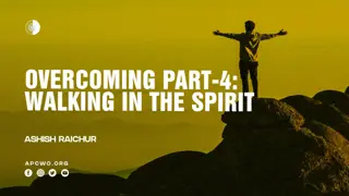
Decoding the Dynamics of Walking: An In-depth Exploration
DR.MURALI KRISHNA P.V
M.S.(ORTHO)
GENERAL HOSPITAL ANEKAL,
DEPARTMENT OF ORTHOPEDICS,
BANGALORE
GOVERNMENT OF KARNATAKA
Walking Theory
Walking Theory
Walking is a complex phenomenon, In this
technological era how the body moves forward has
not been decoded.
Walking as a subject is not taught to children in their
curriculum, even in the medical curriculum walking
or normal gait is not taught.
Eating, Working and Walking are the basic activities
of life. Dynamics of Walking is essential to solve the
life style diseases.
Walking how it is described
Bradford Founder of JBJS in 1897
He describes “by the action of the muscles, the thigh
is advanced forward by the action of the Ileacus,
Tensor Vaginae and rectus muscles. the body’s
weight is thrown on the Heel and the body is brought
forward by the push of the foot of the rear of the leg.
in a slow gait, in feeble persons and in children the
weight of the body is used as a means of propulsion”.
He classifies the Gait into Erect gait which is the
active Gait and uses the muscles for the forward
push and the Forward Gait which uses the body
weight for the forward movement of the body.
Walking how it is described by
Gait analysts
“Normal walking is defined as a
highly controlled, coordinated,
repetitive series of limb movements
whose function is to advance the
body safely from place to place with
a
minimum expenditure of energy”.
Walking- Action of Muscles how it is described in
Anatomy
Gray’s Anatomy describes action of the Lower limb
muscles as abduction, adduction, flexion, extension,
internal rotation and external rotation
Gluteus maximus, Medius and Minimus abductors of
Hip – How many times we abduct during walking?
Adductor muscles – How many times we adduct
during walking?
Walking how it is described in Anatomy
Plantar flexion – gastro soleus plantar flexes the
ankle but in reality plantar flexion is a passive
phenomenon. Actively during walking we can not
plantar flex the foot.
Dorsiflexors – Tibialis anterior, EHL, EDL are
plantar flexors why do we need so powerfull plantar
flexors, what are their exact roles in walking?
Muscular action with reference to walking is not
described in any Anatomy or any other books.
PET-CT Gold standard in radiology
PET-CT analyses the uptake of radio labeled
glucose.
Radio labeled glucose is injected into the body
person is made to walk for one hour and the uptake
of glucose by the muscles after the activity is
assessed
PET-CT Gold standard in radiology- used in
quantification of muscular activity a first time in
the world
PET-CT analysis also provides the quantitative
uptake of glucose by individual muscles.
PET-CT analyses full stretch of muscles,
activity along the full length of muscles can be
analysed.
muscular activity of muscles across the length
of the muscles from origin to insertion can be
analyzed and uptake of radio labeled glucose is
quantified.
Walking Theory
Dr.Murali Krishna P.V
Findings of the PET-CT analysis are usefull to
analyse the walking in detail.
Some of the questions I have raised previously were
not answered in any of the textbooks and teachings.
Lot of researchers have made their contributions in
analysing the individual muscles.
My walking theory is a culmination of all those
dedicated researchers who have studied individual
muscles in detail and my PET-CT analysis
Walking Theory
Dr.Murali Krishna P.V.
“Human Walking is defined as the
Propulsion of the body forward as a result
of series of movement of lower limbs by the
systematic contraction of the muscles of the
lower limb over the adoptive bony frame
work, in a specified way, with specific
contraction of hip, thigh, leg and foot
muscles to produce forward movement of
the body, Maintaining stability, agility and
speed.
Walking Theory
Walking is divided into phases depending on the
contraction of group of muscles and the action it brings
in moving the body forward, Here I have described how
body propels forward by individual set of action of
muscles. Each group of muscles plays an important role
in moving the body forward.
1. Movement of the Lower limb
2.Movement of the Trunk
3.Stabilisation Muscles
1. Movement of the Lower limb
This phase is akin to swing phase, we explain the
individual group of muscles activity to bring the
movement of the lower limb forward
Movement of the lower limb is divided into two
phases
1. A. Movement of the lower limb from Toe off phase
to Foot flat phase
1.B. Movement of the lower limb from Foot flat
phase to Just before Heel strike phase
1. A. Movement of the lower limb-
from Toe off phase to Foot flat phase
This phase extends from Toe off phase to Foot flat
phase of walking in the swing Phase,
Ileo Psoas Muscles Pull the Femur forward internally
Tenosor fascia latae, Sartorius muscles pull the
femur and tibia externally.
Tenosor fascia latae with its unique attachment to
the Ileo tibial band assist the pulling of the femur.
1.B. Movement of the lower limb from Foot flat
phase to Just before Heel strike phase
This phase extends from Foot flat phase to just
before Heel strike Phase of walking in the swing
Phase
Quadriceps Muscles flexes the at the Hip and moves
the lower limb forward
2.Movement of the Trunk
Human body is designed extensively to move the
body forward, every bone, muscle, joints and actions
assist the forward movement of the trunk in a
systematic way..
Foot is the only contact point of the body to the
ground.
Bony design provide sufficient leverage actions for
efficiency of energy.
the origin and insertion of the muscles assist specific
activity and moves the body forward systematically
effortlessly.
2.Movement of the Trunk
This Stage is devided into several phases solely
depending on the action of group of muscles.
2.A.- Heel strike Phase
2.B.- Heel strike to foot flat Phase
2.C.- Initial Heel off Phase
2.D- Late Heel off Phase
2.E- Extension of Knee
2.F-Pushing Phase of the Pelvis
2.G-Pulling Phase of the Pelvis
2.A.- Heel strike Phase
Tibialis anterior, Extensor Hallusis Longus and
Extensor digtorum Longus dorisflexes the ankle.
Quadriceps muscles flexes at Hip and extneds at
Knee dorsiflexion of the ankle fixes the foot to the
ankle so much so that at the Heel strike phase the
whole lower limb acts as a single Bony column, This
facilitates the Heel strike.
Striking the Heel by the contraction of the
Dorsiflexors fixes the body at the Calcaneum, this
Pulls the Pelvis at the top of the Bony column.
2.B.- Heel strike to foot flat Phase
Heel strike Moves the Pelvis forward, but the Pelvis is
heavy with all the organs of the body
, Pelvis need to be
pulled forward.
Foot is brought down to the foot flat phase by Plantar
flexors.
Adductor group of muscles pull the pelvis forward
Gracilis, Adductor Magnus, Adductor Brevis, Adductor
longus, Pectineus muscles with their unique attachment
along the Linea aspera of the femur to Pubic rami pulls
the whole trunk smoothly.
Adductor group of Muscles Pull the Pubic Rami forward,
Lower limb glides over the Head of Smooth Talus.
2.C.- Initial Heel off Phase
Plantar Flexors Gastro soleus Muscle plantar flexes
the ankle.
In reality the Heel lifts the whole body from the
ground.
Body weight shifts to the fore foot by the actions of
the Tendo achilis,
This lifting of the body at the ankle Lengthens the
Lower limb this causes Passive flexion at Knee.
This also raises the pelvis, raised Pelvis assist the free
movement of the opposite limb.
2.D- Late Heel off Phase
Action of the Tendoachilis do not lift the body
sufficently, there is limitation of Plantar flexion
because tendo achilis attaches to the calcaneum
Tendo achilis can not lift the Mid foot, this needs an
assistance to lift the hind foot and mid foot.
Peroneus Longus and Peroneus Brevis attaches to
the cuneifrom, and both ends of metatarsal bones,
tendons runs in the Plantar surface of the foot, Thus
the action of these muscles lifts the body further
By the action of this muscles flexes the Knee further.
2.E-Extension of the Knee
At the end of the Heel off phase, Knee is flexed
Passively, Knee needs to be extended so that the
whole weight of the body is shifted to the foot, this
provides stability and agility.
Hamstring Muscle pull the Knee back and extends
the Knee,
Gluteus Maximus attaches to the Ileo Tibial Band
helps to extend the Knee
2.F-Pushing Phase of the Pelvis
At the end of the Heel off phase after the Knee is
extended, Lower limb is lengthened due to Full
plantar flexion
The lengthened Lower limb provides a lever
mechnaism for the action of the Gluteus Maximus,
Gluteus maximus extends the Hip by pulling the Ileo
Tibial band.
Lower limb is lengthened, Gluteus contraction
results in Pelvis being pushed forward.
Thus The whole trunk moves forward
Ileo Tibial Band
Ileo tibial Band
originates at
the anterolateral Ileac
tubercle portion of the
external lip of the Iliac crest
and inserts at the Lateral
condyle of the Tibia. Gluteus
Maximus attaches posteriorly
and Tensor facia latae
attaches anteriorly.
ITB moves the Lower
limb forward and
backward.
2.G-Pulling Phase of the Pelvis
Earth surface is not Plain it is uneven, walking on a
hilly surface requires further pulling of the pelvis.
Body needs to be upright at all times during walking.
Gluteus Medius and minimus attaches from dorsal
Ileum and greater trochanter, the muscle tissue are
arranged in group of fibers. Each Groups of fibers
pull one after the other which in turn pulls the Pelvis
forward in a systematic way.
This action Helps to walk in the slopy areas easily.
Martin Beck, John B. Sledge, Emmanuel Gautier, Claudio F. Dora The anatomy
and function of the gluteus medius, minimus muscle:
J Bone Joint Surg [Br]
2000;82-B:358-63
.
Foot is the only contact point of the body
Newton’s Law of motion
Walking in straight line
provides least resistance.
Hence Foot should move
in a straight line from
Heel to fore foot
This achieves the goal of
energy efficient movement
This is achieved by
fallowing the rules of
Newton’s Law of motion.
3.Stabilisation Muscles- stabilisation of all the
joints are necessary for walking in straight line
Newton’s 1
st
law- states Every object in a state of
uniform motion tends to remain in that state of
motion unless an external force is applied to it.
In walking To reduce the resistance and wastage of
muscular forces body needs to move in straight line.
Foot moves should move in straight line, over which
body moves, All joints of the lower limb requires to
be stabilised to achieve this goal.
3.A Stabilisation at the Hip
Obturator Internus,
Obturator externus,
Gamelli, Piriformis
muscles stabilise the
Hip during walking.
Ligamentum teres
Holds the femur in the
Acetabulam
3.B.Stabilisation of the Femur and Knee
Semi membranosus,
semi tendinosus holds
the femur in straight
line.
Popliteus muscle hold
the knee joint firmly.
3.C
Stabilisation of the
Ankle
Ankle is the most complex weight bearing joint, it has to
withstand the rotational forces.
Tibialis posterior Holds the foot in straight line during
the whole of stance phase.
Ankle is stabilised during Heel strike phase by Tibialis
anterior, Extensor Hallusis longus and Extensor
digitorum Longus muscles anteriorly.
Posteriorly passive stretching of the Tendoachilis tendon
provides support
Eversion of the foot: strong Tibialis anterior Peroneus
Brevis and Peroneus Longus muscles contract and bring
the foot to normal level and prevent injury to the ankle.
Tibialis Posterior
By its Unique
attachment of Tibia,
fibula, Intermuscualar
septum
Inserts into the
navicualar, cuneiform,
metatarsals.
Holds the ankle in
straight line.
3.D. Stabilisation at the foot
Foot is stabilised In the Heel strike phase up
to foot flat phase by the
Adductor digiti
minimi.
The tough
Plantaris fascia
across the plantar
aspect of the foot keeps integrity of the foot
in carrying the load during the transition of
the foot from dorsiflexion to plantar flexion,
Plantaris fascia keeps the integrity of the
shape of the foot at this high voltage phase.
Stabilisation at the foot
In the Toe off phase stability of the foot is
maintained by the
Abductor Hallusis longus
,
abductor hallusis longus helps the foot to maintain
the shape of full plantar flexion.
other foot muscles
firmly hold the foot, they not only
assist and maintain the integrity of the foot in
withstanding the tremendous load but also assist in
smooth transition of the body.
Walking Theory
Dr. P.V. Murali Krishna
Walking is a complex phenomenon, each group of
muscles play an important role, Each joint is adopted
to the walking, Ligaments provide stability to each
joint.
Bones provide transmission of weight, levers to the
action of the muscles.
By walking in the way I have explained provides
mechanical stimulus to all muscles of the lower limb.
Walking is the only solution for the life style
diseases.
Walking Theory- Future
Walking is life, life is walking
Walking the way explained provides mechanical
stimulus to all groups of muscles.
Muscle bulk of the body increases,
Increased muscular activity increases the glucose
and fat expenditure.
Increased muscle bulk store the circulating glucose
and fat.
Right walking keeps the body into
“Virtuous cycle of Health”
Walking Theory
Questions Please
Thank you
drpvmkm@gmail.com
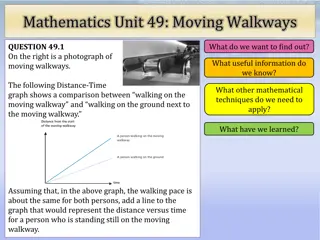
Walking is a fundamental yet complex phenomenon that plays a crucial role in our daily lives. Despite its significance, the science behind how we walk is often overlooked in education and healthcare. This article delves into the various aspects of walking, from muscle actions to gait analysis, shedding light on the mechanics of this essential human activity.

Download Presentation

Please find below an Image/Link to download the presentation.
The content on the website is provided AS IS for your information and personal use only. It may not be sold, licensed, or shared on other websites without obtaining consent from the author.If you encounter any issues during the download, it is possible that the publisher has removed the file from their server.
You are allowed to download the files provided on this website for personal or commercial use, subject to the condition that they are used lawfully. All files are the property of their respective owners.
The content on the website is provided AS IS for your information and personal use only. It may not be sold, licensed, or shared on other websites without obtaining consent from the author.
E N D
Presentation Transcript
Walking Theory DR.MURALI KRISHNA P.V M.S.(ORTHO) GENERAL HOSPITAL ANEKAL, DEPARTMENT OF ORTHOPEDICS, BANGALORE GOVERNMENT OF KARNATAKA
Walking Theory Walking is a complex phenomenon, In this technological era how the body moves forward has not been decoded. Walking as a subject is not taught to children in their curriculum, even in the medical curriculum walking or normal gait is not taught. Eating, Working and Walking are the basic activities of life. Dynamics of Walking is essential to solve the life style diseases.
Walking how it is described Bradford Founder of JBJS in 1897 He describes by the action of the muscles, the thigh is advanced forward by the action of the Ileacus, Tensor Vaginae and rectus muscles. the body s weight is thrown on the Heel and the body is brought forward by the push of the foot of the rear of the leg. in a slow gait, in feeble persons and in children the weight of the body is used as a means of propulsion . He classifies the Gait into Erect gait which is the active Gait and uses the muscles for the forward push and the Forward Gait which uses the body weight for the forward movement of the body.
Walking how it is described by Gait analysts Normal walking is defined as a highly controlled, coordinated, repetitive series of limb movements whose function is to advance the body safely from place to place with a minimum expenditure of energy .
Walking- Action of Muscles how it is described in Anatomy Gray s Anatomy describes action of the Lower limb muscles as abduction, adduction, flexion, extension, internal rotation and external rotation Gluteus maximus, Medius and Minimus abductors of Hip How many times we abduct during walking? Adductor muscles How many times we adduct during walking?
Walking how it is described in Anatomy Plantar flexion gastro soleus plantar flexes the ankle but in reality plantar flexion is a passive phenomenon. Actively during walking we can not plantar flex the foot. Dorsiflexors Tibialis anterior, EHL, EDL are plantar flexors why do we need so powerfull plantar flexors, what are their exact roles in walking? Muscular action with reference to walking is not described in any Anatomy or any other books.
PET-CT Gold standard in radiology PET-CT analyses the uptake of radio labeled glucose. Radio labeled glucose is injected into the body person is made to walk for one hour and the uptake of glucose by the muscles after the activity is assessed
PET-CT Gold standard in radiology- used in quantification of muscular activity a first time in the world PET-CT analysis also provides the quantitative uptake of glucose by individual muscles. PET-CT analyses full stretch of muscles, activity along the full length of muscles can be analysed. muscular activity of muscles across the length of the muscles from origin to insertion can be analyzed and uptake of radio labeled glucose is quantified.
Glucose Uptake Value right side Left side Name of the Muscle Muscles of the Thigh: Psoas major Psoas minor Iliacus The Anterior Femoral Muscles Sartorius Quadriceps femoris Rectus femoris Vastus lateralis Vastus medialis Vastus intermedius 1.5 1.5 1.5 1.4 1.4 1.4 0.8 0.6 1.3 1.52 o.8 2.05 1.4 1.22 0.7 1.5
The Medial Femoral Muscles Gracilis Pectineus Adductor longus Adductor brevis Adductor magnus The Muscles of the Gluteal Region Glut us maximus Glut us medius Glut us minimus Piriformis Tensor fasci lat The Posterior Femoral Muscles (Hamstring Muscles) Biceps femoris Semitendinosus Semimembranosus 0.6 1.1 1.5 1.8 1.4 0.6 0.9 1.4 1.6 1.9 1.5 2.1 1.9 1.6 1.8 1.6 0.7 0.85 1.2 1.4 1.4. 1.6 1.6 1.2
Tibialis anterior Extensor hallucis longus Extensor digitorum longus Peron us tertius Tibialis posterior Gastrocnemius Soleus Popliteus Flexor hallucis longus Flexor digitorum longus Peron us longus Peron us brevis Abductor hallucis Abductor digiti quinti 2.71 2.5 2.6 1.8 2.3 2.2 2.8 1.9 2.5 1.3 1.9 1.6 3 1.9 2.1 1.6 2.64 1.5 1.4 15.2 4.1 2.31 1.7 1.2 13 4.8
Walking Theory Dr.Murali Krishna P.V Findings of the PET-CT analysis are usefull to analyse the walking in detail. Some of the questions I have raised previously were not answered in any of the textbooks and teachings. Lot of researchers have made their contributions in analysing the individual muscles. My walking theory is a culmination of all those dedicated researchers who have studied individual muscles in detail and my PET-CT analysis
Walking Theory Dr.Murali Krishna P.V. Human Walking is defined as the Propulsion of the body forward as a result of series of movement of lower limbs by the systematic contraction of the muscles of the lower limb over the adoptive bony frame work, in a specified way, with specific contraction of hip, thigh, leg and foot muscles to produce forward movement of the body, Maintaining stability, agility and speed.
Walking Theory Walking is divided into phases depending on the contraction of group of muscles and the action it brings in moving the body forward, Here I have described how body propels forward by individual set of action of muscles. Each group of muscles plays an important role in moving the body forward. 1. Movement of the Lower limb 2.Movement of the Trunk 3.Stabilisation Muscles
1. Movement of the Lower limb This phase is akin to swing phase, we explain the individual group of muscles activity to bring the movement of the lower limb forward Movement of the lower limb is divided into two phases 1. A. Movement of the lower limb from Toe off phase to Foot flat phase 1.B. Movement of the lower limb from Foot flat phase to Just before Heel strike phase
1. A. Movement of the lower limb- from Toe off phase to Foot flat phase This phase extends from Toe off phase to Foot flat phase of walking in the swing Phase, Ileo Psoas Muscles Pull the Femur forward internally Tenosor fascia latae, Sartorius muscles pull the femur and tibia externally. Tenosor fascia latae with its unique attachment to the Ileo tibial band assist the pulling of the femur.
1.B. Movement of the lower limb from Foot flat phase to Just before Heel strike phase This phase extends from Foot flat phase to just before Heel strike Phase of walking in the swing Phase Quadriceps Muscles flexes the at the Hip and moves the lower limb forward
2.Movement of the Trunk Human body is designed extensively to move the body forward, every bone, muscle, joints and actions assist the forward movement of the trunk in a systematic way.. Foot is the only contact point of the body to the ground. Bony design provide sufficient leverage actions for efficiency of energy. the origin and insertion of the muscles assist specific activity and moves the body forward systematically effortlessly.
2.Movement of the Trunk This Stage is devided into several phases solely depending on the action of group of muscles. 2.A.- Heel strike Phase 2.B.- Heel strike to foot flat Phase 2.C.- Initial Heel off Phase 2.D- Late Heel off Phase 2.E- Extension of Knee 2.F-Pushing Phase of the Pelvis 2.G-Pulling Phase of the Pelvis
2.A.- Heel strike Phase Tibialis anterior, Extensor Hallusis Longus and Extensor digtorum Longus dorisflexes the ankle. Quadriceps muscles flexes at Hip and extneds at Knee dorsiflexion of the ankle fixes the foot to the ankle so much so that at the Heel strike phase the whole lower limb acts as a single Bony column, This facilitates the Heel strike. Striking the Heel by the contraction of the Dorsiflexors fixes the body at the Calcaneum, this Pulls the Pelvis at the top of the Bony column.
2.B.- Heel strike to foot flat Phase Heel strike Moves the Pelvis forward, but the Pelvis is heavy with all the organs of the body, Pelvis need to be pulled forward. Foot is brought down to the foot flat phase by Plantar flexors. Adductor group of muscles pull the pelvis forward Gracilis, Adductor Magnus, Adductor Brevis, Adductor longus, Pectineus muscles with their unique attachment along the Linea aspera of the femur to Pubic rami pulls the whole trunk smoothly. Adductor group of Muscles Pull the Pubic Rami forward, Lower limb glides over the Head of Smooth Talus.
2.C.- Initial Heel off Phase Plantar Flexors Gastro soleus Muscle plantar flexes the ankle. In reality the Heel lifts the whole body from the ground. Body weight shifts to the fore foot by the actions of the Tendo achilis, This lifting of the body at the ankle Lengthens the Lower limb this causes Passive flexion at Knee. This also raises the pelvis, raised Pelvis assist the free movement of the opposite limb.
2.D- Late Heel off Phase Action of the Tendoachilis do not lift the body sufficently, there is limitation of Plantar flexion because tendo achilis attaches to the calcaneum Tendo achilis can not lift the Mid foot, this needs an assistance to lift the hind foot and mid foot. Peroneus Longus and Peroneus Brevis attaches to the cuneifrom, and both ends of metatarsal bones, tendons runs in the Plantar surface of the foot, Thus the action of these muscles lifts the body further By the action of this muscles flexes the Knee further.
2.E-Extension of the Knee At the end of the Heel off phase, Knee is flexed Passively, Knee needs to be extended so that the whole weight of the body is shifted to the foot, this provides stability and agility. Hamstring Muscle pull the Knee back and extends the Knee, Gluteus Maximus attaches to the Ileo Tibial Band helps to extend the Knee
2.F-Pushing Phase of the Pelvis At the end of the Heel off phase after the Knee is extended, Lower limb is lengthened due to Full plantar flexion The lengthened Lower limb provides a lever mechnaism for the action of the Gluteus Maximus, Gluteus maximus extends the Hip by pulling the Ileo Tibial band. Lower limb is lengthened, Gluteus contraction results in Pelvis being pushed forward. Thus The whole trunk moves forward
Ileo Tibial Band Ileo tibial Band originates at the anterolateral Ileac tubercle portion of the external lip of the Iliac crest and inserts at the Lateral condyle of the Tibia. Gluteus Maximus attaches posteriorly and Tensor facia latae attaches anteriorly. ITB moves the Lower limb forward and backward.
2.G-Pulling Phase of the Pelvis Earth surface is not Plain it is uneven, walking on a hilly surface requires further pulling of the pelvis. Body needs to be upright at all times during walking. Gluteus Medius and minimus attaches from dorsal Ileum and greater trochanter, the muscle tissue are arranged in group of fibers. Each Groups of fibers pull one after the other which in turn pulls the Pelvis forward in a systematic way. This action Helps to walk in the slopy areas easily.
Martin Beck, John B. Sledge, Emmanuel Gautier, Claudio F. Dora The anatomy and function of the gluteus medius, minimus muscle: J Bone Joint Surg [Br] 2000;82-B:358-63.
Foot is the only contact point of the body Newton s Law of motion Walking in straight line provides least resistance. Hence Foot should move in a straight line from Heel to fore foot This achieves the goal of energy efficient movement This is achieved by fallowing the rules of Newton s Law of motion.
3.Stabilisation Muscles- stabilisation of all the joints are necessary for walking in straight line Newton s 1st law- states Every object in a state of uniform motion tends to remain in that state of motion unless an external force is applied to it. In walking To reduce the resistance and wastage of muscular forces body needs to move in straight line. Foot moves should move in straight line, over which body moves, All joints of the lower limb requires to be stabilised to achieve this goal.
3.A Stabilisation at the Hip Obturator Internus, Obturator externus, Gamelli, Piriformis muscles stabilise the Hip during walking. Ligamentum teres Holds the femur in the Acetabulam
3.B.Stabilisation of the Femur and Knee Semi membranosus, semi tendinosus holds the femur in straight line. Popliteus muscle hold the knee joint firmly.
3.C Stabilisation of the Ankle Ankle is the most complex weight bearing joint, it has to withstand the rotational forces. Tibialis posterior Holds the foot in straight line during the whole of stance phase. Ankle is stabilised during Heel strike phase by Tibialis anterior, Extensor Hallusis longus and Extensor digitorum Longus muscles anteriorly. Posteriorly passive stretching of the Tendoachilis tendon provides support Eversion of the foot: strong Tibialis anterior Peroneus Brevis and Peroneus Longus muscles contract and bring the foot to normal level and prevent injury to the ankle.
Tibialis Posterior By its Unique attachment of Tibia, fibula, Intermuscualar septum Inserts into the navicualar, cuneiform, metatarsals. Holds the ankle in straight line.
3.D. Stabilisation at the foot Foot is stabilised In the Heel strike phase up to foot flat phase by the Adductor digiti minimi. The tough Plantaris fascia across the plantar aspect of the foot keeps integrity of the foot in carrying the load during the transition of the foot from dorsiflexion to plantar flexion, Plantaris fascia keeps the integrity of the shape of the foot at this high voltage phase.
Stabilisation at the foot In the Toe off phase stability of the foot is maintained by the Abductor Hallusis longus, abductor hallusis longus helps the foot to maintain the shape of full plantar flexion. other foot muscles firmly hold the foot, they not only assist and maintain the integrity of the foot in withstanding the tremendous load but also assist in smooth transition of the body.
Walking Theory Dr. P.V. Murali Krishna Walking is a complex phenomenon, each group of muscles play an important role, Each joint is adopted to the walking, Ligaments provide stability to each joint. Bones provide transmission of weight, levers to the action of the muscles. By walking in the way I have explained provides mechanical stimulus to all muscles of the lower limb. Walking is the only solution for the life style diseases.
Walking Theory- Future Walking is life, life is walking Walking the way explained provides mechanical stimulus to all groups of muscles. Muscle bulk of the body increases, Increased muscular activity increases the glucose and fat expenditure. Increased muscle bulk store the circulating glucose and fat. Right walking keeps the body into Virtuous cycle of Health
Walking Theory Questions Please Thank you drpvmkm@gmail.com