Comprehensive Overview of Thigh Anatomy and Functions
Explore the detailed anatomy of the thigh, focusing on the medial compartment and structures like Gracilis, Adductor Magnus, and more. Understand the innervation, functions, and vascular supply of these thigh muscles. Dive into the complexities of the obturator artery and its branches as they relate to the muscles in the thigh region.
Download Presentation

Please find below an Image/Link to download the presentation.
The content on the website is provided AS IS for your information and personal use only. It may not be sold, licensed, or shared on other websites without obtaining consent from the author. Download presentation by click this link. If you encounter any issues during the download, it is possible that the publisher has removed the file from their server.
E N D
Presentation Transcript
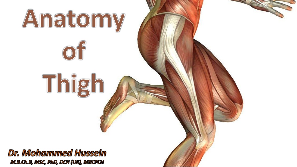
Anatomy of Thigh Dr. Mohammed Hussein M.B.Ch.B, MSC, PhD, DCH (UK), MRCPCH
Medial compartment Medial compartment 1. Gracilis 2. Pectineus 3. Obturator externus 4. Adductor longus 5. Adductor brevis 6. Adductor magnus
Obturator externus Pectineus Adductor brevis Adductor longus Adductor magnus Adductor magnus Gracilis Adductor magnus
Adductor Magnus Adductor Magnus
Adductor magnus (adductor part) Adductor magnus (hamstring part) Adductor tubercle
Adductor magnus (adductor part) Adductor magnus (hamstring or ischial part) Adductor hiatus Adductor tubercle
Adductor Magnus Adductor Magnus Innervation Innervation The adductor part is innervated by the obturator nerve The hamstring part by the tibial division of the sciatic nerve.
Functions Functions 1. Obturator externus Laterally rotates thigh at hip joint 2. Gracilis Adducts thigh at hip joint + Flexes leg at knee joint Adducts thigh at hip joint + Flexes thigh at hip joint 3. Pectineus 4. Adductor longus Adducts thigh at hip joint + Adducts thigh at hip joint + 5. Adductor brevis Medially rotates thigh at hip joint 6. Adductor magnus Adducts thigh at hip joint +
The Obturator Artery The Obturator Artery
Internal iliac artery Obturator artery
Obturator artery Posterior branch Anterior branch Obturator externus
Obturator artery Artery of ligament of head of femur Acetabular branch Posterior branch
The Obturator Nerve The Obturator Nerve
Obturator nerve Posterior branch Adductor brevis Anterior branch
Obturator externus Posterior branch Adductor brevis Adductor magnus
Adductor brevis Anterior branch Pectineus Cutaneous branch Adductor lognus Gracilis
The Adductor Canal The Adductor Canal ( Subsartorial Canal) ( Subsartorial Canal) (Hunter s Canal) (Hunter s Canal)
Femoral triangle Sartorius Femoral triangle Adductor magnus Adductor canal Adductor canal Adductor hiatus
Walls of the Adductor Canal Walls of the Adductor Canal The anteromedial wall is formed by the sartorius muscle and fascia. The anterolateral wall is formed by the vastus medialis. The posterior wall is formed by the adductor longus and magnus.
Contents of the Adductor Canal Contents of the Adductor Canal The adductor canal contains 1. The terminal part of the femoral artery 2. The femoral vein 3. The deep lymph vessels 4. The saphenous nerve 5. The nerve to the vastus medialis 6. The terminal part of the obturator nerve
Dr. Mohammed Hussein M.B.Ch.B, MSC, PhD, DCH (UK), MRCPCH


