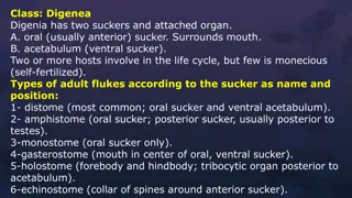
Comparative Vertebrate Anatomy - Alimentary System Overview

ZOO 366-Comparative Vertebrate
Anatomy
By
Dr. O.A Oke
Department of Biological Sciences
University of Agriculture, Abeokuta,
Nigeria.
THE ALIMENTARY SYSTEM
INTRODUCTION
The alimentary canal is a tube
beginning at the mouth and passing
through the body to the anus. The
greater part of the tube is lined by
endoderm, forming a glandular
epithelial lining of a variable nature
termed the mucosa, but at the anterior
and posterior ends ectoderm has been
invaginated to form a stomodeum and
proctodeum respectively. The wall of
the alimentary canal is completed by
tissue of mesodermal (splanchnic)
origin and consisits mainly of muscle
and connective tissue together with the
blood vessels and nerves. Outwardly
the tube is invested by the peritoneum (
coelomic epithelium), which also
covers the mesenteries by which it is
suspended in the body.
Primarily, the alimentary canal is for
the reception of food, but in all
vertebrates part of the anterior region
plays a part in respiration. It originates
in the embryo as a simple straight tube,
but in the adult it is differentiated into
regions which differ according to the
particular work carried out therein. The
tube is usually much longer than the
body, so that since the two ends fixed,
it is thrown into coils.
The main regions of the alimentary
canal are the buccal cavity, pharynx,
esophagus, stomach and intestine.
The Buccal Cavity- This is derived
from the stomodeum and consequently
is lined by ectoderm and the mouth
aperture opens directly into it. As
derivatives of the skin covering the
jaws, teeth are present, the from and
functions of which differ in different
vertebrates. In the higher craninates a
variable shaped, movable, muscular
tongue is found on the floor of the
buccal cavity. In many instances, even
in the lower vertebrates, there is no
clear line of demarcation between the
buccal cavity and the next region, the
pharynx, but developmentally the
former is lined with ectoderm and the
latter by endoderm. The two regions
can be called the bucco-pharyngeal
region.
The Pharynx- It is this region that, in
the embryo, the visceral clefts make
their appearance. In the fishes they
persist in the adult as branchial clefts,
but in the higher vertebrates for the
most part they close up and disappear.
It is from the hinder end of this region
that the lungs, the respiratory organs
found in air-breathing vertebrates,
originate. The lungs may arise directly
from a layngcal chamber or when a
neck is present, as in the mammals,
they are situated at the end of a tubular
trachea. In those vertebrates where the
majority of the visceral clefts have
been lost, the pharyngeal part of the
first cleft is retained as part of the
auditory apparatus to form Eustachian
tube and tympanic chamber.
The Oesphagus, Following the pharynx
is the oesophagus in which, either by
the flattening of the tube or the folding
of its lining, the lumen is considerably
reduced. The oesophagus forms a
connecting channel between the
pharynx and the next region, the
stomach.
The Stomach: In this region the tube is
dialated to form a receptacle in which
the food can accumulate during
feeding and also where some digestive
processes can take place. The form of
the stomach varies in different animals,
according to their type of food and
mode of life, but usually regurgitation
is prevented by a valvular arrangement,
the cardia, where the oesophagus
enters the stomach. The premature
escape of the food from the stomack
into the intestine is guarded against by
the development in the wall of the
distal (pyloric) end of the stomach of a
ring of muscle termed a sphincter, the
pyloric sphincter, which closes the
aperture and only opens under certain
conditions. The position of this
sphincter is usually marked externally
by a constriction, the pyloric
constriction. In the wall of the stomach
numerous simple glands, the peptic or
gastric glands, are present which
secrete the gastric juice.
The Intestine, - Following the stomach
comes the longest part of the
alimentary canal, the intestine. In most
vertebrates the intestine can be divided
into regions distinguishable by
morphological and historogical
leatures. Into the first part, which
immediately follows the stomach, open
the ducts of the two important
accessory glands, the liver and
pancreas, and this region is called the
duodenum. The next part of the
intestine is the region where a great
part of digestion and most of the
aborption of the digested food takes
place. And is appropriately modified in
different vertebrates. The lining
epithelium provides numerous glands,
the products of which are poured out
on to the food. The form of this region
varies considerably in different
vertebrates as also does its
terminology, any details must be left to
the description of individual examples.
The terminal portion of the intestinal
region is the rectum, which includes
the proctodeum and terminates at the
anus.
The passage of the food through the
alimentary tube is caused by waves of
muscular contraction of a rhymical
character which pass along the length
of the tube, pushing the food in front of
the constricted region. This rhythmical
contraction is termed peristalsis.
The Accessory Glands. In the higher
vertebrates salivary gland are present.
The salivary secretion or saliva is
poured on to the food during
mastication. In other instances, where
salivary glands are absent, the lining
epithelium of the buccal cavity and
pharynx contains many muscus-
sereting glands which help to lubricate
the food during its passage through
these regions. The liver and pancreas,
already mentioned as opening by their
ducts into the duodenum, arise in
some animals 9e.g chick0 as
outgrowths of the endoderm of the
alimentary canal, but soon mesodermal
derivatives become incorporated with
these outgrowths to form the adult
glands, the connection between the
gland and the intestine being retained
as the duct. The secretions of these
glands, bile and pancreatic juice, play
an important part in the digestive
processes.
In addition to these special glands, the
lining epithelium of all regions of the
alimentary canal contains numerous
mucous glands.
VILL. THE URINARY ORGANS
The toad’s kidney is formed of a
complex mass of hephric units among
which lie blood vessels and
capillaries. The adrenal gland lies on
its ventral surface. The kindney is
bathed in the lymph which is contained
in the cisteria magna and its drained
into the veins of the kidney. These are
the renal veins and the renal portal
vein. Renal arteries supply the kidney.
You ought to know the structure of the
nephric unit in orer toi understand well
the section of the kidney. A nephric
unit consist of a long trubule which
begins by a peculiar structure called
the Malpighian body or corpuscle.
This is formed of a thin double-walled
Bowman’s capsule into which pushes
an affercent arteriole that branches off
then leaves away the corpuscle as an
efferent arteriole. The tuft of vessels
thus formed insiude the corpuscle is
called the glomerulus. (The efferent
arteriole breaks up, outside the
glomerulus, into capillaries which
connect with those of the renal portal
vein).
The Browman’s capsule leads into a
uriniferous tu bule which is much
convoluted and ultimately opens into a
collecting tubule. The collecting
tubules pour the urine into the Wolffian
duct which extends along the outer
lateral border of the kidney.
Search for the above mentioned
structure in the section. These are:
-
The Wolffian duct is seen on
thelateral edge of the organ, lined by a
simple cubical epithelium and
surrounded by connective tissue and
unstriated muscle fibres.
-
The renal portal vein lies next to
the Wolffian duct. It may contain
blood corpuscles and its wall consists
of the usual layers characteristic of
veins.
-
The renal artery an renal are
conspicuous on the ventral side of the
kidney
-
The adrenal gland lies on the
ventral surface, and consists of
glandular cells which are surrounded
by numerous blood vessels.
-
The wall of the cistern magna and
the nephrostomes or peritoneal funnels
are found on the ventral side of the
section. Each peritoneal funnel is lined
largely with ciliated cuboidal cells.
-
The Malpighian bodies or
corpuscles are found each of the
glomerulus in the middle, and of the
Bowman’s capsule to the outside. The
wall of the latter is built up of a simple
squanmous epithelium.
-
The uriniferous or convoluted
tubules are lined by large granular cells
and each have a narrow lumen. They
are the greatest elements of the kidney
in number.
-
The collecting tubules appear in
the section lined by cuboidal cells
which contain but few granules and
have a wide lumen. They are mush
fewer in number that the uriniferous
tubules.
-
A network of blood vessels and
capillaries is held by connective tissue
among the tubules.
THE GENITAL GLANDS (GONADS)
The genital glands differ according to
sex, thus the testis in the male
produces the spermatozoa, while the
ovary in the female product the ova
(sing.ovum).
THE TESTIS
The testis is build up of a large number
of seminiferous tubules. The
spermatozoa are formed in the walls of
these tubules in the mature testis. The
tubules are held together by an
intertubular connective tissue which
contains particular interstitial cells that
secrete certain hormones which are
responsible for the appearance of the
secondary sexual characters.
Since the spermatozoon passes through
a series of phase till it reaches its final
form, the wall of the seminferous
tubule thus contains all that represent
these phases. The process is known as
spermatogenesis.
Examine and note:
-
The seminiferous tubules appear
rounded or oval in section, each
surround by a thin basement
membrane and contains in its wall
several layers of cells representing
(from outside inwards)
(i)
The spermatogonia lie along
the periphery of the tubule and
appear closely packed together..
(ii)
The primary spermatocytes are
the largest of the cells and have
large nuclei.
(iii)
The secondary spermatocytes
are smaller than the preceding cells,
about half size, and their nuclei
stain deeply.
(iv)
The spermatids are still smaller
than the preceding cells and their
nuclei are more condense. They
aggregate in clusters.
The spermatozoa lei in the cavity of
the tubule. They are always
grouped in clusters and some
appear connected with peculiar
large cells which lie at the
periphery of the tubule. These are
cells of Sertoli.
A spermatozoon (or sperm) has an
elongated head and a long delicate
tail. Its nucleus lies in the head
which is pointed at the acrosome.
Identify all the above mentioned
stages in the seminiferous tubule
with the help of the H.P. and
proceed studying other structures of
the testis:
-The intertubular tissue is formed
of connective tissue which holds
the tubules together and contains
blood vessels. It also contains cells
of endocrine secretion, the
interstitial cells.
-The tunica albuginea is built up of
fibrous connective tissue and
surrounds the testis. The
intertubular tissue extends to the
periphery of the testis where it is
connected with this sheath.
-The peritoneal epithelium is the
outermost covering of the testis.
3
THE OVARY
The ovary is concerned with the
formation of eggs. The process of
formation of the eggs, or oogenesis,
closely resembles the process of
formation of the spermatozoa.
However, the ova (eggs) are very
much larger in size than the sperms,
since the ova represent the non-
motile and food storing gametes.
The sperms, on the other hand,
ought to be very small because they
are motile. They are also formed in
much greater numbers than the ova.
104
I.T.S of the ovary of the toad
Here the ovary consist of a number
of hallow lobules in which the ova
are formed. Each lobule is
surrounded externally by the theca
externa, which corresponds to the
peritoneal epithelium of the testis.
Thousands of sacs of various sizes
are connected to the theca externa,
depending on the size of the ovum
that each sac contains. Each sac is
surrounded by the theca interna, an
envelope which contains unstriated
muscle fibres, blood vessels and
nerves. The theca interna,
however,is incomplete where the
sac is connected to the outer wall of
the ovary. It is this place at which
the ovum, when fully mature,
bursts out to fall into the body
cavity.
The ovum is also surrounded by a
number of cells form the ovarian
stroma which is responsible for the
secretion of the ovarian hormones.
The ovum passes through a number
of phases following the oogonium
stage. First is the primary oocyte
which increases in size gradually.
Its nucleus also undergoes certain
changes and shows several
nucleoli.
Finally a vitelline membrance is
formed around the primary oocyte,
which separates it from the
follicular cells.
The first reduction division usually
occurs when the ovum reaches the
oviduct, thus becoming the
secondary oocyte. The second
maturation division occurs on
fertilization, that is, externally in
water.87
The Mammalian Circulation
Basically the muscular heart pumps
the blood into the system of
arteries, which split up within the
tissues into capillaries. Here
exchange of materials between
blood and cells takes place. From
the capillaries blood is collected up
into a series of veins by which it is
returned to the heart. The heart is
divided into four chambers: right
and left atria (auricles), and right
and left ventricles. Blood returning
to the heart enters the right atrium,
whence it passes into the right
ventricle, and then via the
pulmonary artery to the lungs. This
is deoxygenated blood, oxygen
having been removed from it and
carbon dioxide added to it during
its passage through the tissues. As
the blood flows through the
capillaries in the lungs it shed its
carbon dioxide and take up oxygen.
The oxygenated blood now returns
via the pulmonary veins to the
heart, entering the left atrium. From
this chamber it passes into the left
ventricle, and thence to the dorsal
aorta, the main artery of the body.
From this numerous arteries, some
median and paired, convey blood to
the capillary systems in the organs
and tissues, where gaseous
exchange takes place.
Corresponding veins convey the
deoxygenated blood to the venae
cavae (great veins) by which it is
returned to the right atrium. The
walls of the arteries and veins are
elastic, and the heart and the veins
are equipped with valves which
prevent blood flowing in the wrong
direction. At one time it was
thought that blood was pumped
from the heart and subsequently
drawn back into it in the same
vessels, a sort of ebb-and-flow
system. It is interesting to note that
this kind of thing does happen in
certain primitive animals, but not
vertebrates. That the blood
circulates was first discovered by
the seventeenth century physician,
William Harvey. By meticulous
dissection and ingenious
experiments, Harvey showed
beyond all reasonable doubt that
blood flows away from the heart in
certain vessels (arteries) and returns
to it in different vessels ( veins).
From a functional point of view
the two most important parts of the
circulatory system are the heart and
capillaries. As the organ
responsible for pumping the blood,
the heart is of the utmost
importance in maintaining the
tissues in a state of health and
efficiency. The capillaries represent
the place where exchange of
materials takes place, and as such
provide the raison d’etre
for the circulatory system. We shall
deal with these two parts of the
circulatory in turn.
The alimentary system in vertebrates undergoes invagination to form a stomodeum and proctodeum, with tissue of mesodermal origin forming the wall of the alimentary canal. Differentiated into regions, the canal includes the buccal cavity, pharynx, esophagus, stomach, and intestine. The system is crucial for digestion and nutrient absorption.

Uploaded on Oct 06, 2024 | 1 Views
Download Presentation

Please find below an Image/Link to download the presentation.
The content on the website is provided AS IS for your information and personal use only. It may not be sold, licensed, or shared on other websites without obtaining consent from the author.If you encounter any issues during the download, it is possible that the publisher has removed the file from their server.
You are allowed to download the files provided on this website for personal or commercial use, subject to the condition that they are used lawfully. All files are the property of their respective owners.
The content on the website is provided AS IS for your information and personal use only. It may not be sold, licensed, or shared on other websites without obtaining consent from the author.
E N D
Presentation Transcript
ZOO 366-Comparative Vertebrate Anatomy
University of Agriculture, Abeokuta, Nigeria.
invaginated to form a stomodeum and proctodeum respectively. The wall of the alimentary canal is completed by tissue of mesodermal (splanchnic) origin and consisits mainly of muscle and connective tissue together with the blood vessels and nerves. Outwardly the tube is invested by the peritoneum ( coelomic epithelium), which also covers the mesenteries by which it is suspended in the body.
in the embryo as a simple straight tube, but in the adult it is differentiated into regions which differ according to the particular work carried out therein. The tube is usually much longer than the body, so that since the two ends fixed, it is thrown into coils.
The main regions of the alimentary canal are the buccal cavity, pharynx, esophagus, stomach and intestine.
variable shaped, movable, muscular tongue is found on the floor of the buccal cavity. In many instances, even in the lower vertebrates, there is no clear line of demarcation between the buccal cavity and the next region, the pharynx, but developmentally the former is lined with ectoderm and the latter by endoderm. The two regions can be called the bucco-pharyngeal region.
found in air-breathing vertebrates, originate. The lungs may arise directly from a layngcal chamber or when a neck is present, as in the mammals, they are situated at the end of a tubular trachea. In those vertebrates where the majority of the visceral clefts have been lost, the pharyngeal part of the first cleft is retained as part of the auditory apparatus to form Eustachian tube and tympanic chamber.
the flattening of the tube or the folding of its lining, the lumen is considerably reduced. The oesophagus forms a connecting channel between the pharynx and the next region, the stomach.
escape of the food from the stomack into the intestine is guarded against by the development in the wall of the distal (pyloric) end of the stomach of a ring of muscle termed a sphincter, the pyloric sphincter, which closes the aperture and only opens under certain conditions. The position of this sphincter is usually marked externally by a constriction, the pyloric constriction. In the wall of the stomach numerous simple glands, the peptic or
intestine is the region where a great part of digestion and most of the aborption of the digested food takes place. And is appropriately modified in different vertebrates. The lining epithelium provides numerous glands, the products of which are poured out on to the food. The form of this region varies considerably in different vertebrates as also does its terminology, any details must be left to the description of individual examples.
muscular contraction of a rhymical character which pass along the length of the tube, pushing the food in front of the constricted region. This rhythmical contraction is termed peristalsis.
these regions. The liver and pancreas, already mentioned as opening by their ducts into the duodenum, arise in some animals 9e.g chick0 as outgrowths of the endoderm of the alimentary canal, but soon mesodermal derivatives become incorporated with these outgrowths to form the adult glands, the connection between the gland and the intestine being retained as the duct. The secretions of these glands, bile and pancreatic juice, play
In addition to these special glands, the lining epithelium of all regions of the alimentary canal contains numerous mucous glands.
capillaries. The adrenal gland lies on its ventral surface. The kindney is bathed in the lymph which is contained in the cisteria magna and its drained into the veins of the kidney. These are the renal veins and the renal portal vein. Renal arteries supply the kidney.
Bowmans capsule into which pushes an affercent arteriole that branches off then leaves away the corpuscle as an efferent arteriole. The tuft of vessels thus formed insiude the corpuscle is called the glomerulus. (The efferent arteriole breaks up, outside the glomerulus, into capillaries which connect with those of the renal portal vein).
convoluted and ultimately opens into a collecting tubule. The collecting tubules pour the urine into the Wolffian duct which extends along the outer lateral border of the kidney.
Search for the above mentioned structure in the section. These are:
thelateral edge of the organ, lined by a simple cubical epithelium and surrounded by connective tissue and unstriated muscle fibres.
the Wolffian duct. It may contain blood corpuscles and its wall consists of the usual layers characteristic of veins.
The renal artery an renal are conspicuous on the ventral side of the kidney -
The adrenal gland lies on the ventral surface, and consists of glandular cells which are surrounded by numerous blood vessels. -
the nephrostomes or peritoneal funnels are found on the ventral side of the section. Each peritoneal funnel is lined largely with ciliated cuboidal cells.
corpuscles are found each of the glomerulus in the middle, and of the Bowman s capsule to the outside. The wall of the latter is built up of a simple squanmous epithelium.
tubules are lined by large granular cells and each have a narrow lumen. They are the greatest elements of the kidney in number.
the section lined by cuboidal cells which contain but few granules and have a wide lumen. They are mush fewer in number that the uriniferous tubules.
A network of blood vessels and capillaries is held by connective tissue among the tubules. -