Case Study: Cesarean Myomectomy Conducted by Dr. Alisha Das
A 25-year-old female primigravida underwent a cesarean myomectomy procedure at Dr. D. Y. Patil Medical College and Research Centre. The patient had a normal pregnancy history with no significant medical issues. Detailed history, examination findings, and abdominal assessment were reported in preparation for the caesarean delivery. The surgery was performed successfully, highlighting the collaboration between Dr. Alisha Das and Dr. Hemant Deshpande from the Department of Obstetrics & Gynaecology.

Download Presentation

Please find below an Image/Link to download the presentation.
The content on the website is provided AS IS for your information and personal use only. It may not be sold, licensed, or shared on other websites without obtaining consent from the author.If you encounter any issues during the download, it is possible that the publisher has removed the file from their server.
You are allowed to download the files provided on this website for personal or commercial use, subject to the condition that they are used lawfully. All files are the property of their respective owners.
The content on the website is provided AS IS for your information and personal use only. It may not be sold, licensed, or shared on other websites without obtaining consent from the author.
E N D
Presentation Transcript
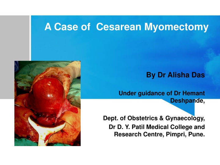
A Case of Cesarean Myomectomy By Dr Alisha Das Under guidance of Dr Hemant Deshpande, Dept. of Obstetrics & Gynaecology, Dr D. Y. Patil Medical College and Research Centre, Pimpri, Pune.
HISTORY OF PRESENTING COMPLAINTS A 25 year old female Primigravida came on 30th March 2020 admitted under Unit-1 OBGY for regular ANC visit. No H/O Pain abdomen, PV leak, PV bleed, decreased fetal movements, B/B complaints Last menstrual period 12th July,2019 Expected Date of Delivery - 17th April, 2020 Corresponding to 38 weeks on the day of caesarean section by dates, and 37.1 weeks by the first trimester ultrasonography of 8.2 weeks.
PAST MENSTRUAL HISTORY: 4 to 5 days / 28-32 days / normal flow (2 to 3 pads per day)/ not associated with passage of clots and dysmenorrhoea OBSTETRIC HISTORY: Married life 1 year , Prmigravida PAST MEDICAL AND SURGICAL HISTORY: No history of Diabetes mellitus , hypertension, tuberculosis , thyroid disorder. No history of intake of OC pills routinely No major surgery in the past and no prior blood transfusion. PERSONAL HISTORY: Normal sleep and bowel bladder habits No history of smoking or alcohol consumption
GENERAL PHYSICAL EXAMINATION Moderately built and nourished Comfortable and co-operative at the time of examination Height-165 cm Weight- 65 kg BMI 23.875 kg/m Skin normal in texture and colour Pulse 76 bpm regular in rate, rhythm and normal volume measured in left radial artery. No apex pulse deficit and radio radial delay BP 110/70 mmHg taken in left brachial artery in supine position Breasts - clinically normal Thyroid - clinically normal Spine - clinically normal No pallor, icterus, edema, cyanosis, lymphadenopathy, clubbing CVS S1 S2 normal . No murmurs RS air entry bilaterally equal . No crepts , rales , rhonchi or abnormal breath sounds No cervical lymphadenopathy
ABDOMINAL EXAMINATION Inspection: Umbilicus central and everted, flanks full All quadrants move equally with respiration. Linea nigra and striae gravidarum visible. No striae , dilated veins, sinuses Palpation : Uterus fundal height corresponding to full term Relaxed, cephalic, head floating, FHS + 142bpm Back to maternal left, liquor adequate, EFW~2.5 kg 1st Leopold(Fundal grip): broad, soft and irregular structure s/o fetal buttocks 2nd Leopold(Lateral grip): Left: smooth, curved, resilient feel s/o fetal back Right: irregular, knob like structures s/o fetal limbs 3rd Leopold(Pawlik's grip): smooth, hard and globular structure s/o fetal head 4th Leopold (Pelvic grip) : convergent fingers- not engaged.
PER VAGINAL EXAMINATION: Cervix os admits tip Cervix uneffaced Station (-2) Membranes (+) Pelvis adequate
INVESTIGATIONS Hemogram, blood sugar levels and liver and renal function test with blood electrolytes : WNL HIV , HbsAg negative USG : 1st Trimester Scan: 37.1 weeks Single Live Intrauterine gestaiton, of 8.2 weeks , FHs+154bpm 2nd trimester Scan: 1.) 38.1 weeks: (24/10)SLIUG of 15 weeks , FHS +148bpm, NT- 2.8 mm 2.) 37.6 weeks: (30/11)SLIUG of 20 weeks , FHS +152bpm, Normal anomaly scan
3rd trimester Scan: 1.)37.1 weeks : (08/02) SLIUG of 29.3 weeks, FHS +150bpm, placenta- fundoposterior, AFI - adequate, with -A subserosal exophytic uterine fibroid of size 4.7 *2.3 cm arising from lower anterior uterine wall - Another 4.2 * 3.6 cm subserosal fibroid noted along the lower segment in the right lateral wal' cystic degeneration. 2.)34.4 weeks: (30/3) SLIUG of 34.1 weeks, FHS +146bpm, cephalic, placenta - fundoposterior, AFI- 9 cm, BPP-8/8, EFW- 2,67kg, with multiple uterine fibroids in the anterior wall of uterus along the lower uterine segment.
MANAGEMENT In view of full term pregnancy with mild decrease in AFI and multiple anterior wall uterine fibroids, patient was planned for Elective LSCS with - Arrangement of adequate blood - High risk consent for Myomectomy/ Hysterectomy SOS - Consent for Surgery The patient was taken up for definitive surgery
Patient was taken for Lower Segment Caesarean Section and further myomectomy was done for both anterior wall fibroids through the tunnel formed via the lower segment incision (Kehr incision)
Specimen was resected post delivery of the baby and further steps of LSCS were continued. Specimen was sent for histopathological examination
Patient withstood the prodedure well and delivered a healthy female child on 3rd April, 2020 at 9.29 am of 2.7kg birth weight, shifted to mother side Patient was monitored in recovery then shifted to ward and post-op analgesics and antibiotics covered.
The patient had an uneventful post- operative period and was discharged after 8 days with full recovery. Histopathology report was suggestive of uterine myoma
DISCUSSION A leiomyoma, also known as fibroids, is a benign smooth muscle tumour. Uterine fibroids are leiomyomata of the uterine smooth muscle. Described based on location in the uterus: Intramural: develop from within uterine wall, do not distort uterine cavity, <50% protruding into serosal surface Submucosal: develop from myometrial cells just below endometrium, often protrude into and distort uterine cavity Subserosal: originate from serosal surface of uterus, >50% protrudes out of serosal surface Cervical: located in the cervix, rather than uterine corpus
Uterine myomas are observed in pregnancy more frequently now than in the past Because many women are delaying child bearing till their late thirties, which is the time for greatest risk of myoma growth. Also the use of ultrasonography has improved the diagnostic capability of detecting small myomas and has increased our knowledge of myomas in pregnancy.
PREVALENCE The prevalence of uterine fibroids in pregnancy varies between 1.6 and 10.7 percent depending upon the trimester of assessment and the size threshold
CHANGES IN SIZE DURING PREGNANCY The growth of fibroids during pregnancy using ultrasonography have refuted the commonly held belief that fibroids increase in size throughout gestation Indeed, the majority of uterine fibroids (49 to 60 percent) have negligible (defined as <10 percent) change in volume across gestation, while 22 to 32 percent increase in growth, and 8 to 27 percent decrease in size In those fibroids that do increase in size, most of the growth occurs in the first trimester, with little if any further increase in size during the second and third trimesters Larger fibroids (>5 cm in diameter) are more likely to grow, whereas smaller fibroids are more likely to remain stable in size The mean increase in fibroid volume during pregnancy is 12 percent, and very few fibroids increase by more than 25 percent
TIME OF DIAGNOSIS Simultaneous surgical removal of a previously diagnosed myoma during cesarean section Simultaneous surgical removal of a accidentally diagnosed myoma during cesarean section Unavoidable caesarean myomectomy with a huge uterine fibroid located in the lower segment Avoidable caesarean myomectomy
SELECTIVE REMOVAL OF FIBROIDS Myomectomy during caesarean delivery can be safely effective procedure in safe hands. The main principles of myomectomy during pregnancy are; 1. It should be done by an experience person. 2. It should be done in in a well equipped tertiary institution 3. With the advent of better anesthesia and availability of blood 4. It should be done with full consent of the patient. 5. It should be done in selected patients according to site and size of myoma 6. It should be done with techniques that decrease blood loss( UAL ,UAE ,High Oxytocin ) 7 - It is however important that baby must be delivered prior to attempting myomectomy Selection of patients for caesarean myomectomy reduces blood loss, anaemia and other complications.
TIME OF REMOVAL OF FIBROIDS Myomectomy was done after delivering the baby. This was followed by suturing with adequate hemostasis Large myoma situated over lower segment needing myomectomy before and for delivery of the fetus during LSCS Sufficient grouped and matched blood must be made available With prophylactic broad spectrum antibiotics to combat infection and Analgesics were given in the postoperative period
FUTURE OF FERTILITY The future fertility and or subsequent pregnancy outcome was unaffected by caesarean myomectomy (Evidence level III)
FINAL SUMMARY Management of fibroids during antenatal period is never surgical except torsion of pendculated fibroid. Fibroids in pregnancy are common and incidence will increase in future. Myomectomy at CS is not absolutely CONTRAINDICATED as believed earlier. Decision depends on location, size and number.
FINAL SUMMARY Enucleation is easier. Preoperative counseling and consent. Decision should be case to case basis. Management at tertiary care hospital.
FINAL MESSAGE Risk and Benefits to be evaluated in every case and decision individualized.