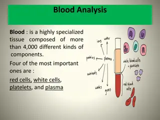
Blood: Composition and Functions
Blood is a vital fluid tissue in the human body, classified as a connective tissue. It consists of living cells known as formed elements suspended in a non-living matrix called plasma. The physical characteristics of blood, such as color range, pH level, and temperature, play crucial roles in maintaining homeostasis. Plasma, which makes up about 90% of blood, contains various dissolved substances essential for bodily functions. The formed elements in blood include erythrocytes (red blood cells), leukocytes (white blood cells), and platelets. Each component of blood serves unique functions, such as oxygen transport, defense against disease, and clotting. Hemoglobin, present in red blood cells, plays a key role in oxygen binding. Understanding the composition and functions of blood is essential for grasping the intricacies of human physiology.

Download Presentation

Please find below an Image/Link to download the presentation.
The content on the website is provided AS IS for your information and personal use only. It may not be sold, licensed, or shared on other websites without obtaining consent from the author.If you encounter any issues during the download, it is possible that the publisher has removed the file from their server.
You are allowed to download the files provided on this website for personal or commercial use, subject to the condition that they are used lawfully. All files are the property of their respective owners.
The content on the website is provided AS IS for your information and personal use only. It may not be sold, licensed, or shared on other websites without obtaining consent from the author.
E N D
Presentation Transcript
General Human Anatomy & Physiology Chapter 5 Blood
Blood The only fluid tissue in the human body Classified as a connective tissue Living cells = formed elements Non-living matrix = plasma
Physical Characteristics of Blood Color range Oxygen-rich blood is scarlet red Oxygen-poor blood is dull red pH must remain between 7.35 7.45 Blood temperature is slightly higher than body temperature
Blood Plasma Composed of approximately 90 percent water Includes many dissolved substances Nutrients Salts (metal ions) Respiratory gases Hormones Proteins Waste products
Plasma Proteins Albumin regulates osmotic pressure Clotting proteins help to stem blood loss when a blood vessel is injured Antibodies help protect the body from antigens
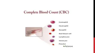
Formed Elements Erythrocytes = red blood cells Leukocytes = white blood cells Platelets = cell fragments
Erythrocytes (Red Blood Cells) The main function is to carry oxygen Anatomy of circulating erythrocytes Biconcave disks Essentially bags of hemoglobin Anucleate (no nucleus) Contain very few organelles
Hemoglobin Iron-containing protein Binds strongly, but reversibly, to oxygen Each hemoglobin molecule has four oxygen binding sites
Leukocytes (White Blood Cells) Crucial in the body s defense against disease These are complete cells, with a nucleus and organelles Able to move into and out of blood vessels) Can move by ameboid motion Can respond to chemicals released by damaged tissues
Leukocyte Levels in the Blood Normal levels are between 4,000 and 11,000 cells per millimeter Abnormal leukocyte levels Leukocytosis Above 11,000 leukocytes/ml Generally indicates an infection Leukopenia Abnormally low leukocyte level Commonly caused by certain drugs
Types of Leukocytes Granulocytes Granules in their cytoplasm can be stained Include neutrophils, eosinophils, and basophils
Types of Leukocytes Agranulocytes Lack visible cytoplasmic granules Include lymphocytes and monocytes
Agranulocytes Lymphocytes Nucleus fills most of the cell Play an important role in the immune response Monocytes Largest of the white blood cells Function as macrophages Important in fighting chronic infection
Platelets Derived from ruptured multinucleate cells (megakaryocytes) Needed for the clotting process Normal platelet count = 300,000/mm3
Hematopoiesis Blood cell formation Occurs in red bone marrow All blood cells are derived from a common stem cell (hemocytoblast) Hemocytoblast differentiation Lymphoid stem cell produces lymphocytes Myeloid stem cell produces other formed elements
Fate of Erythrocytes Unable to divide, grow, or synthesize proteins Wear out in 100 to 120 days When worn out, are eliminated by phagocytes in the spleen or liver Lost cells are replaced by division of hemocytoblasts
Control of Erythrocyte Production Rate is controlled by a hormone (erythropoietin) Kidneys produce most erythropoietin as a response to reduced oxygen levels in the blood Homeostasis is maintained by negative feedback from blood oxygen levels
Control of Erythrocyte Production Figure10.5
Hemostasis Stoppage of blood flow Result of a break in a blood vessel Hemostasis involves three phases Platelet plug formation Vascular spasms Coagulation
Blood Groups and Transfusions Large losses of blood have serious consequences Loss of 15 to 30 percent causes weakness Loss of over 30 percent causes shock, which can be fatal Transfusions are the only way to replace blood quickly Transfused blood must be of the same blood group
Human Blood Groups There are over 30 common red blood cell antigens The most vigorous transfusion reactions are caused by ABO and Rh blood group antigens
ABO Blood Groups Based on the presence or absence of two antigens Type A Type B The lack of these antigens is called type O
ABO Blood Groups The presence of bothAand B is called typeAB The presence of eitherAor B is called typesAand B, respectively
Rh Blood Groups Named because of the presence or absence of one of eight Rh antigens (agglutinogen D) MostAmericans are Rh+ Problems can occur in mixing Rh+ blood into a body with Rh blood
Rh Dangers During Pregnancy Danger is only when the mother is Rh and the father is Rh stirehni eht hR factor + dlihc eht dna , +
Rh Dangers During Pregnancy The mismatch of an Rh mother carrying an Rh+baby can cause problems for the unborn child The first pregnancy usually proceeds without problems The immune system is sensitized after the first pregnancy In a second pregnancy, the mother s immune system produces antibodies to attack the Rh+ blood (hemolytic disease of the newborn)


")

")