Biochemical Experiments and Blood Components
224 PHL
Lab#
1
Spectrophotometer
Blood
Urine
Cerebrospinal fluid (CSF)
Saliva
Diet.
Drugs.
Blood
is a tissue that circulates in the closed
system of blood vessels
.
Respiration.
Nutrition.
Excretion of waste products.
Defense mechanisms.
Regulation of body water.
Regulation of acid base balance.
Regulation of body temperature.
1)
Diagnostic.
2)
Prognostic (investigation).
If whole blood is allowed to clot and the clot
is removed, the remaining fluid is called
Serum.
Serum = Plasma – Blood clotting factor.
1. Heparin:
Mode of action:
Anti –thrombin i.e. :prevents the conversion of prothrombin to
thrombin.
2. E.D.T.A :
Mode of action:
Binds to Ca
++
3. Oxalates:
Mode of action:
Forms insoluble salts with Ca
++
4. Na Fluoride
:
(enzyme poison)
It inhibits glycolysis; therefore it is used in determination of blood sugar.
It inhibits urease enzyme, therefore it is not used in determination of blood
urea.
5. Tri Na Citrate
:
To determine E.S.R
6. Acid Citrate Dextrose Solution:
Used mainly to study platelets and to preserve
blood.
Purpose of deproteinization is to precipitate
the proteins. This is necessary because:
proteins have a certain U-V absorption and
could give false reading.
Proteins are colloids which make the solution
turbid and difficult to read on the
spectrophotometer.
1. Trichloroacetic Acid &Tungestic Acid
Mode of action:
Proteins at a pH lower than their
isoelectric pH become cations and are precipitated as
insoluble salts of the acids.
2. Zinc Hydroxide
Mode of action:
Proteins at a pH higher than their
isoelectric pH become anions and are precipitated as
salts of the heavy metals.
3.Organic Substance :
remove water and extract some
blood constituents e.g. ethanol, ether precipitate
proteins and extract fat and cholesterol.
Serum glucose is determined by two methods :
Oxidation method (Enzymatic method).
Alkaline copper reduction method (Asatoor
and King method).
Principle:
Glucose + O
2
+ H
2
O gluconic acid +
H
2
O
2
2H
2
O
2
+ phenol + amino-4-antipyrine
quinoneimine + 4H
2
O
Glucose oxidase
Peroxidase
1- Bring two
dried
test tubes (
sample & standard
).
2- Incubate for
10 minutes
at
37ºC
in water bath.
3- Measure the absorbance for the sample & standard against
blank at
ℷ= 546 nm.
Calculation:
Serum glucose (mg/dl)= Absorbance (sample)
Normal value:
75 – 110 mg/dl
Absorbance (standard)
×
Standard
Concentration
(100 mg/dl)
Hyperglycemia is blood glucose level
›
110 mg/dl
Hypoglycemia is blood glucose level
‹
75 mg/dl
Causes
:
1. Diabetes mellitus.
2. Hyperactivity of thyroid,
adrenal, pituitary gland.
3.
↑
secretion of GH
(Acromegaly)
4. Asphyxia causing acidosis
and increased mobilization of
glucose from glycogen in the
liver.
5. Exercise or stress causing
increased secretion of
adrenaline.
6. Pancreatic carcinoma or
acute pancreatitis.
Causes:
1. Overdose of insulin.
2. Hypoactivity of thyroid,
adrenal, or pituitary
gland.
3. Glycogen storage
disease in which there is
deficiency of G6P
→
inability to produce
glucose
from glycogen.
Normally glucose present in the blood is filtered
out in the renal glomeruli but reabsorbed back
into the blood by the kidney tubules.
The limit to the ability of the tubules to reabsorb
glucose is 180 mg/ dl.
If
blood sugar
level rises above this value glucose
appears in the urine
(Glucosurea).
The blood glucose level of 180 mg/dl is therefore
called the renal glucose threshold.
This content delves into various aspects of biochemical experiments, including qualitative and quantitative methods for substance identification and concentration measurement. It also covers the use of a spectrophotometer and different bodily fluids for analysis. Additionally, it explores the components of blood, the difference between serum and plasma, and the modes of action of common anticoagulants used in laboratory procedures.

Download Presentation

Please find below an Image/Link to download the presentation.
The content on the website is provided AS IS for your information and personal use only. It may not be sold, licensed, or shared on other websites without obtaining consent from the author.If you encounter any issues during the download, it is possible that the publisher has removed the file from their server.
You are allowed to download the files provided on this website for personal or commercial use, subject to the condition that they are used lawfully. All files are the property of their respective owners.
The content on the website is provided AS IS for your information and personal use only. It may not be sold, licensed, or shared on other websites without obtaining consent from the author.
E N D
Presentation Transcript
224 PHL Lab#1
for a qualitative purposes to identify the presence or absence of a substance Chemical method Chemical method Types of biochemical experiments Types of biochemical experiments for a quantitative purposes to measure the concentration of a substance Colorimetric method Colorimetric method
Spectrophotometer Spectrophotometer
Blood Urine Cerebrospinal fluid (CSF) Saliva
Diet. Diet. Drugs. Drugs.
Blood system of blood vessels. Blood is a tissue that circulates in the closed
Respiration. Nutrition. Excretion of waste products. Defense mechanisms. Regulation of body water. Regulation of acid base balance. Regulation of body temperature.
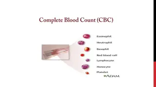
Blood Cellular elements Cellular elements Plasma Plasma RBCs, WBCs, Platelets Water Solids Diffusible constituents Non diffusible constituents e.g. albumin, globulin, fibrinogen. Anabolic Catabolic
1) Diagnostic. 2) Prognostic (investigation).
If whole blood is allowed to clot and the clot is removed, the remaining fluid is called Serum. Serum. Serum = Plasma Serum = Plasma Blood clotting factor. Blood clotting factor.
1. Heparin: thrombin. 1. Heparin: Mode of action: Mode of action: Anti thrombin i.e. :prevents the conversion of prothrombin to 2. E.D.T.A : 2. E.D.T.A : Mode of action: Mode of action: Binds to Ca++ 3. Oxalates: 3. Oxalates: Mode of action: Mode of action: Forms insoluble salts with Ca++ 4. Na Fluoride It inhibits glycolysis; therefore it is used in determination of blood sugar. It inhibits urease enzyme, therefore it is not used in determination of blood urea. 4. Na Fluoride:(enzyme poison) 5. Tri Na Citrate 5. Tri Na Citrate: To determine E.S.R 6. Acid Citrate Dextrose Solution: blood. 6. Acid Citrate Dextrose Solution: Used mainly to study platelets and to preserve
Purpose of the proteins. This is necessary because: proteins have a certain U-V absorption and could give false reading. Purpose of deproteinization the proteins. This is necessary because: deproteinization is to precipitate is to precipitate Proteins are colloids which make the solution turbid and difficult to read on the spectrophotometer.
1. Mode of action: isoelectric pH become cations and are precipitated as insoluble salts of the acids. 1. Trichloroacetic Mode of action: Proteins at a pH lower than their Trichloroacetic Acid & Acid &Tungestic Tungestic Acid Acid 2. Zinc Hydroxide Mode of action: isoelectric pH become anions and are precipitated as salts of the heavy metals. 2. Zinc Hydroxide Mode of action: Proteins at a pH higher than their 3.Organic Substance : blood constituents e.g. ethanol, ether precipitate proteins and extract fat and cholesterol. 3.Organic Substance : remove water and extract some
Serum glucose is determined by two methods : Oxidation method (Enzymatic method). Serum glucose is determined by two methods : Alkaline copper reduction method (Asatoor and King method).
Principle: Glucose + O2 + H2O gluconic acid + H2O2 Principle: Glucose Glucose oxidase oxidase Peroxidase Peroxidase 2H2O2 + phenol + amino-4-antipyrine quinoneimine + 4H2O
Sample Sample Standard Standard Blank Blank Serum 0.1 ml - - Standard - 0.1 ml - Working reagent 1 ml 1 ml 1 ml 1- Bring two dried 2- Incubate for 10 minutes 3- Measure the absorbance for the sample & standard against blank at = 546 nm. dried test tubes (sample & standard 10 minutes at 37 C sample & standard). 37 C in water bath. = 546 nm.
Calculation: Calculation: Standard Concentration (100 mg/dl Standard Concentration (100 mg/dl) ) Serum glucose (mg/dl)= Absorbance (sample) Serum glucose (mg/dl)= Absorbance (sample) Absorbance (standard) Absorbance (standard) Normal value: Normal value: 75 110 mg/dl
Hyperglycemia is blood glucose level 110 mg/dl Hypoglycemia is blood glucose level 75 mg/dl 110 mg/dl 75 mg/dl Causes: 1. Overdose of insulin. 2. Hypoactivity of thyroid, adrenal, or pituitary gland. 3. Glycogen storage disease in which there is deficiency of G6P inability to produce glucose from glycogen. Causes: Causes 1. Diabetes mellitus. 2. Hyperactivity of thyroid, adrenal, pituitary gland. 3. secretion of GH (Acromegaly) 4. Asphyxia causing acidosis and increased mobilization of glucose from glycogen in the liver. 5. Exercise or stress causing increased secretion of adrenaline. 6. Pancreatic carcinoma or acute pancreatitis. Causes:
Normally glucose present in the blood is filtered out in the renal glomeruli but reabsorbed back into the blood by the kidney tubules. The limit to the ability of the tubules to reabsorb glucose is 180 mg/ dl. If blood sugar level rises above this value glucose appears in the urine ( (Glucosurea Glucosurea). ). The blood glucose level of 180 mg/dl is therefore called the renal glucose threshold.


 Diagnostic.")