AVIAN INFLUENZA

Avian influenza, caused by RNA viruses in the family Orthomyxoviridae, poses significant challenges to poultry worldwide. The virus can range from mildly pathogenic to highly virulent, leading to severe respiratory disease and high mortality rates. Learn about the etiology, subtypes, nomenclature, and key differences between low and highly pathogenic avian influenza viruses.

Download Presentation

Please find below an Image/Link to download the presentation.
The content on the website is provided AS IS for your information and personal use only. It may not be sold, licensed, or shared on other websites without obtaining consent from the author.If you encounter any issues during the download, it is possible that the publisher has removed the file from their server.
You are allowed to download the files provided on this website for personal or commercial use, subject to the condition that they are used lawfully. All files are the property of their respective owners.
The content on the website is provided AS IS for your information and personal use only. It may not be sold, licensed, or shared on other websites without obtaining consent from the author.
E N D
Presentation Transcript
AVIAN INFLUENZA 8th Semester Dept. of VCC, BVC, Patna Dr. Anil Kumar Asst. Professor
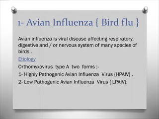
Avian Influenza Problem for poultry around the world. It can cause a range of disease symptoms from-- subclinical infection to ----highly virulent with 100% mortality. A single amino acid change in the hemagglutinin gene make difference between mild pathogenic to highly pathogenic form. Complete eradication is not possible because of wildlife reservoirs of the virus. Because of its broad host range, can represent a zoonotic risk. Etiology: RNA viruses in the family Orthomyxoviridae Influenza viruses can be subdivided into three different antigenic types, A, B and C. Type A influenza viruses are of veterinary importance, since type B and C influenza viruses are human pathogens that rarely infects other species
Type A influenza viruses have 8 different gene segments that encode 10 different viral proteins. They have two surface glycoproteins, the hemagglutinin (HA) and neuraminidase (NA) proteins, and important in regard to virulence of the virus and the antibody response. The HA protein has 16 defined antigenic subtypes, H1-H16, and the NA has 9 antigenic subtypes, N1-N9. The virus can be generally divided into viruses that cause a localized infection, often primarily restricted to the respiratory or enteric tract, and viruses that cause systemic infections. The viruses that cause localized infections are usually referred to as low pathogenic avian influenza (LPAI) viruses, and typically these viruses do not cause high mortality in affected flocks. The viruses that cause systemic infections usually cause high mortality and are referred to as highly pathogenic avian influenza (HPAI) viruses, or historically as fowl plague virus.
The standard system of nomenclature proposes that the name should include: Antigenic type (A, B or C), Host of origin, Geographical location, Strain reference number, Year of isolation, and The HA and NA subtypes, i.e., antigenic description of the HA (H) and NA (N) in parenthesis (brackets). For example, a type A influenza virus isolated from turkey in Wisconsin (USA) in 1968, a nd classified as H8N4, is designated as A/turkey/Wisconsin/1/68 (H8N4). The first highly pathogenic outbreak of avian influenza (H5N1 subtype) was recorded in 1959 in Scotland, showing classical signs of "fowl plague. All the outbreaks have been of either H5 or H7 subtype.
The LPAI viruses: mild to severe respiratory disease, that in conjunction with secondary pathogens can on rare occasions cause high mortality in a flock. The LPAI viruses can be of many different hemagglutinin and neuraminidase subtypes The HPAI viruses are restricted to the H5 and H7 subtypes and generally they are low pathogenic, but some time they can mutate into the highly pathogenic forms of the virus. HPAI is not believed to be normally present in the natural wild bird host reservoir. The hemagglutinin cleavage site is the most important virulence trait and cleaved into the HA1 and HA2 subunits by ubiquitous proteases on the inside of the host cell before it can become infectious.
Trypsin-like proteases (Lungs and enteric tract) cleave the hemagglutinin protein in the extracellular environment, that s why virus replication is restricted to these locations. When HA protein is cleaved during the assembly stage of virus replication, it become infectious after released from the cell. The damage to critical organs (brain, heart, pancreas, and skeletal muscle) or to endothelial cells lining blood vessels can cause a variety of disease symptoms that often leads to the death of the bird. Host Range: Wide (humans, swine, horses, chickens, turkeys and wild birds). Wild ducks, gulls, and shorebirds are the natural and reservoir for influenza virus In the wild bird reservoir, it causes an asymptomatic infection and genetically stable in this group.
They are generally species specific, but this general rule of host adapted influenza viruses staying within a single species can be broken. Avian influenza viruses can present a public health threat as a zoonotic pathogen, although the risk is considered to be small. Transmission: Airborne virus particles from the respiratory tract, droppings, and People-carrying virus on their clothing and equipment are the main routes of transmission. Clinical Signs: Extremely variable and depend on the: species affected, age sex concurrent infections type of the virus environmental factors etc.
The first sign of highly pathogenic avian influenza (HPAI)in chickens or turkeys is the sudden onset of high mortality, which may approach 100% within a few days. Other clinical signs include (as single or in combination): Cessation of egg Laying Respiratory signs (coughing, sneezing, lachrymation) Pronounced depression and decreased activity; Decreased feed consumption and emaciation; Oedema of head and face Cyanosis of unfeathered skin Nervous disorder and Diarrhoea. The less virulent viruses(LPAI) may cause: Drop in egg production or complete cessation Respiratory disease, anorexia, depression, sinusitis, and low mortality rales, excessive
DIAGNOSIS: Virus isolation Agar gel immune-diffusion test (AGID). The commercial ELISA tests The AGID test detects antibody to both the nucleoprotein and matrix 1 (M1) proteins that are conserved for all type A influenza viruses. The commercial ELISA tests detect only antibody to the nucleoprotein. TREATMENT & CONTROL: Quarantine of the infected flocks and usually a quarantine zone around the infected flocks. Increased biosecurity by greatly restricting access of personnel and equipment to and from farms in the quarantine zone. Increased surveillance on surrounding farms to monitor for evidence of spread of influenza infections.
For outbreaks of HPAI, infected flocks are depopulated, often with disposal of the birds by burning or burial on the farm Vaccination: Two types of vaccines, a killed whole virus adjuvanted vaccine administered either intramuscularly or subcutaneously and a live vectored recombinant vaccine for poultry. Killed vaccine produces good humoral immunity, but no cellular immunity The recombinant vaccines are vaccines using a Fowl pox virus vaccine strain or the Herpes virus turkey (HVT) as a vector in which the hemagglutinin gene from influenza virus is inserted. It produces both cellular and humoral immunity AND vaccinated birds can be distinguished from naturally infected birds since vaccinated birds will not have NP antibody because the vector only contains the hemagglutinin gene.


 cleave")