Applied Nerve & Muscle Physiology
Applied Nerve & Muscle
Physiology
:
Nerve
Conduction Study
( NCS)
)and
Electromyography
(
EMG
)
Objectives
•
Define what
is
nerve
conduction study
(NCS)
and
electromyography
( emg)
.
•
Explain
the
procedure
of
NCS
using Abductor
Pollicicis Brevis
muscle
.
•
Define
the
normal
conduction
velocity
in
upper
limb and
lower
limb
nerves
.
•
Define
the
motor
unit
potentials
(
MUPs)
and
how
they
are
changed
in muscle and
nervediseases
.
Nerve Conduction
Study
(
NCS)
•
A nerve conduction study (NCS) is an
electrophysiology
test test
commonly
used to
evaluate
the function
of peripheral
nerves
of the
human
body.
•
It
could
be motor
nerve
conduction study
( motor
NCS) , sensory
nerve
conduction study or mixed
nerve
conduction study
.
•
In this lecture,
because
of
time
constraint,
only
motor nerve conduction study will be
discussed
•
In the motor
test the
recorded response is
the
muscle CMAP ( compound muscle action potential
)
3
Procedure
•
An electrical stimulus is applied over
a
nerve
( e.g.,
median nerve
) and a recording
electrode is place over the muscle
suppllied
by that motor nerve
.
•
The
stimulus is
applied at
two
sites :
a
distal
site
(
wrist
) and a
proximal
one (
antecubital
fossa
, elbow)
.
•
The
muscle usually
chosen
in this
routine
test is the Abductor
Pollicis
Brevis
•
The
active recording electrode
(G1)
is
place
over the thenar
eminence
which overlies
the muscle
.
•
And the
reference
recording
electrode
(G2) about
3 cm
away
.
•
The
oscilloscope
( CRO) sweep speed
is
4
•
The
stimulus duration
used
is
0.2
ms
and
stimulus
frequency
to
1 /
sec.
•
Apply the stimulus
and
record the response from
stimulation
at
the wrist
.
•
Store the
CMAP (
compound muscle
action potential )
in the
first
channel
of
the
oscilloscope
.
•
Change
the stimulating site from wrist to antecubital fossa
(
elbow
)
.
•
Stimulate the nerve
& record
the
CMAP
for
median
nerve
stimulation
at
the elbow
.
6
L
1
L
a
t
e
n
c
y
A
t
w
r
i
s
t
=
3
.
5
m
s
L
2
L
a
t
e
n
c
y
A
t
e
l
b
o
w
=
8
.
5
m
s
D
i
s
t
a
n
c
e
d
=
2
8
4
m
m
•
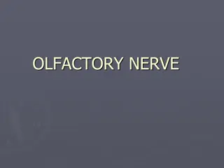
Measure the distance from elbow to
wrist with
a
measuring
tape.
•
Measure the latency in
first
CMAP & in the next
CAMP.
•
Enter
the
distance
between the elbow and
wrist
•
.
7
Motor conduction study:
Median
nerve
Nerve
conduction
velocity
D
1
D
2
Latency
(s)
L
1
L
2
NCV
=
D1-D2
L1
-
L
2
L
2
L
1
MNCV
•
MNCV
will
appear.
•
It
can
also be
calculated
by
formula
•
MNCV
(m/sec)=
•
L1 =
latency
at
wrist
•
L2 =
latency
at
elbow
11
D
i
s
t
a
n
c
e
(
m
m
)
-
-
-
-
-
-
-
-
-
-
-
-
-
-
-
-
-
-
-
-
-
-
-
-
-
-
L
2
-
L
1
(
m
s
)
Normal
values
for
conduction
velocity
In
arm
–
50 – 70 m /
sec.
In
leg
–
40 – 60 m /
sec.
12
Electromyography
(
EMG)
•
Electromyography
(EMG)
is a
technique
for
evaluating
and
recording
physiologic properties
of muscles
at
rest
and while
contracting.
•
It’s a recording of electrical activity of the
muscle by inserting needle electrode in
the
belly
of
the
muscles ( needle emg ) or
by
applying
the
surface electrodes ( surface emg
)
•
The potentials recorded in needle emg are
derived
from
motor
units
of the muscle, hence
known
as motor
unit
potentials
(MUPs).
•
Q: Define what is
a “ motor
unit
”?
14
•
A
motor
unit
is
defined as one
motor
neuron
and all of
the
muscle
fibers it
innervates.
15
•
•
Normal
MUPs
Amplitude : 300 μV (
microvolt)
– 5 mV (
millivolts)
Duration
: 3 – 15
ms(milliseconds
)
16
Electromyography
(
EMG)
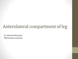
During
Moderate
Effort
note
recruitment
of additional
motoneurons
During Full Voluntary Effort
.
There
is
full
recruitment
(
you
can not see the
baseline
)
MUPs
(2)
During
Mild
Effort
Examples
of
Abnormalities
of
MUPs
•
In
nerve
diseases
:
Giant
MUPs
due
to reinnervation
> 5
mV
•
In
muscle
disease
:
Small
MUPs <
300μV
Clinical Application
Carpal
tunnel
syndrome
Nerve
injury
Myasthenia
gravis
Thanks
A nerve conduction study (NCS) is an electrophysiology test used to evaluate peripheral nerves. This article explains the procedures of NCS and electromyography (EMG), focusing on motor nerve conduction studies, muscle action potentials, and more. Explore the normal conduction velocities in upper and lower limb nerves, along with changes in motor unit potentials in muscle and nerve diseases.

Download Presentation

Please find below an Image/Link to download the presentation.
The content on the website is provided AS IS for your information and personal use only. It may not be sold, licensed, or shared on other websites without obtaining consent from the author.If you encounter any issues during the download, it is possible that the publisher has removed the file from their server.
You are allowed to download the files provided on this website for personal or commercial use, subject to the condition that they are used lawfully. All files are the property of their respective owners.
The content on the website is provided AS IS for your information and personal use only. It may not be sold, licensed, or shared on other websites without obtaining consent from the author.
E N D
Presentation Transcript
Applied Nerve & Muscle Physiology : Nerve Conduction Study ( NCS) )and Electromyography ( EMG)
Objectives Define what is nerve conduction study (NCS) and electromyography ( emg). Explain the procedure of NCS using Abductor Pollicicis Brevis muscle. Define the normal conduction velocity in upper limb and lower limb nerves. Define the motor unit potentials ( MUPs) and how they are changed in muscle and nervediseases.
Nerve ConductionStudy ( NCS) A nerve conduction study (NCS) is an electrophysiology test test commonly used to evaluate the function of peripheral nerves of the human body. It couldbe motor nerve conduction study ( motor NCS) , sensory nerve conduction study or mixed nerve conduction study . In this lecture, because of time constraint, only motor nerve conduction study will be discussed In the motor test the recorded response is the muscle CMAP ( compound muscle action potential ) 3
Procedure An electrical stimulus is applied over a nerve ( e.g., median nerve ) and a recording electrode is place over the muscle suppllied by that motor nerve . The stimulus is applied at two sites : a distal site ( wrist ) and a proximal one ( antecubital fossa , elbow) . The muscle usually chosen in this routine test is the Abductor Pollicis Brevis The active recording electrode (G1) is place over the thenar eminence which overlies the muscle . And the reference recording electrode (G2) about 3 cm away . The oscilloscope ( CRO) sweep speed is 4
The stimulus duration used is 0.2 msand stimulus frequency to 1 / sec. Apply the stimulus and record the response from stimulation at the wrist . Store the CMAP ( compound muscle action potential ) in the first channel of the oscilloscope . Change the stimulating site from wrist to antecubital fossa ( elbow ) . Stimulate the nerve & record the CMAP for median nerve stimulation at the elbow .
Distance d = 284 mm L2 LatencyAt elbow L1 LatencyAt wrist = 8.5 ms 6 = 3.5 ms
Measure the distance from elbow to wrist with a measuring tape. Measure the latency in first CMAP & in the next CAMP. Enter the distance between the elbow and wrist . 7
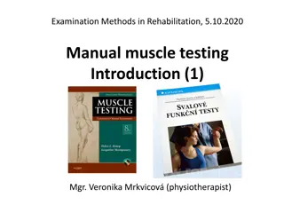
Motor conduction study: Median nerve
Nerve conduction velocity D2 NCV =D1-D2 L1-L2 D1 Latency (s) L1 L2
L1 L2
MNCV MNCV will appear. It can also be calculated byformula Distance (mm) -------------------------- L2-L1 (ms) MNCV (m/sec)= L1 = latency atwrist L2 = latency at elbow 11
Normal values for conduction velocity In arm 50 70 m / sec. In leg 40 60 m / sec. 12
Electromyography (EMG) is a technique for evaluating and recording physiologic properties of muscles at rest and while contracting. It s a recording of electrical activity of the muscle by inserting needle electrode in the belly of the muscles ( needle emg ) or by applying the surface electrodes ( surface emg ) The potentials recorded in needle emg are derived from motor units of the muscle, hence known as motor unit potentials (MUPs). Q: Define what is a motor unit ? 14
A motor unit is defined as one motor neuron and all of the muscle fibers it innervates. 15
Normal MUPs Amplitude : 300 V ( microvolt) 5 mV ( millivolts) Duration : 3 15 ms(milliseconds ) Electromyography ( EMG) 16
MUPs (2) During Mild Effort During Full Voluntary Effort . There is fullrecruitment ( you can not see the baseline ) During ModerateEffort recruitment of additional motoneurons note
Examples of Abnormalities of MUPs In nerve diseases : Giant MUPs due to reinnervation > 5 mV In muscle disease : Small MUPs < 300 V


")
 is a technique for evaluating")


")