Anatomy of GI Rectum and Anal Canal: A Detailed Visual Guide
This visual guide showcases detailed images of the gastrointestinal (GI) rectum and anal canal anatomy, including structures such as the perineum, pelvic diaphragm, external and internal anal sphincters, and various junctions within the rectum. Each image provides a clear depiction of the anatomical features and their relationships, making it a valuable resource for understanding this region of the digestive system.
Uploaded on Sep 13, 2024 | 0 Views
Download Presentation

Please find below an Image/Link to download the presentation.
The content on the website is provided AS IS for your information and personal use only. It may not be sold, licensed, or shared on other websites without obtaining consent from the author.If you encounter any issues during the download, it is possible that the publisher has removed the file from their server.
You are allowed to download the files provided on this website for personal or commercial use, subject to the condition that they are used lawfully. All files are the property of their respective owners.
The content on the website is provided AS IS for your information and personal use only. It may not be sold, licensed, or shared on other websites without obtaining consent from the author.
E N D
Presentation Transcript
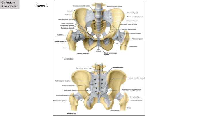
GI: Rectum & Anal Canal Figure 1
GI: Rectum & Anal Canal Figure 1.2
GI: Rectum & Anal Canal Figure 2.2 Figure 2.1 Piriformis Coccygeus Figure 2.3 Iliococcygeus Rectum Obturator Internus Vagina/ Uterus Tendinous Arch Urethra Bladder Pubococcygeus Puborectalis
GI: Rectum & Anal Canal Figure 3.1 Figure 3.2 1= Sigmoid Colon 2 = Rectum 3 =Anal Canal = Rectosigmoid Junction = Rectoanal Junction 4 = External Anal Sphincter 5 = Internal Anal Sphincter 6 = Transverse Rectal Fold 1 2 6 5 4 4 3
GI: Rectum & Anal Canal Figure 4.1 Blue + Red = Perineum Blue = Anal Triangle Red = Genitofemoral Triangle Yellow = Ischioanal Fossa Levator Ani A = Iliococcygeus B = Pubococcygeus C = Puborectalis A B C Anterior
GI: Rectum & Anal Canal Figure 4.2 Figure 4.3 Pelvic diaphragm (pelvic floor) External anal sphincter 1= Sigmoid Colon 2 = Rectum 3 =Anal Canal = Rectosigmoid Junction = Rectoanal Junction 4 = External Anal Sphincter 5 = Internal Anal Sphincter 6 = Transverse Rectal Fold Inferior rectal nn. Internal Pudendal a. Pudendal n. 1 Piriformis m. 2 Sacrospinous ligament 6 Sacrotuberous ligament 5 4 4 3
