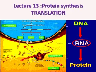
Deciphering the Genetic Code: Translation and Complexity
Translation, a key process in genetics, involves decoding the RNA alphabet into the language of proteins. This intricate process requires understanding codons, redundancies, and non-overlapping triplet codons. Early experiments by researchers like Marshall Nirenberg provided essential insights into the genetic code, leading to the present-day interpretation. Discover the fascinating world of genetic translation and the complexities involved.

Download Presentation

Please find below an Image/Link to download the presentation.
The content on the website is provided AS IS for your information and personal use only. It may not be sold, licensed, or shared on other websites without obtaining consent from the author. Download presentation by click this link. If you encounter any issues during the download, it is possible that the publisher has removed the file from their server.
E N D
Presentation Transcript
Translation is a more complex process than transcription. This would, of course, be expected. After all, the coded messages produced by the German Enigma machine could be copied easily, but required a considerable decoding effort before they could be read with understanding. In a similar sense, DNA replication is simply a complementary base pairing exercise, but the translation of the four letter (bases) alphabet code of RNA to the twenty letter (amino acids) alphabet of protein literature is far from trivial. Clearly, there could not be a direct one-to-one correlation of bases to amino acids, so the nucleotide letters must form short words or codons that define specific amino acids
Many questions pertaining to this genetic code were posed in the late 1950's: How many RNA nucleotide bases designate a specific amino acid? If separate groups of nucleotides, called codons, serve this purpose, at least three are needed. There are 43= 64 different nucleotide triplets, compared with 42 = Are the codons linked separately Sequentially joined triplet codons will result in a nucleotide chain three times longer than the protein it describes. If overlapping codons are used then fewer total nucleotides If triplet segments of mRNA designate specific amino acids in the protein, how are For the sequence ~CUAGGU~ are the codons CUA& GGU or ~C, UAG & GU~ or ~CU,AGG & U~? 16 possible do pairs. or they overlap? would be required. the codons identified?
Are all the codon words the same size? In Morse code the most widely used letters are shorter than less common letters. Perhaps nature employs a similar scheme. Physicists and mathematicians, as well as chemists and microbiologists all contributed to unravelling the genetic code. Although earlier proposals assumed efficient relationships that correlated the nucleotide codons uniquely with the twenty fundamental amino acids, it is now apparent that there is considerable redundancy in the code as it now operates. Furthermore, the code consists exclusively of non-overlapping triplet codons.
Clever experiments provided some of the earliest breaks in deciphering the genetic code. Marshall Nirenberg found that RNA from many different organisms could initiate specific protein synthesis when combined with broken E.coli cells (the enzymes remain active). A synthetic polyuridine RNA phenylalanine, so the UUU codon designated phenylalanine. Likewise an alternating ~CACA~ RNA led to synthesis of a ~His-Thr-His-Thr~ polypeptide. The following table presents the present day interpretation of the genetic code. Note that this is the RNA alphabet, and an equivalent DNA codon table would have all the U nucleotides replaced by T. Methionine and tryptophan are uniquely represented by a single codon. At the other extreme, leucine is represented by eight codons. The average redundancy for the twenty amino acids is about three. Also, there are three stop codons that terminate polypeptide synthesis. induced synthesis of poly-
https://www2.chemistry.msu.edu/faculty/reusch/VirtTxtJml/Images3/trna1.gifhttps://www2.chemistry.msu.edu/faculty/reusch/VirtTxtJml/Images3/trna1.gif
The translation process is fundamentally straightforward. The mRNA strand bearing the transcribed code for synthesis of a protein interacts with relatively small RNA molecules (about 70- nucleotides) to which individual amino acids have been attached by an ester bond at the 3'-end. These transfer RNA's (tRNA) have distinctive three- dimensional structures consisting of loops of single-stranded RNA connected by double stranded segments. This cloverleaf secondary structure is further wrapped into an "L-shaped" assembly, having the amino acid at the end of one arm, and a characteristic anti-codon region at the other end. The anti- codon consists of a nucleotide triplet that is the complement of the amino acid's codon(s). Models of two such tRNA molecules are shown to the right. When read from the top to the bottom, the anti-codons depicted here should complement a codon in the previous table
A cell's protein synthesis takes place in organelles called ribosomes. Ribosomes are complex structures made up of two distinct and separable subunits (one about twice the size of the other). Each subunit is composed of one or two RNA molecules (60-70%) associated with 20 to 40 small proteins (30-40%). The ribosome accepts a mRNA molecule, binding initially to a characteristic nucleotide sequence at the 5'-end (colored light blue in the following diagram). This unique binding assures that polypeptide synthesis starts at the right codon. A tRNA molecule with the appropriate anti-codon then attaches at the starting point and this is followed by a series of adjacent tRNA attachments, peptide bond formation and shifts of the ribosome along the mRNA chain to expose new codons to the ribosomal chemistry.
The following diagram is designed as a slide show illustrating these steps. The outcome is synthesis of a polypeptide chain corresponding to the mRNA blueprint. A "stop codon" at a designated position on the mRNA terminates the synthesis by introduction of a "Release Factor".
https://www2.chemistry.msu.edu/faculty/reusch/VirtTxtJml/Images3/protsyn1.gifhttps://www2.chemistry.msu.edu/faculty/reusch/VirtTxtJml/Images3/protsyn1.gif
Post-translational Modification Once a peptide or protein has been synthesized and released from the ribosome it often undergoes further chemical transformation. This post-translational modification may involve the attachment of other moieties such as acyl groups, alkyl groups, phosphates, sulfates, lipids and carbohydrates. Functional changes such as dehydration, amidation, hydrolysis and oxidation (e.g. disulfide bond formation) are also common. In this manner the limited array of twenty amino acids designated by the codons may be expanded in a variety of ways to enable proper functioning of the resulting protein. Since these post-translational reactions are generally catalyzed by enzymes, it may be said: "Virtually every molecule in a cell is made by the ribosome or by enzymes made by the ribosome."
Modifications, like phosphorylation and citrullination, are part of common mechanisms for controlling the behavior of a protein. As shown on the left below, citrullination is the post- translational modification of the amino acid arginine into the amino acid citrulline. Arginine is positively charged at a neutral pH, whereas citrulline is uncharged, so this change increases the hydrophobicity of a protein. Phosphorylation of serine, threonine or tyrosine residues renders them more hydrophilic, but such changes are usually transient, serving to regulate the biological activity of the protein. Other important functional changes include iodination of tyrosine residues in the peptide thyroglobulin by action of the enzyme thyroperoxidase
formed in this manner are then linked to form the thyroid hormones T3and T4, shown on the right below. Amino acids may be enzymatically removed from the amino end of the protein. Because the "start" codon on mRNA codes for the amino acid methionine, this amino acid is usually removed from the resulting protein during post-translational modification
Peptide chains may also be cut in the middle to form shorter strands. Thus, insulin is initially synthesized as a 105 residue preprotein. The 24- amino acid signal peptide is removed, yielding a proinsulin peptide. This folds and forms disulfide bonds between cysteines 7 and 67 and between 19 and 80. Such dimeric cysteines, joined named cystine. A protease then cleaves the peptide at arg31 and arg60, with loss of the 32-60 sequence (chain C). Removal of arg31 yields mature insulin, with the A and B chains held together by disulfide bonds and a third cystine moiety in chain A. The following cartoon illustrates this chain of events. by a disulfide bond, are
Nisin is a polypeptide (34 amino acids) bacterium Lactococcus lactis. Nisin kills gram positive bacteria by binding to their membranes and targeting lipid II, an essential precursor of cell wall synthesis. Such antimicrobial peptides are a growing family of compounds which have received the name lantibiotics due to the presence of lanthionine, a nonproteinogenic amino acid with the chemical formula HO2C-CH(NH2)-CH2-S-CH2-CH(NH2)-CO2H. Lanthionine is composed of two alanine residues that are crosslinked on their -carbon atoms by a thioether linkage (i.e. it is the monosulfide analog of the disulfide cystine). Lantibiotics are unique in that they are ribosomally synthesized as prepeptides, followed by post-translational processing of a number of amino acids (e.g. serine, threonine and cysteine) into dehydro residues and thioether crossbridges. Nisin is the only bacteriocin that is accepted as a food preservative. Several nisin subtypes that differ in amino acid composition and biological activity are known. A typical structure is drawn below, and a Jmol model will be presented by clicking on the diagram. made by the
https://www2.chemistry.msu.edu/faculty/reusch/VirtTxtJml/Images3/nisin.gifhttps://www2.chemistry.msu.edu/faculty/reusch/VirtTxtJml/Images3/nisin.gif
The bacterial cell wall is a cross- linked glycan polymer that surrounds bacterial cells, dictates their cell shape, and prevents them from breaking due to changes in osmotic pressure. This wall consists mainly of peptidoglycan or murein, a polymer of sugars and amino acids located on the exterior of the cytoplasmic membrane . environmental three-dimensional
The monomer units are composed of two amino sugars, N- acetylglucosamine (NAG) and N-acetylmuramic acid (NAM), shown on the right. Transglycosidase enzymes join these units by glycoside bonds, and they are further interlinked to each other via peptide cross-links between the pentapeptide moieties that are attached to the NAM residues. Peptidoglycan subunits are assembled on the cytoplasmic side of the bacterial membrane from a polyisoprenoid anchor. Lipid II, a membrane-anchored cell-wall precursor that is essential for bacterial cell-wall biosynthesis, is one of the key components in the synthesis of peptidoglycan. Peptidoglycan synthesis via polymerization of Lipid II is illustrated in the following diagram Cross-linking of the peptide side chains is then effected by transpeptidase enzymes. A model of Lipid II complexed with nisin may be examined as part of the previous Jmol display.
In order for bacteria to divide by binary fission and increase their size following division, links in the peptidoglycan must be broken, new peptidoglycan monomers must be inserted, and the peptide cross links must be resealed. Transglycosidase enzymes catalyze the formation of glycosidic bonds between the NAM and NAG of the peptidoglycan monomers and the NAG and NAM of the existing peptidoglycan. Finally, transpeptidase enzymes reform the peptide cross-links between the rows and layers of pepti oglycan making the wall strong
Many antibiotic drugs, including penicillin, target the chemistry of cell wall formation. The effectiveness of choosing Lipid II for an antibacterial strategy is highlighted by the fact that it is the target for at least four different classes of antibiotic, including the clinically important glycopeptide antibiotic vancomycin. The growing problem of bacterial resistance to many current drugs, including vancomycin, has led to increasing interest in the therapeutic potential of other classes of compound that target Lipid II. Lantibiotics such as nisin are part of this interest.


")