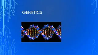
Understanding Genetics: The Key Concepts and Applications

Genetics is a vital field of biology focusing on genes, heredity, and genetic variation. This branch of science has deep roots in human history, influencing agriculture and shaping biological disciplines like evolution and developmental biology. Key subdisciplines include transmission genetics, molecular genetics, and population genetics, each exploring different aspects of genetic inheritance and variation.

Download Presentation

Please find below an Image/Link to download the presentation.
The content on the website is provided AS IS for your information and personal use only. It may not be sold, licensed, or shared on other websites without obtaining consent from the author. Download presentation by click this link. If you encounter any issues during the download, it is possible that the publisher has removed the file from their server.
E N D
Presentation Transcript
genetic course lecture (1) The chemical and engineering basis of heredity Dr. israa hussein hamzah email: esraa_hassan17@yahoo.com szsh@uomustansiriyah.edu.iq Reference book: genetic (Conceptual- Approach) fourth edition (2010) Author: Benjamin C. Pierce
Genetic define Genetics is a branch of biology concerned with the study of genes, genetic variation, and heredity in organisms The process of transmission of characters from one generation to next, either by gametes sperms and ova in sexual reproduction or by the asexual reproductive bodies in asexual reproduction, is called inheritance or heredity. It is the cause of similarities between individuals. This is the reason that brothers and sisters with the same parents resemble each other and with their parents. While the Variation is the cause of differences who do resemble each other are still unique individuals. Thus, we have no trouble in recognizing the differences between sisters, for example, and even identical twins are recognized as distinctive individuals by their parents and close friends.
The role of genetic in biology Although the science of genetics is relatively new compared with sciences such as astronomy and chemistry, people have understood the hereditary nature of traits and have practiced genetics for thousands of years. The rise of agriculture began when people started to apply genetic principles to the domestication of plants and animals. In spite of an understanding of genetics is important to all people, it is critical to the student of biology. Genetics provides one of biology s unifying principles: all organisms use genetic systems that have a number of features in common. Genetics also undergirds the study of many other biological disciplines. Evolution, for example, is genetic change taking place through time; so the study of evolution requires an understanding of genetics. Developmental biology relies heavily on genetics: tissues and organs develop through the regulated expression of genes . Even such fields as taxonomy, ecology, and animal behavior are making increasing use of genetic methods. The study of almost any field of biology or medicine is incomplete without a thorough understanding of genes and genetic methods.
Division of genetic The study of genetics consists of three major subdisciplines: transmission genetics, molecular genetics, and population genetics . classical genetics Also known as transmission genetics encompasses the basic principles of heredity and how traits are passed from one generation to the next. This area addresses the relation between chromosomes and heredity, the arrangement of genes on chromosomes, and gene mapping. Here, the focus is on the individual organism how an individual organism inherits its genetic makeup and how it passes its genes to the next generation. Molecular genetics concerns the chemical nature of the gene itself: how genetic information is encoded, replicated, and expressed. It includes the cellular processes of replication, transcription, and translation (by which genetic information is transferred from one molecule to another) and gene regulation (the processes that control the expression of genetic information). Population genetics explores the genetic composition of groups of individual members of the same species (populations) and how that composition changes geographically
Prokaryotic and eukaryotic cell differ in number of genetic characteristics Biologists traditionally classify all living organisms into two major groups, the prokaryotes and the eukaryotes A prokaryote is a unicellular organism with a relatively simple cell structure. lack a nuclear membrane and possess no membrane-bounded cell organelles whereas eukaryotic cells are more complex , it has a compartmentalized cell structure with components bounded by intracellular membranes; eukaryotes are either unicellular or multicellular ,it has a nuclear envelope, which surrounds the genetic material to form a nucleus and separates the DNA from the other cellular contents., Genetic information is carried in DNA and RNA. Genetic information is encoded in the molecular structure of nucleic acids, which come in two types: deoxyribonucleic acid (DNA) and ribonucleic acid (RNA). Nucleic acids are polymers consisting of repeating units called nucleotides; each nucleotide consists of a sugar, a phosphate, and a nitrogenous base. The nitrogenous bases in DNA are of four types: adenine (A), cytosine (C), guanine (G), and thymine (T). The sequence of these bases encodes genetic information. DNA consists of two complementary nucleotide strands. Most organisms carry their genetic information in DNA, but a few viruses carry it in RNA. The four nitrogenous bases of RNA are adenine, cytosine, guanine, and uracil (U).
The vehicles of genetic information within a cell are chromosomes which consist of DNA and associated proteins. DNA is closely associated with a special class of proteins, the histones, to form tightly packed chromosomes. This complex of DNA and histone proteins is termed chromatin The cells of each species have a characteristic number of chromosomes; for example, bacterial cells normally possess a single chromosome; human cells possess 46; pigeon cells possess 80. Each chromosome carries a large number of genes. Genes are located on chromosomes The gene is the fundamental unit of heredity. It is a Segment of DNA that has the information (the code) for a protein or RNA. these unit of information that encodes a genetic characteristic come in multiple forms called alleles. A single molecule of DNA has thousands of genes on the molecule
Chromosome structure Each eukaryotic species has a characteristic number of chromosomes per cell: potatoes have 48 chromosomes, fruit flies have 8, and humans have 46. There appears to be no special relation between the complexity of an organism and its number of chromosomes per cell The chromosomes of eukaryotic cells are larger and more complex than those found in prokaryotes, but each unreplicated chromosome nevertheless consists of a single molecule of DNA. Although linear, the DNA molecules in eukaryotic chromosomes are highly folded and condensed To package such a tremendous length of DNA into this small volume, each DNA molecule is coiled again and again and tightly packed around histone proteins, forming a rod-shaped chromosome. Most of the time, the chromosomes are thin and difficult to observe but, before cell division, they condense further into thick, readily observed structures; it is at this stage that chromosomes are usually studied. A functional chromosome has three essential elements: a centromere, a pair of telomeres, and origins of replication. The centromere is the attachment point for spindle microtubules the filaments responsible for moving chromosomes in cell division
Figure each eukaryotic chromosome has a centromere and telomeres.
On the basis of the location of the centromere, chromosomes are classified into four types: metacentric, submetacentric, acrocentric, and telocentric . One of the two arms of a chromosome (the short arm of a submetacentric or acrocentric chromosome) is designated by the letter p and the other arm is designated by q.
Types of Chromosomes: Autosomes = Body chromosomes or non sex chromosomes ( humans have 44 or 22 pairs) Sex Chromosomes = XX or XY (23rd pair for humans) determines the sex of the offspring
The first 22 pairs of homologous chromosomes are called autosomes or autosomal chromosomes. The 23rd pair of chromosomes determines the sex of the individual and are called sex chromosomes. The sex chromosomes of a female are XX. The sex chromosomes of a male are XY
In most eukaryotic cells, there are two sets of chromosomes. The presence of two sets is a consequence of sexual reproduction: one set is inherited from the male parent and the other from the female parent. Each chromosome in one set has a corresponding chromosome in the other set, together constituting a homologous pair . Human cells, for example, have 46 chromosomes, constituting 23 homologous pairs. The two chromosomes of a homologous pair are usually alike in structure and size, and each carries genetic information for the same set of hereditary characteristics. (The sex chromosomes are an exception ) For example, if a gene on a particular chromosome encodes a characteristic such as hair color, another copy of the gene (each copy is called an allele) at the same position on that chromosome s homolog also encodes hair color. However, these two alleles need not be identical: one might encode brown hair and the other might encode blond hair. Thus, most cells carry two sets of genetic information; these cells are diploid. But not all eukaryotic cells are diploid: reproductive cells (such as eggs, sperm, and spores) and even nonreproductive cells of some organisms may contain a single set of chromosomes. Cells with a single set of chromosomes are haploid. A haploid cell has only one copy of each gene
Mitosis and Meiosis Cell Division
Chromosomes separate through the processes of mitosis and meiosis. The processes of mitosis and meiosis ensure that a complete set of an organism s chromosomes exists in each cell resulting from cell division. Mitosis is the separation of chromosomes in the division of somatic (nonsex) cells. Meiosis is the pairing and separation of chromosomes in the division of sex cells to produce gametes (reproductive cells). Origins of replication are the sites where DNA synthesis begins; they are not easily observed by microscopy. In preparation for cell division, each chromosome replicates, making a copy of itself, as already mentioned. These two initially identical copies, called sister chromatids, are held together at the centromere . Each sister chromatid consists of a single molecule of DNA
The cell cycle and mitosis The cell cycle is the life story of a cell, the stages through which it passes from one division to the next . This process is critical to genetics because, through the cell cycle, the genetic instructions for all characteristics are passed from parent to daughter cells. A new cycle begins after a cell has divided and produced two new cells. The cell cycle consists of two major phases. The first is interphase, the period between cell divisions, in which the cell grows, develops, and functions. In interphase, critical events necessary for cell division also take place. The second major phase is the M phase (mitotic phase), the period of active cell division. The M phase includes mitosis, the process of nuclear division, and cytokinesis, or cytoplasmic division. Let s take a closer look at the details of interphase and the M phase.
Interphase Interphase is the extended period of growth and development between cell divisions. Although little activity can be observed with a light microscope, the cell is quite busy: DNA is being synthesized, RNA and proteins are being produced, and hundreds of biochemical reactions necessary for cellular functions are taking place. interphase is divided into three subphases: G1, S, and G2 M phase The M phase is the part of the cell cycle in which the copies of the cell s chromosomes (sister chromatids) separate and the cell undergoes division. The separation of sister chromatids in the M phase is a critical process that results in a complete set of genetic information for each of the resulting cells. Biologists usually divide the M phase into six stages: the five stages of mitosis (prophase, prometaphase, metaphase, anaphase, and telophase), illustrated in Figure 2.10, and cytokinesis. It s important to keep in mind that the M phase is a continuous process, and its separation into these six stages is somewhat arbitrary.
Prophase. As a cell enters prophase, the chromosomes become visible under a light microscope. Because the chromosome was duplicated in the preceding S phase, each chromosome possesses two chromatids attached at the centromere. The mitotic spindle, an organized array of microtubules that move the chromosomes in mitosis, forms. In animal cells, the spindle grows out from a pair of centrosomes that migrate to opposite sides of the cell. Within each centrosome is a special organelle, the centriole, which also is composed of microtubules. Some plant cells do not have centrosomes or centrioles, but they do have mitotic spindles. Prometaphase. Disintegration of the nuclear membrane marks the start of prometaphase. Spindle microtubules, which until now have been outside the nucleus, enter the nuclear region. The ends of certain microtubules make contact with the chromosomes. For each chromosome, a microtubule from one of the centrosomes anchors to the kinetochore of one of the sister chromatids; a microtubule from the opposite centrosome then attaches to the other sister chromatid, and so the chromosome is anchored to both of the centrosomes. The microtubules lengthen and shorten, pushing and pulling the chromosomes about. Some microtubules extend from each centrosome toward the center of the spindle but do not attach to a chromosome.
Metaphase. During metaphase, the chromosomes become arranged in a single plane, the metaphase plate, between the two centrosomes. The centrosomes, now at opposite ends of the cell with microtubules radiating outward and meeting in the middle of the cell, center at the spindle poles. A spindle-assembly checkpoint ensures that each chromosome is aligned on the metaphase plate and attached to spindle fibers from opposite poles. The passage of a cell through the spindle-assembly checkpoint depends on tension generated at the kinetochore as the two conjoined chromatids are pulled in opposite directions by the spindle fibers. This tension is required for the cell to pass through the spindle-assembly checkpoint. If a microtubule attaches to one chromatid but not to the other, no tension is generated and the cell is unable to progress to the next stage of the cell cycle. The spindle-assembly checkpoint is able to detect even a single pair of chromosomes that are not properly attached to microtubules. The importance of this checkpoint is illustrated by cells that are defective in their spindle-assembly checkpoint; these cells often end up with abnormal numbers of chromosomes. Anaphase. After the spindle-assembly checkpoint has been passed, the connection between sister chromatids breaks down and the sister chromatids separate. This chromatid separation marks the beginning of anaphase, during which the chromosomes move toward opposite spindle poles. The microtubules that connect the chromosomes to the spindle poles are composed of subunits of a protein
called tubulin . Chromosome movement is due to the disassembly of tubulin molecules at both the kinetochore end (called the end) and the spindle end (called the end) of the spindle fiber. Special proteins called molecular motors disassemble tubulin molecules from the spindle and generate forces that pull the chromosome toward the spindle pole. Telophase. After the chromatids have separated, each is considered a separate chromosome. Telophase is marked by the arrival of the chromosomes at the spindle poles. The nuclear membrane re-forms around each set of chromosomes, producing two separate nuclei within the cell. The chromosomes relax and lengthen, once again disappearing from view. In many cells, division of the cytoplasm (cytokinesis) is simultaneous with telophase. The major features of the cell cycle are summarized in following figure
Table :Features of the cell cycle Stage Major Features Stage Major Features G0 phase Stable, Non dividing period of variable length. Interphase G1 phase Growth and development of the cell; G1/S checkpoint. S phase Synthesis of DNA. Synthesis of DNA. G2 phase Preparation for division; G2/M checkpoint. M phase Prophase. Chromosomes condense and mitotic spindle forms Nuclear envelope disintegrates, and spindle microtubules anchor to kinetochores. Chromosomes align on the metaphase plate; spindle-assembly checkpoint. Sister chromatids separate, becoming individual chromosomes that migrate toward spindle poles Chromosomes arrive at spindle poles, the nuclear envelope re-forms, and the condensed chromosomes relax. Prometaphase Metaphase Anaphase. Telophase Cytokinesis Cytoplasm divides; cell wall forms in plant cells
Miosis The words mitosis and meiosis are sometimes confused. They sound a bit alike, and both refer to chromosome division and cytokinesis. But don t be deceived. The outcomes of mitosis and meiosis are radically different, and several unique events that have important genetic consequences take place only in meiosis. How does meiosis differ from mitosis? Mitosis consists of a single nuclear division and is usually accompanied by a single cell division. Meiosis, on the other hand, consists of two divisions. After mitosis, chromosome number in newly formed cells is the same as that in the original cell, whereas meiosis causes chromosome number in the newly formed cells to be reduced by half. Finally, mitosis produces genetically identical cells, whereas meiosis produces genetically variable cells. Let s see how these differences arise. Like mitosis, meiosis is preceded by an interphase stage that includes G1, S, and G2 phases. Meiosis consists of two distinct processes: meiosis I and meiosis II, each of which includes a cell division. The first division, which comes at the end of meiosis I, is termed the reduction division because the number of chromosomes per cell is reduced by half .The second division, which comes at the end of meiosis II, is sometimes termed the equational division. The events of meiosis II are similar to those of mitosis. However, meiosis II differs from mitosis in that chromosome number has already been halved in meiosis I, and the cell does not begin with the same number of chromosomes as it does in mitosis