Understanding Pharyngeal Suppuration and Abscesses
Pharyngeal suppuration and abscesses, such as peritonsillar abscess (Quinsy), parapharyngeal abscess, and retropharyngeal abscess, can present with symptoms like pain, fever, trismus, and neck swelling. Complications may include laryngeal edema and mediastinitis. Treatment involves systemic antibiot
1 views • 4 slides
Management of Quittor in Equine Medicine
Quittor is a condition in horses characterized by necrosis of lateral cartilage, leading to sinus openings in the coronet region. It can result from infections, pastern region necrosis, or suppuration from neighboring lesions. Symptoms include swollen coronet with sinus openings, potentially leading
0 views • 26 slides
Anatomy of the Pharynx: Structure and Function
The pharynx is a musculomembranous tube that connects the nasal and oral cavities to the larynx and esophagus. It is divided into three parts: nasopharynx, oropharynx, and laryngopharynx, each with unique features and functions. Muscles like pharyngeal constrictors and palatopharyngeus play a crucia
0 views • 31 slides
Perianal Suppuration: Abscess & Fistula Overview
Anorectal suppuration can lead to acute anal sepsis (abscess) or chronic anal fistula. This condition involves glandular secretions, infection, and suppuration, often originating from cryptoglandular sources. It can be caused by various specific conditions like Crohn's disease, ulcerative colitis, T
0 views • 47 slides
Development of Fetal Head and Neck Structures in Week 12
The fetal head and neck structures in week 12 exhibit a complex formation process involving contributions from all three embryonic layers and the neural crest. Neural crest plays a significant role in developing jaw skeletal elements, connective tissues, and tendons. The pharynx, starting at the buc
0 views • 30 slides
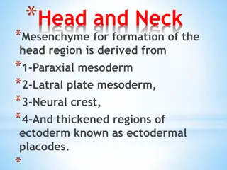
Development of Head and Neck Mesenchyme in Embryonic Formation
The formation of the head and neck region in embryonic development involves mesenchyme derived from paraxial mesoderm, lateral plate mesoderm, neural crest, and ectodermal placodes. Paraxial mesoderm contributes to brain case and muscle formation, while lateral plate mesoderm forms laryngeal cartila
0 views • 34 slides
