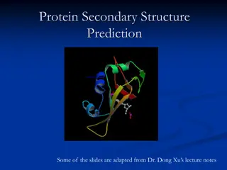
Understanding Protein Structure and Function in Biochemistry
Proteins play vital roles in the body as large, complex molecules made up of amino acids. Understanding peptide bonding, levels of protein structure, and forces stabilizing proteins is crucial. Explore how protein misfolding can lead to diseases like Alzheimer's. Learn about the primary structure and functions of proteins, including structural components and transport/storage roles.

Download Presentation

Please find below an Image/Link to download the presentation.
The content on the website is provided AS IS for your information and personal use only. It may not be sold, licensed, or shared on other websites without obtaining consent from the author. Download presentation by click this link. If you encounter any issues during the download, it is possible that the publisher has removed the file from their server.
E N D
Presentation Transcript
Protein structure Color Index: Important. Extra Information. 436 Biochemistry team Doctors slides.
Objectives By the end of this lecture, the students should be able to: Understand the peptide bonding between amino acids. Explain the different levels of protein structure and the forces stabilizing these structures and what happens when the protein is denatured. Define the -helix and -sheet as the most commonly encountered secondary structures in a protein molecule. Correlate the protein structure with function with hemoglobin as an example. Understand how the misfolding of proteins may lead to diseases like Alzheimer s or prion disease.
What are proteins? Play many critical roles in the body They are large, complex molecules They do most of work in the cell They are required for the structure, function and regulation of the body s tissues and organs they are made up of hundreds or thousands of smaller units called amino acids which are attached to one another in long chains by a peptide bond.
What are proteins? Proteins can be described according to their large range of functions in the body: There are mainly 20 different types of amino acids that can be combined to make a protein. The sequence of amino acids determines : 1- each protein s unique three- dimensional (3D) structure Antibody Enzyme Messenger 2- its specific function. Structural component Transport/storage
Peptide Bond (amide bond) : Amide linkage that is formed between -carboxyl group of an amino acid and -amino group of the other amino acid. Each amino acid (in) a chain makes two peptide bonds. Covalent bond formed by: Removal of water : 1-OH from COOH 2-H from NH3 Group. By : (dehydration) The amino acids (at the two ends) of a chain make only one peptide bond. The amino acid with a free amino group is called amino terminus or NH2-terminus. The amino acid with a free carboxylic group is called carboxyl terminus or COOH-terminus.
Peptide bond (amide bond): water is eliminated O O H2N CH C OH H2N CH C OH two amino acids condense to form... R1 R2 C or carboxy terminus N or amino terminus O O H2N CH C NH CH C OH ...a dipeptide. If there are more it becomes a polypeptide. Short polypeptide chains are usually called peptides while longer ones are called proteins. + HOH R1 R2 peptide bond is formed residue 2 residue 1 : Residues : amino acid in a peptide chain We always start from N terminus to C terminus
1.Primary structure It is the linear sequence of amino acids in a protein **Primary structure proteins are not functional Covalent bonds in the primary structure of protein: 1.Peptide bond 2.Disulfide bond (if any) (it is not always present) Which is the SS bond , It links two residues of cysteine NEAR TO EACH OTHER as shown in the picture. : Peptide bond are not broken by conditions that denature proteins, such as heat They can break by prolonged exposure to a strong acid or base at elevated temperatures to hydrolyze (break) these bond or by using enzymes How to determine the primary structure sequence? 1. DNA sequencing. 2.Direct amino acids sequencing.
peptides Amino acids can be polymerized to form chains: Two amino acids One peptide bond dipeptide Three amino acids Two peptide bonds tripeptide Four amino acids Three peptide bonds tetrapeptide Few (2-20)amino acids oligopeptide More(>20 amino acids) polypeptide
2.Secondary structure *It is also not functional It is regular arrangements of amino acids that are located near to each other in the linear sequence. Examples of secondary structures frequently found in proteins are: -helix -sheet -bends Excluding the conformations (3D arrangements) of its side chains. *Bonds that are found in it: Hydrogen bonds : for the secondary structure , we do not look at the R side chains nor do we look at the hydrophobic interactions that give it its 3D structure so we exclude that.. We only look at the hydrogen bonds
Secondary structure -helix: It is a right-handed spiral 1. side chains of amino acids extended outward. 2. Hydrogen bonds are what Stabilize the -helix. (Hydrogen bonds form between the peptide bond carbonyl oxygen and amide hydrogen) 3. Amino acids per turn: Each turn contains 3.6 amino acids. 3.6 ) 4. ) ) 3 (
-helix : -Amino acids that disrupt an -helix: Proline imino group, interferes with the smooth helical structure. ) ( Glutamate, aspartate, histidine, lysine or arginine form ionic bonds. ( these are all polar CHARGED amino acids so they would form ionic bonds thus it would change the shape) Bulky side chain such as tryptophan. ) ( Branched amino acids at the -carbon, such as valine or isoleucine. * : https://www.youtube.com/watch?v=V 3DgrOG1exY
Secondary structure -sheet (Composition of a -sheet) Two or more polypeptide chains make hydrogen bonding with each other. (beta sheet could be a long polypeptide) (the helix is just one polypeptide chain) Also called pleated sheets: because they appear as folded structures with edges Hydrogen bonds: Stabilize the -sheet. * : https://www.youtube.co m/watch?v=koyE9Nplacc
Secondary structure -sheet (Antiparallel and parallel sheets) Antiparallel: N C C N Parallel: N C N C Hydrogen bonds in the parallel direction are less stable than in the antiparallel (notice the dotted lines in the picture..in the antiparallel the lines are straight but in the parallel , they aren t)
Secondary structure Other secondary structure examples: -bends (reverse turns): Reverse the direction of a polypeptide chain. Usually found on the surface of the molecule and often include charged residues. The name comes because they often connect successive strands of antiparallel -sheets. -bends are generally composed of four amino acid residues, proline or glycine are frequently found in -bends. Nonrepetitive secondary structure: e.g. loop or coil conformation.
Secondary structure Other secondary structure examples: Supersecondary structures (motifs): A combination of secondary structural elements (that is, -helices, -sheets, and coils) . These form primarily the core (interior) region of the molecule. They are connected by loop regions . motif: two helices together motif: a helix connects two sheets hairpin: reverse turns connect antiparallel sheets barrels: rolls of sheets
Tertiary structure It is the three-dimensional (3D) structure of an entire polypeptide chain including side chains. What is it? The fundamental functional and 3D structural units of a polypeptide, >200 amino acids fold into two or more clusters. Domains are The core of a domain is built from combinations of supersecondary structural elements (motifs) and their side chains. The tertiary structure of a proteins is the functional protein. a protein with three domains Domains can be combined to form tertiary structure. supersecondary structural elements (motifs) When combined The core of a domain Domains tertiary structure.
Interactions stabilizing tertiary structure: Disulfide bonds. Hydrophobic interactions. Hydrogen bonds. Ionic interactions. Negatively charged groups can interact with positively charged groups such as : carboxylate group ( COO ) in the side chain of aspartate and amino group ( NH3+) Amino acid side chains containing oxygen- or nitrogen- bound hydrogen a covalent linkage formed from the sulfhydryl group ( SH) of each of two cysteine residues to produce a cystine residue Amino acids with nonpolar side chains tend to be located in the interior of the polypeptide molecule, where they associate with other hydrophobic amino acid
Tertiary structure Protein folding: Interactions between the side chains of amino acids determine how a long polypeptide chain folds into the intricate three- dimensional shape of the functional protein. : https://www.youtube.co m/watch?v=QSyCPD2qlPs
Tertiary structure are a specialized group of proteins, required for the proper folding of many species of proteins. Chaperons Role of chaperons, also known as heat chock , in protein folding: They interact with polypeptide at various stages during the folding process. Examples of chaperons: Hsp60 and Hsp70.
Quaternary structure Hemoglobin Some proteins contain two or polypeptide chains that may be more structurally identical or totally unrelated. Hemoglobin is a globular protein. spherical ("globe-like") A multisubunit protein is called oligomer ( an oligomer usually refers to a macromolecular complex ) Composed of 2 2 subunits (4 subunits). Each chain forms a 3D structure called subunit. According to the number of subunits: dimeric, trimeric, or multimeric. Two same subunits are called protomers. (a protomer is the structural unit of an oligomeric protein) . Subunits may either function independently of each other, or work cooperatively, hemoglobin. e.g. Example:
Denaturation of proteins unfolding and disorganization of the protein s secondary and tertiary structures. when a protein is denatured, it results in the Denaturation agents Organic solvents Mechanical mixing. Strong acids or bases Ions of heavy metals (e.g. lead and mercury) Heat Detergents Most proteins, once denatured, remain permanently disordered. Denatured proteins are therefore, precipitate from solution. often insoluble and,
Protein misfolding Every protein must fold to achieve its normal conformation and function. Abnormal folding of proteins leads to a number of diseases in humans. Alzheimer s disease Creutzfeldt-Jacob or prion disease amyloid protein is a misfolded protein. Amyloid is aggregates of misfolded proteins outside neurons, it interfere with neurons ability of sending massages. Prion protein is present in normal brain tissue , in diseased brains, the same protein is misfolded. It, therefore, forms insoluble fibrous aggregates that damage brain cells. It forms fibrous deposits or plaques in the brains of Alzheimer s patients.
Takehome messages Native conformation of the protein is the functional, fully folded protein structure The unique 3D structure of the native conformation is determined by its primary structure, i.e. the amino acid sequence Interactions of between the amino acid side chains guide the folding of the polypeptide chain to form secondary, tertiary and sometimes quaternary structures that cooperate in stabilizing the native conformation of the protein. Protein denaturation results in unfolding and disorganization of of the protein s structure, which are not accompanied by hydrolysis of peptide bonds. Disease can occur when an apparently normal protein assumes a conformation that is cytotoxic, as in the case of Alzheimer disease and Prion disease.
MCQs https://www.onlineexambuilder.com/bio/exa m-98731 Video: Overview of protein Protein structure
Girls team members: Boys team members: . - 1 . - 1 . - 2 . - 2 . - 3 . - 3 . - 4 . - 5 . - 6 . - 7 -Team leaders: -Contact us: . . Biochemistryteam436@gmail.com twitter.com/436biochemteam


 :")
:")