Understanding Kidney Physiology and Function in the Human Body
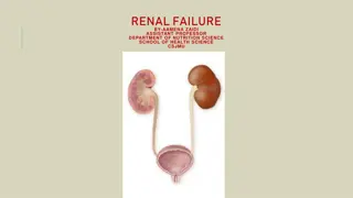
Kidneys play a crucial role in regulating arterial blood pressure, erythrocyte production, glucose synthesis, and filtration through nephrons. They cannot regenerate nephrons, leading to a gradual decrease in function with age. Key structures like the hilum, renal artery, and vein, as well as the nephron's glomerulus and tubule, contribute to kidney function. Understanding these processes is essential for grasping kidney physiology and its impact on overall health.
- Kidney physiology
- Arterial blood pressure regulation
- Erythropoietin production
- Glomerulus filtration
- Renal anatomy
Download Presentation

Please find below an Image/Link to download the presentation.
The content on the website is provided AS IS for your information and personal use only. It may not be sold, licensed, or shared on other websites without obtaining consent from the author. Download presentation by click this link. If you encounter any issues during the download, it is possible that the publisher has removed the file from their server.
E N D
Presentation Transcript
Extra Notes from Extra Notes from Reference Books Reference Books 1. Guyton and Hall 12th Edition 2. Linda 5th Edition If you do not understand a certain point in any of the lectures just read its explanation from the reference books. Good luck! Done by: Amal Alqarni Najd Altheeb Reema Alotaibi Amal Alshaibi Elham Alamy Ghada Alhadlaq Ruba Barnawi - Ghada Alskait Leena Alwakeel Laila Mathkour Heba Alnasser 1
Lecture 1 Chapter 26 page 304 Guyton corner : remember from CVS block - Regulation of arterial blood pressure : the kidneys play a dominant role in long-term regulation of arterial pressure by excreting variable amounts of sodium and water. The kidneys also contribute to short-term arterial pressure regulation by secreting hormones and vasoactive factors or substances (e.g., renin) that lead to the formation of vasoactive products (e.g., angiotensin II). Guyton corner : remember from Foundation block - Regulation of Erythrocyte Production : The kidneys secrete erythropoietin, which stimulates the production of blood cells by hematopoietic stem cells in the bone marrow. One important stimulus for erythropoietin secretion by the kidneys is hypoxia. The kidneys normally account for almost all the erythropoietin secreted into the circulation. In people with severe kidney disease or who have had their kidneys removed and have been placed on hemodialysis, severe anemia develops as a result of decreased erythropoietin production. 2
Cont. Guyton corner : remember from MSK block - Glucose synthesis : The kidneys capacity to add glucose to the blood during prolonged periods of fasting rivals that of the liver. Physiology and anatomy of kidneys Guyton corner : ( page 304 ) - General Organization of the Kidneys and Urinary Tract The medial side of each kidney contains an indented region called the hilum through which pass the renal artery and vein, lymphatics, nerve supply, and ureter, which carries the final urine from the kidney to the bladder, where it is stored until emptied. The kidney is surrounded by a tough, fibrous capsule that protects its delicate inner structures. Guyton corner : ( page 305 ) - The Nephron Is the Functional Unit of the Kidney: The kidney cannot regenerate new nephrons. Therefore, with renal injury, disease, or normal aging, there is a gradual decrease in nephron number. After age 40, the number of functioning nephrons usually decreases about 10 percent every 10 years; thus, at age 80, many people have 40 percent fewer functioning nephrons than they did at age 40. This loss is not life threatening because adaptive changes in the remaining nephrons allow them to excrete the proper amounts of water, electrolytes, and waste products. 3
Cont.(Important Notes from 435 ) Note that the pressures along the afferent arteriole, glumerulus and efferent arteriole are high. Why? To increase filtration. Note that the afferent arteriole is larger than the efferent arteriole Nephrons Vessels Tubes (5) 4
Cont. Guyton corner : ( page 305 ) - The Nephron Is the Functional Unit of the Kidney: Each nephron contains (1) a tuft of glomerular capillaries called the glomerulus, through which large amounts of fluid are filtered from the blood, and (2) a long tubule in which the filtered fluid is converted into urine on its way to the pelvis of the kidney 5
Cont.(Structure of a Nephron) Guyton corner : Each nephron has some differences from the others, depending on how deep the nephron lies within the kidney mass. Those nephrons that have glomeruli located in the outer cortex are called cortical nephrons; they have short loops of Henle that penetrate only a short distance into the medulla About 20 to 30 percent of the nephrons have glomeruli that lie deep in the renal cortex near the medulla and are called juxtamedullary nephrons. These nephrons have long loops of Henle that dip deeply into the medulla, in some cases all the way to the tips of the renal papillae. The vascular structures supplying the juxtamedullary nephrons also differ from those supplying the cortical nephrons. For the cortical nephrons, the entire tubular system is surrounded by an extensive network of peritubular capillaries. For the juxtamedullary nephrons, long efferent arterioles extend from the glomeruli down into the outer medulla and then divide into specialized peritubular capillaries called vasa recta that extend downward into the medulla, lying side by side with the loops of Henle. Like the loops of Henle, the vasa recta return toward the cortex and empty into the cortical veins. This specialized network of capillaries in the medulla plays an essential role in the formation of a concentrated urine 6
Cont.(Structure of a Nephron) Linda corner: Bowman s space is continuous with the first portion of the nephron. Blood is ultra filtered across the glomerular capillaries into Bowman s space, which is the first step in urine formation. The remainder of the nephron is a tubular structure lined with epithelial cells, which serve the functions of reabsorption and secretion. The nephron or renal tubule comprises the following segments (beginning with Bowman s space): the proximal convoluted tubule, the proximal straight tubule, the loop of Henle (which contains a thin descending limb, a thin ascending limb, and a thick ascending limb), the distal convoluted tubule, and the collecting ducts. Each segment of the nephron is functionally distinct, and the epithelial cells lining each segment have a different ultrastructure. For example, the cells of the proximal convoluted tubule are unique in having an extensive development of microvilli, called a brush border, on their luminal side. The brush border provides a large surface area for the major reabsorptive function of the proximal convoluted tubule. There are two types of nephrons, superficial cortical nephrons and juxtamedullary nephrons, which are distinguished by the location of their glomeruli. The superficial cortical nephrons have their glomeruli in the outer cortex. These nephrons have relatively short loops of Henle, which descend only into the outer medulla. The juxtamedullary nephrons have their glomeruli near the corticomedullary border. The glomeruli of the juxtamedullary nephrons are larger than those of the superficial cortical nephrons and, accordingly, have higher glomerular filtration rates. The juxtamedullary nephrons are characterized by long loops of Henle that descend deep into the inner medulla and papilla and are essential for the concentration of urine. 7
Cont. Linda corner: Blood enters each kidney via a renal artery, which branches into interlobar arteries, arcuate arteries, and then cortical radial arteries. The smallest arteries sub- divide into the first set of arterioles, the afferent arterioles. The afferent arterioles deliver blood to the first capillary network, the glomerular capillaries, across which ultrafiltration occurs. Blood leaves the glomerular capillaries via a second set of arterioles, the efferent arterioles, which deliver blood to a second capillary network, the peritubular capillaries. The peritubular capillaries surround the nephrons. Solutes and water are reabsorbed into the peritubular capillaries, and a few solutes are secreted from the peritubular capillaries. Blood from the peritubular capillaries flows into small veins and then into the renal vein. The blood supply of superficial cortical nephrons differs from that of juxtamedullary nephrons. In the superficial nephrons, peritubular capillaries branch off the efferent arterioles and deliver nutrients to the epithelial cells. These capillaries also serve as the blood supply for reabsorption and secretion. In the juxtamedullary nephrons, the peritubular capillaries have a specialization called the vasa recta, which are long, hairpinshaped blood vessels that follow the same course as the loop of Henle. The vasa recta serve as osmotic exchangers for the production of concentrated urine 8
Cont.(Renal blood flow ) Guyton corner : -In an average 70-kilogram man, the combined blood flow percent of the cardiac output. Considering that the two kidneys constitute only about 0.4 percent of the total body weight, one can readily see that they receive an extremely high blood flow compared with other organs. As with other tissues, blood flow supplies the kidneys with nutrients and removes waste products. However, the high flow to the kidneys greatly exceeds this need. -the kidneys normally consume oxygen at twice the rate of the brain but have almost seven times the blood flow of the brain. Thus, the oxygen delivered to the kidneys far exceeds their metabolic needs, and the arterial-venous extraction of oxygen is relatively low compared with that of most other tissues. -Most of the renal vascular resistance resides in three major segments: interlobular arteries, afferent arterioles, controlled by the sympathetic nervous system, various hormones, and local internal renal control mechanisms. An increase in the resistance of any of the vascular segments of the kidneys tends to reduce the renal blood flow, whereas a decrease in vascular resistance increases renal blood flow if renal artery and renal vein pressures remain constant. -The outer part of the kidney, the renal cortex, receives most of the kidney s blood flow. Blood flow in the renal medulla accounts for only 1 to 2 percent of the total renal blood flow. Flow to the renal medulla is supplied by a specialized portion of the peritubular capillary system called the vasa recta. Linda corner: As with blood flow in any organ, RBF (Q) is directly proportional to the pressure gradient ( P) between the renal artery and the renal vein, and it is inversely proportional to the resistance (R) of the renal vasculature. (Recall that Q = P/R. Recall, also, that resistance is provided mainly by the arterioles.) The kidneys are unusual, however, in that there are two sets of arterioles, the afferent and the efferent. The major mechanism for changing blood flow is by changing arteriolar resistance. In the kidney, this can be accomplished by changing afferent arteriolar resistance and/ or efferent arteriolar resistance. 9
Cont.(Urine formation ) Guyton corner : The rates at which different substances are excreted in the urine represent the sum of three renal processes, (1) glomerular filtration, (2) reabsorption of substances from the renal tubules into the blood, and (3) secretion of substances from the blood into the renal tubules. Expressed mathematically , Urinary Excretion Rate = Filtration Rate Reabsorption Rate + Secretion Rate Urine formation begins when a large amount of fluid that is virtually free of protein is filtered from the glomerular capillaries into Bowman s capsule. Most substances in the plasma, except for proteins, are freely filtered, so their concentration in the glomerular filtrate in Bowman s capsule is almost the same as in the plasma. As filtered fluid leaves Bowman s capsule and passes through the tubules, it is modified by reabsorption of water and specific solutes back into the blood or by secretion of other substances from the peritubular capillaries into the tubules In general, tubular reabsorption is quantitatively more important than tubular secretion in the formation of urine, but secretion plays an important role in determining the amounts of potassium and hydrogen ions and a few other substances that are excreted in the urine. Most substances that must be cleared from the blood, especially the end products of metabolism such as urea, creatinine, uric acid, and urates, are poorly reabsorbed and are therefore excreted in large amounts in the urine. Certain foreign substances and drugs are also poorly reabsorbed but, in addition, are secreted from the blood into the tubules, so their excretion rates are high. Conversely, electrolytes, such as sodium ions, chloride ions, and bicarbonate ions, are highly reabsorbed, so only small amounts appear in the urine. Certain nutritional substances, such as amino acids and glucose, are completely reabsorbed from the tubules and do not appear in the urine even though large amounts are filtered by the glomerular capillaries. 10
Cont. Glumerular membrane is made up of: - Endothelial cells - Basement membrane - Podocytes (visceral layer of the capsule) Passage of molecules through the membrane depends on: 1. Molecular weight 2. Electrical charge Gluemerular filtration rate depends on: 1. Net Filtration pressure: (Which depends on) A. Hydrostatic pressure of the capillary: - Arterial blood pressure - Afferent Vasoconstriction (sympathetic stimulation) Reduced hydrostatic capillary pressure Less GFR Vasodilation (parasympathetic stimulation) More hydrostatic capillary pressure More GFR - Efferent Vasoconstriction (Angiotensin II or strong sympthetic stimulation) More hydrostatic capillary pressure More GFR hydrostatic pressure Less GFR Severe vasoconstriction More hydrostatic capillary pressure and even more oncotic capillary pressure Less GFR B. Oncotic pressure of the capillary: C. Hydrostatic pressure of the capsule (affected by obstruction): - Tumors (e.g. prostatic cancer) - Prostatic hyperplasia/hypertrophy 2 . Capillary filtration coefficient: (Which depends on) A. Filtration barrier permeability B. Surface area of the filtration barrier Vasodilation Less 12
Cont. Note that, as the afferent arteriole constricts in response to sympathetic stimulation, prostaglandins are released to cause vasodilation to prevent further damage and renal failure. This explains why NSAIDs should not be given at this point. Why is the oncotic pressure of the capsule zero? Because usually, plasma proteins do not cross the filtration barrier. Early in diabetes, GFR is high. It drops later due to membrane thickening. Diabetes and hypertension thicken the membrane, reducing permeability and filtration coefficient. 13
Cont. Guyton corner : The glomerular capillary membrane is similar to that of other capillaries, except that it has three (instead of the usual two) major layers: (1) the endothelium of the capillary, (2) a basement membrane, and (3) a layer of epithelialcells (podocytes) surrounding the outer surface of the capillary basement membrane Together, these layers make up the filtration barrier, which, despite the three layers, filters several hundred times as much water and solutes as the usual capillary membrane. Even with this high rate of filtration, the glomerular capillary membrane normally prevents filtration of plasma proteins. The high filtration rate across the glomerular capillary membrane is due partly to its special characteristics. The capillary endothelium is perforated by thousands of small holes called fenestrae, similar to the fenestrated capillaries found in the liver. Although the fenestrations are relatively large, endothelial cells are richly endowed with fixed negative charges that hinder the passage of plasma proteins. Surrounding the endothelium is the basement membrane, which consists of a meshwork of collagen and proteoglycan fibrillae that have large spaces through which large amounts of water and small solutes can filter. The basement membrane effectively prevents filtration of plasma proteins, in part because of strong negative electrical charges associated with the proteoglycans. The final part of the glomerular membrane is a layer of epithelial cells that line the outer surface of the glomerulus. These cells are not continuous but have long footlike processes (podocytes) that encircle the outer surface of the capillaries The foot processes are separated by gaps called slit pores through which the glomerular filtrate moves. The epithelial cells, which also have negative charges, provide additional restriction to filtration of plasma proteins. Thus, all layers of the glomerular capillary wall provide a barrier to filtration of plasma proteins. 14
Cont. Guyton corner : The glomerular capillary membrane is similar to that of other capillaries, except that it has three (instead of the usual two) major layers: (1) the endothelium of the capillary, (2) a basement membrane, and (3) a layer of epithelial cells (podocytes) surrounding the outer surface of the capillary basement membrane. Together, these layers make up the filtration barrier, which, despite the three layers, filters several hundred times as much water and solutes as the usual capillary membrane. Even with this high rate of filtration, the glomerular capillary membrane normally prevents filtration of plasma proteins. The high filtration rate across the glomerular capillary membrane is due partly to its special characteristics. The capillary endothelium is perforated by thousands of small holes called fenestrae, similar to the fenestrated capillaries found in the liver, although smaller than the fenestrae of the liver. Although the fenestrations are relatively large, endothelial cell proteins are richly endowed with fixed negative charges that hinder the passage of plasma proteins. Surrounding the endothelium is the basement membrane, which consists of a meshwork of collagen and proteoglycan fibrillae that have large spaces through which large amounts of water and small solutes can filter. The basement membrane effectively prevents filtration of plasma proteins, in part because of strong negative electrical charges associated with the proteoglycans. The final part of the glomerular membrane is a layer of epithelial cells that line the outer surface of the glomerulus. These cells are not continuous but have long footlike processes (podocytes) that encircle the outer surface of the capillaries .The foot processes are separated by gaps called slit pores through which the glomerular filtrate moves. The epithelial cells, which also have negative charges, provide additional restriction to filtration of plasma proteins. Thus, all layers of the glomerular capillary wall provide a barrier to filtration of plasma proteins. 15
Cont. Linda corner: Layers of the Glomerular Capillary ENDOTHELIUM The endothelial cell layer has pores 70 to 100 nanometers (nm) in diameter. Because these pores are relatively large, fluid, dissolved solutes, and plasma proteins all are filtered across this layer of the glomerular capillary barrier. On the other hand, the pores are not so large that blood cells can be filtered. BASEMENT MEMBRANE The basement membrane has three layers. The lamina rara interna is fused to the endothelium; the lamina densa is located in the middle of the basement membrane; and the lamina rara externa is fused to the epithelial cell layer. The multilayered basement membrane does not permit filtration of plasma proteins and, therefore, constitutes the most significant barrier of the glomerular capillary. EPITHELIUM The epithelial cell layer consists of specialized cells called podocytes, which are attached to the basement membrane by foot processes. Between the foot processes are filtration slits, 25 to 60 nm in diameter, which are bridged by thin diaphragms. Because of the relatively small size of the filtration slits, the epithelial layer (in addition to the basement membrane) 16
Cont. 17
Cont.(GFR) Linda corner: Kf, filtration coefficient, is the water permeability or hydraulic conductance of the glomerular capillary wall. The two factors that contribute to Kfare the water permeability per unit of surface area and the total surface area. Kffor glomerular capillaries is more than 100-fold that for systemic capillaries (e.g., skeletal muscle capillaries) because of the combination of a higher total surface area and a higher intrinsic water permeability of the barrier. The consequence of this extremely high Kfis that much more fluid is filtered from glomerular capillaries than from other capillaries (i.e., GFR is 180 L/day) Guyton corner : [ Forces favouring filtration ] : 1) Glomerular hydrostatic pressure= 60 mmHg. 2) Bowman's capsule colloid osmotic pressure = zero mmHg [ Forces opposing filtration ] : 1) Bowman's capsule hydrostatic pressure = 18 mmHg. 2) Glomerular colloid osmotic pressure = 32 mmHg - NFP = 60-18-32 = +10 mmHg 18
Cont.(GFR) Linda corner: GC, oncotic pressure in glomerular capillaries, is another force opposing filtration. GC is determined by the protein concentration of glomerular capillary blood. GC does not remain constant along the capillary length; rather, it progressively increases as fluid is filtered out of the capillary. GC eventually increases to the point where net ultrafiltration pressure becomes zero and glomerular filtration stops (called filtration equilibrium). Figure 6-10B shows the three Starling pressures at the end of the glomerular capillary. At this point, the blood has been extensively filtered and is about to leave the glomerular capillary to enter the efferent arteriole. The sum of the three Starling pressures now is zero. Because net ultrafiltration is zero, no filtration can occur, a point called filtration equilibrium. Conveniently, filtration equilibrium normally occurs at the end of the glomerular capillary. An important question to ask is What causes filtration equilibrium to occur? Stated differently, Which Starling pressure has changed to make the net ultrafiltration pressure zero? To answer this question, compare the Starling pressures at the beginning of the glomerular capillary with those at the end of the capillary. The only pressure that changes is GC, the oncotic pressure of glomerular capillary blood. As fluid is filtered out of the glomerular capillary, protein is left behind and the protein concentration and GC increase. By the end of the glomerular capillary, GC has increased to the point where the net ultrafiltration pressure becomes zero. (A related point is that this blood leaving the glomerular capillaries will become peritubular capillary blood. The peritubular capillary blood will, therefore, have a high oncotic pressure [ c], which becomes a driving force for reabsorption in the proximal tubule of the nephron.) There is no decrease in PGC along the length of the glomerular capillaries, as occurs in systemic capillaries. The difference for glomerular capillaries is the presence of a second set of arterioles, the efferent arterioles. Constriction of efferent arterioles prevents the decline in PGC that would otherwise occur as fluid is filtered out along the length of the glomerular capillaries. 19
Cont. This slide has questions that the doctor gave 435 as homework, have a look at them. Q1 : Read more about Renin; where is it synthesized ? Guyton corner : The Renin-Angiotensin System : Its Role in Arterial Pressure Control Aside from the capability of the kidneys to control arterial pressure through changes in extracellular fluid volume, the kidneys also have another powerful mechanism for controlling pressure. It is the renin-angiotensin system. Renin is a protein enzyme released by the kidneys when the arterial pressure falls too low. In turn, it raises the arterial pressure in several ways, thus helping to correct the initial fall in pressure. Renin is synthesized and stored in an inactive form called prorenin in the juxtaglomerular cells (JG cells) of the kidneys. The JG cells are modified smooth muscle cells located in the walls of the afferent arterioles immediately proximal to the glomeruli. When the arterial pressure falls, intrinsic reactions in the kidneys themselves cause many of the prorenin molecules in the JG cells to split and release renin. Most of the renin enters the renal blood and then passes out of the kidneys to circulate throughout the entire body. However, small amounts of the renin do remain in the local fluids of the kidney and initiate several intrarenal functions. Page 220 20
Cont. Q2 : Why does GFR increase in high protein diet and hyperglycemia ? Guyton corner : Page 321 Other Factors That Increase Renal Blood Flow and GFR: High Protein Intake and Increased Blood Glucose Although renal blood flow and GFR are relatively stable under most conditions, there are circumstances in which these variables change significantly. For example,a high protein intake is known to increase both renal blood flow and GFR.With a chronic high-protein diet, such as one that contains large amounts of meat, the increases in GFR and renal blood flow are due partly to growth of the kidneys. However,GFR and renal blood flow increase 20 to 30 percent within 1 or 2 hours after a person eats a high-protein meal. One likely explanation for the increased GFR is the following: A high-protein meal increases the release of amino acids into the blood, which are reabsorbed in the proximal tubule. Because amino acids and sodium are reabsorbed together by the proximal tubules,increased amino acid reabsorption also stimulates sodium reabsorption in the proximal tubules. This decreases sodium delivery to the macula densa which elicits a tubuloglomerular feedback mediated decrease in resistance of the afferent arterioles, as discussed earlier. The decreased afferent arteriolar resistance then raises renal blood flow and GFR. This increased GFR allows sodium excretion to be maintained at a nearly normal level while increasing the excretion of the waste products of protein metabolism, such as urea. 21
Cont. A similar mechanism may also explain the marked increases in renal blood flow and GFR that occur with large increases in blood glucose levels in uncontrolled diabetes mellitus. Because glucose, like some of the amino acids, is also reabsorbed along with sodium in the proximal tubule,increased glucose delivery to the tubules causes them to reabsorb excess sodium along with glucose. This, in turn, decreases delivery of sodium chloride to the macula densa, activating a tubuloglomerular feedback- mediated dilation of the afferent arterioles and subsequent increases in renal blood flow and GFR. These examples demonstrate that renal blood flow and GFR per se are not the primary variables controlled by the tubuloglomerular feedback mechanism. The main purpose of this feedback is to ensure a constant delivery of sodium chloride to the distal tubule, where final processing of the urine takes place. Thus, disturbances that tend to increase reabsorption of sodium chloride at tubular sites before the macula densa tend to elicit increased renal blood flow and GFR, which helps return distal sodium chloride delivery toward normal so that normal rates of sodium and water excretion can be maintained . An opposite sequence of events occurs when proximal tubular reabsorption is reduced. For example, when the proximal tubules are damaged (which can occur as a result of poisoning by heavy metals, such as mercury, or large doses of drugs, such as tetracyclines), their ability to reabsorb sodium chloride is decreased. As a consequence, large amounts of sodium chloride are delivered to the distal tubule and, without appropriate compensations, would quickly cause excessive volume depletion. One of the important compensatory responses appears to be a tubuloglomerular feedback mediated renal vasoconstriction that occurs in response to the increased sodium chloride delivery to the macula densa in these circumstances. These examples again demonstrate the importance of this feedback mechanism in ensuring that the distal tubule receives the proper rate of delivery of sodium chloride, other tubular fluid solutes, and tubular fluid volume so that appropriate amounts of these substances are excreted in the urine. 22
Lecture 2 Inulin Clearance Can Be Used to Estimate GFR : If a substance is freely filtered (filtered as freely as water) and is not reabsorbed or secreted by the renal tubules, then the rate at which that substance is excreted in the urine is equal to the filtration rate of the substance by the kidneys. A substance that fits these criteria is inulin, a polysaccharide molecule with a molecular weight of about 5200. Inulin, which is not produced in the body, is found in the roots of certain plants and must be administered intravenously to a patient to measure GFR. Creatinine Clearance and Plasma Creatinine Concentration Can Be Used to Estimate GFR : Creatinine is a by-product of muscle metabolism and is cleared from the body fluids almost entirely by glomerular filtration. Therefore, the clearance of creatinine can also be used to assess GFR. Because measurement of creatinine clearance does not require intravenous infusion into the patient, this method is much more widely used than inulin clearance for estimating GFR clinically. However, creatinine clearance is not a perfect marker of GFR because a small amount of it is secreted by the tubules, so the amount of creatinine excreted slightly exceeds the amount filtered. There is normally a slight error in measuring plasma creatinine that leads to an overestimate of the plasma creatinine concentration, and fortuitously, these two errors tend to cancel each other. Therefore, creatinine clearance provides a reasonable estimate of GFR 23
Cont. Guyton corner : Feedback mechanisms intrinsic to the kidneys normally keep the renal blood flow and GFR relatively constant, despite marked changes in arterial blood pressure. These mechanisms still function in blood-perfused kidneys that have been removed from the body, independent of systemic influences. This relative constancy of GFR and renal blood flow is referred to as auto regulation (Figure 26-17) The major function of auto regulation in the kidneys is to maintain a relatively constant GFR and to allow precise control of renal excretion of water and solutes. The GFR normally remains auto regulated (that is, remains relatively constant), despite considerable arterial pressure fluctuations that occur during a person's usual activities. For instance, a decrease in arterial pressure to as low as 75 mm Hg or an increase to as high as 160 mm Hg usually changes GFR less than 10 percent. In general, renal blood flow is auto regulated in parallel with GFR, but GFR is more efficiently auto regulated under certain conditions. Importance of GFR Auto regulation in Preventing Extreme Changes in Renal Excretion Linda corner: The only way to maintain this constancy of blood flow in the phase of changing arterial pressure is by varying the resistance of the arterioles. Thus, as renal arterial pressure increases or decreases, renal resistance must increase or decrease proportionately (recall that Q = P/R). 24
Cont. Guyton corner : The juxtaglomerular complex consists of macula densa cells in the initial portion of the distal tubule and juxtaglomerular cells in the walls of the afferent and efferent arterioles. The macula densa is a specialized group of epithelial cells in the distal tubules that comes in close contact with the afferent and efferent arterioles. The macula densa cells contain Golgi apparatus, which are intracellular secretory organelles directed toward the arterioles, suggesting that these cells may be secreting a substance toward the arterioles. If we are in stressful condition our blood pressure increases the blood flow increase which result in increased hydrostatic pressure NFP increase GFR increase. But even though we don t urinate, why? Because kidney has mechanism to regulate by itself (auto regulation ) within certain blood pressure range . If the BP within 75-160 the kidney is auto regulated which mean GFR is constant . And its auto regulated by 2 mechanism THE 1ST one is myogenic mechanism : if the BP increases the renal blood flow increases which will stretch the A.A this will stimulate Ca+2 channels to open Ca+2 influx to smooth muscle cells in the wall of A.A leading to vasoconstriction decreased blood flow decreased Pg decreased NFP decreased GFR . Remember :GFR in addition to renal plasma flow are constant due to autoregulation At the beginning of distal convoluted tubules ( between A.A & E.A ) there are cells called macula densa cells. The cells opposite to macula densa are called juxtaglomerular cells. Macula densa is sensitive to NaCl (salt). Let s say BP is increased, fluid going to A.A increases therefore increased GFR, and the filtrated fluid in proximal convoluted tubules increases and moves very fast which make no time for reabsorption in PCT. When this fluid reach to macula densa in DCT it will sense high sodium chloride and send signals that stimulate vasoconstriction of A.A . In addition, it prevents release of renin by juxtaglomerular cells and prevent production of Ang II, no vasoconstriction of E.A. Low BP Low blood flow low Pg low GFR Fluids enter the proximal convoluted tubule are low and move slowly which make them reabsorbed Decrease delivery of NaCl to the macula densa cells vasodilatation of Afferentarterioles IncreaseinReninreleasefromthejuxtaglomerularcellsto AngII cause vasoconstriction of EFFERNT Arteriole. Inaddition,itstimulatesjuxtaglomerularcellstoreleaserenin produceAngII causing vasoconstriction of E.A. 25
Cont. Guyton corner : Tubuloglomerular Feedback and Autoregulation of GFR Decreased Macula Densa Sodium Chloride Causes Dilation of Afferent Arterioles and Increased Renin Release. The macula densa cells sense changes in volume delivery to the distal tubule by way of signals that are not completely understood. Experimental studies suggest that decreased GFR slows the flow rate in the loop of Henle, causing increased reabsorption of sodium and chloride ions in the ascending loop of Henle, thereby reducing the concentration of sodium chloride at the macula densa cells. This decrease in sodium chloride concentration initiates a signal from the macula densa that has two effects (Figure 26-19): (1) It decreases resistance to blood flow in the afferent arterioles, which raises glomerular hydrostatic pressure and helps return GFR toward normal, and (2) it increases renin release from the juxtaglomerular cells of the afferent and efferent arterioles, which are the major storage sites for renin. Renin released from these cells then functions as an enzyme to increase the formation of angiotensin I, which is converted to angiotensin II. Finally, the angiotensin II constricts the efferent arterioles, thereby increasing glomerular hydrostatic pressure and helping to return GFR toward normal.These two components of the tubuloglomerular feedback mechanism, operating together by way of the special anatomical structure of the juxtaglomerular apparatus, provide feedback signals to both the afferent and the efferent arterioles for efficient autoregulation of GFR during changes in arterial pressure. When both of these mechanisms are functioning together, the GFR changes only a few percentage points, even with large fluctuations in arterial pressure between the limits of 75 and 160 mm Hg. 26
Cont. Guyton corner pg321 : Another mechanism that contributes to the maintenance of a relatively constant renal blood flow and GFR is the ability of individual blood vessels to resist stretching during increased arterial pressure, a phenomenon referred to as the myogenic mechanism. Studies of individual blood vessels (especially small arterioles) throughout the body have shown that they respond to increased wall tension or wall stretch by contraction of the vascular smooth muscle. Stretch of the vascular wall allows increased movement of calcium ions from the extracellular fluid into the cells, causing them to contract ,This contraction prevents excessive stretch of the vessel and at the same time, by raising vascular resistance, helps prevent excessive increases in renal blood flow and GFR when arterial pressure increases. Although the myogenic mechanism probably operates in most arterioles throughout the body, its importance in renal blood flow and GFR autoregulation has been questioned by some physiologists because this pressure sensitive mechanism has no means of directly detecting changes in renal blood flow or GFR per se. On the other hand, this mechanism may be more important in protecting the kidney from hypertension-induced injury. In response to sudden increases in blood pressure, the myogenic constrictor response in afferent arterioles occurs within seconds and therefore attenuates transmission of increased arterial pressure to the glomerular capillaries Linda corner pg252: The myogenic hypothesis states that increased arterial pressure stretches the blood vessels, which causes reflex contraction of smooth muscle in the blood vessel walls and consequently increased resistance to blood flow The mechanism of stretch- induced contraction involves the opening of stretch-activated calcium (Ca2+) channels in the smooth muscle cell membranes. When these channels are open, more Ca2+ enters vascular smooth muscle cells, leading to more tension in the blood vessel wall. The myogenic hypothesis explains autoregulation of RBF as follows: Increases in renal arterial pressure stretch the walls of the afferent arterioles, which respond by contracting. Afferent arteriolar contraction leads to increased afferent arteriolar resistance. The increase in resistance then balances the increase in arterial pressure, and RBF is kept constant 27
Cont. Guyton corner : The kidneys also contribute to short-term arterial pressure regulation by secreting hormones and vasoactive factors or substances (e.g., renin) that lead to the formation of vasoactive products (e.g., angiotensin II). Hormones that constrict afferent and efferent arterioles, causing reductions in GFR and renal blood flow, include norepinephrine and epinephrine released from the adrenal medulla. In general, blood levels of these hormones parallel the activity of the sympathetic nervous system; thus, norepinephrine and epinephrine have little influence on renal hemodynamics except under extreme conditions, such as severe hemorrhage. Several hormones and autacoids can influence GFR and renal blood flow, as summarized in Table 26-4 28
Cont. Guyton corner : Angiotensin II is a powerful renal vasoconstrictor, Receptors for angiotensin II are present in virtually all blood vessels of the kidneys. However, the preglomerular blood vessels, especially the afferent arterioles, appear to be relatively protected from angiotensin II mediated constriction in most physiologic conditions.The efferent arterioles, however, are highly sensitive to angiotensin II. increased angiotensin II levels raise glomerular hydrostatic pressure while reducing renal blood flow. Linda corner: Angiotensin II is a potent vasocon-strictor of both afferent and efferent arterioles.The effect of angiotensin on RBF is clear: It constricts both sets of arterioles, increases resistance, and decreases blood flow. However, efferent arterioles are more sensitive to angiotensin II than afferent arterioles, and this difference in sensitivity has con-sequences for its effect on GFR. Briefly, low levels of angiotensin II produce an increase in GFR by constricting efferent arterioles, while high levels of angiotensin II produce a decrease in GFR by constricting both afferent and efferent arterioles. 29
Cont. Guyton corner : Essentially all the blood vessels of the kidneys, including the afferent and the efferent arterioles, are richly innervated by sympathetic nerve fibers. Strong activation of the renal sympathetic nerves can constrict the renal arterioles and decrease renal blood flow and GFR. Moderate or mild sympathetic stimulation has little influence on renal blood flow and GFR Linda corner: Both afferent and efferent arterioles are innervated by sympathetic nerve fibers that produce vasoconstriction by activating 1 receptors. However, because there are far more 1 receptors on afferent arterioles, increased sympathetic nerve activity causes a decrease in both RBF and GFR. 30
Lecture 3 (concepts of clearance) Guyton s corner : Cs is the clearance rate of a substance s, Ps is the plasma concentration of the substance, Us is the urine concentration of that substance, and V is the urine flow rate. Rearranging this equation, clearance can be expressed as : Thus, renal clearance of a substance is calculated from the urinary excretion rate (Us V) of that substance divided by its plasma concentration. Guyton corner : The rates at which different substances are cleared from the plasma provide a useful way of quantitating the effectiveness with which the kidneys excrete various substances (Table 27-4). By definition, the renal clearance of a substance is the volume of plasma that is completely cleared of the substance by the kidneys per unit time. This concept is somewhat abstract because there is no single volume of plasma that is completely cleared of a substance. However, renal clearance provides a useful way of quantifying the excretory function of the kidneys and, as discussed later, can be used to quantify the rate at which blood flows through the kidneys, as well as the basic functions of the kidneys: glomerular filtration, tubular reabsorption, and tubular secretion. To illustrate the clearance principle, consider the following example: If the plasma passing through the kidneys contains 1 milligram of a substance in each milliliter and if 1 milligram of this substance is also excreted into the urine each minute, then 1 ml/min of the plasma is cleared of the substance. Thus, clearance refers to the volume of plasma that would be necessary to supply the amount of substance excreted in the urine per unit time. 31
Cont. : 1 clearance : 100% amount filtered = amount excreted 0% reabsorbed 0% secreted GFR . example of substance that help us indicate GFR: inulin&creatinine . . we cannot say that inulin is completely cleared; because there is still some amount of it that is unfiltered. We say that the amount filtered is completely cleared. Steady state . 32
Cont. 2 3 100% reabsorbed 0% clearance Example: glucose glucose max excretion reabsorption reabsorption carriers 350 Reabsorption . Threshold . 350 200 200 Partially reabsorbed Partially cleared Example : Na+ ions , urea 33
Cont. 4 clearance of substance that is completely filtered and secreted = 0% reabsorption Ex: para aminohippuric acid amount entered the nephron = amount excreted + 10% ( 10% 10% ( peritubular arterioles 80% 20% . What can we indicate from this substance? By measuring the clearance of the substance we know the renal plasma flow. Therefore we can calculate the whole blood flow by : RPF (560) = 55% RBF = 100% RBF= 560 x 100 / 55 = 1018 ml/min 34
Cont. Guyton corner : Figure 26-10 shows the renal handling of four hypothetical substances. The substance shown in panel A is freely filtered by the glomerular capillaries but is neither reabsorbed nor secreted. Therefore, its excretion rate is equal to the rate at which it was filtered. Certain waste products in the body, such as creatinine, are handled by the kidneys in this manner, allowing excretion of essentially all that is filtered. In panel B, the substance is freely filtered but is also partly reabsorbed from the tubules back into the blood. Therefore, the rate of urinary excretion is less than the rate of filtration at the glomerular capillaries. In this case, the excretion rate is calculated as the filtration rate minus the reabsorption rate. This is typical for many of the electrolytes of the body such as sodium and chloride ions. In panel C, the substance is freely filtered at the glomerular capillaries but is not excreted into the urine because all the filtered substance is reabsorbed from the tubules back into the blood. This pattern occurs for some of the nutritional substances in the blood, such as amino acids and glucose, allowing them to be conserved in the body fluids. The substance in panel D is freely filtered at the glomerular capillaries and is not reabsorbed, but additional quantities of this substance are secreted from the peritubular capillary blood into the renal tubules. This pattern often occurs for organic acids and bases, permitting them to be rapidly cleared from the blood and excreted in large amounts in the urine. The excretion rate in this case is calculated as filtration rate plus tubular secretion rate.For each substance in the plasma, a particular combination of filtration, reabsorption, and secretion occurs. The rate at which the substance is excreted in the urine depends on the relative rates of these three basic renal processes. 35
Cont. Guyton corner : Inulin Clearance Can Be Used to Estimate GFR If a substance is freely filtered (filtered as freely as water) and is not reabsorbed or secreted by the renal tubules, then the rate at which that substance is excreted in the urine (Us V) is equal to the filtration rate of the substance by the kidneys (GFR Ps). Thus, The GFR, therefore, can be calculated as the clearance of the substance as follows: A substance that fits these criteria is inulin, a polysaccharide molecule with a molecular weight of about 5200. Inulin, which is not produced in the body, is found in the roots of certain plants and must be administered intravenously to a patient to measure GFR. Figure 27-19 shows the renal handling of inulin. In this example, the plasma concentration is 1 mg/ml, urine concentration is 125 mg/ml, and urine flow rate is 1 ml/min. Therefore, 125 mg/min of inulin passes into the urine. Then, inulin clearance is calculated as the urine excretion rate of inulin divided by the plasma concentration, which yields a value of 125 ml/min. Thus, 125 milliliters of plasma flowing through the kidneys must be filtered to deliver the inulin that appears in the urine. Inulin is not the only substance that can be used for determining GFR. Other substances that have been used clinically to estimate GFR include radioactive iothalamateand creatinine. 36
Cont. Guyton corner : Creatinine Clearance and Plasma Creatinine Concentration Can Be Used to Estimate GFR Creatinine is a by-product of muscle metabolism and is cleared from the body fluids almost entirely by glomerular filtration. Therefore, the clearance of creatinine can also be used to assess GFR. Because measurement of creatinine clearance does not require intravenous infusion into the patient, this method is much more widely used than inulin clearance for estimating GFR clinically. However, creatinine clearance is not a perfect marker of GFR because a small amount of it is secreted by the tubules, so the amount of creatinine excreted slightly exceeds the amount filtered. There is normally a slight error in measuring plasma creatinine that leads to an overestimate of the plasma creatinine concentration, and fortuitously, these two errors tend to cancel each other. Therefore, creatinine clearance provides a reasonable estimate of GFR. In some cases, it may not be practical to collect urine in a patient for measuring creatinine clearance (CCr). An approximation of changes in GFR, however, can be obtained by simply measuring plasma creatinine concentration (PCr), which is inversely proportional to GFR: If GFR suddenly decreases by 50%, the kidneys will transiently filter and excrete only half as much creatinine, causing accumulation of creatinine in the body fluids and raising plasma concentration. Plasma concentration of creatinine will continue to rise until the filtered load of creatinine (PCr GFR) and creatinine excretion (UCr ) return to normal and a balance between creatinine production and creatinine excretion is re-established. This will occur when plasma creatinine increases to approximately twice normal, as shown in Figure 27-20. Figure 27-21 Approximate relationship between glomerular filtration rate (GFR) and plasma If GFR falls to one-fourth normal, plasma creatinine would increase to about four times normal and a decrease of GFR to one-eighth normal would raise plasma creatinine to eight times normal. Thus, under steady-state conditions, the creatinine excretion rate equals the rate of creatinine production, despite reductions in GFR. However, this normal rate of creatinine excretion occurs at the expense of elevated plasma creatinine concentration, as shown in Figure 27-21. 37
Cont. What goes into the nephrons = What leaves the nephrons: 38
Cont. Guyton corner : PAH Clearance Can Be Used to Estimate Renal Plasma Flow Theoretically, if a substance is completely cleared from the plasma, the clearance rate of that substance is equal to the total renal plasma flow. In other words, the amount of the substance delivered to the kidneys in the blood (renal plasma flow Ps) would be equal to the amount excreted in the urine (Us ). Thus, renal plasma flow (RPF) could be calculated as : Because the GFR is only about 20 percent of the total plasma flow, a substance that is completely cleared from the plasma must be excreted by tubular secretion, as well as glomerular filtration (Figure 27-22). There is no known substance that is completely cleared by the kidneys. One substance, however, PAH, is about 90 percent cleared from the plasma. Therefore, the clearance of PAH can be used as an approximation of renal plasma flow. To be more accurate, one can correct for the percentage of PAH that is still in the blood when it leaves the kidneys. The percentage of PAH removed from the blood is known as the extraction ratio of PAH and averages about 90 percent in normal kidneys. In diseased kidneys, this extraction ratio may be reduced because of inability of damaged tubules to secrete PAH into the tubular fluid. The calculation of RPF can be demonstrated by the following example: Assume that the plasma concentration of PAH is 0.01 mg/ml, urine concentration is 5.85 mg/ml, and urine flow rate is 1 ml/min. PAH clearance can be calculated from the rate of urinary PAH excretion (5.85 mg/ml 1 ml/min) divided by the plasma PAH concentration (0.01 mg/ml). Thus, clearance of PAH calculates to be 585 ml/min. If the extraction ratio for PAH is 90 percent, the actual renal plasma flow can be calculated by dividing 585 ml/min by 0.9, yielding a value of 650 ml/min. Thus, total renal plasma flow can be calculated as : The extraction ratio (EPAH) is calculated as the difference between the renal arterial PAH (PPAH) and renal venous PAH (VPAH) concentrations, divided by the renal arterial PAH concentration : One can calculate the total blood flow through the kidneys from the total renal plasma flow and hematocrit (the percentage of red blood cells in the blood). If the hematocrit is 0.45 and the total renal plasma flow is 650 ml/min, the total blood flow through both kidneys is 650/(1 to 0.45), or 1182 ml/min. 39
Cont. Guyton corner : Comparisons of Inulin Clearance with Clearances of Different Solutes The following generalizations can be made by comparing the clearance of a substance with the clearance of inulin, a measure of GFR: (1) If the clearance rate of the substance equals that of inulin, the substance is only filtered and not reabsorbed or secreted (2) if the clearance rate of a substance is less than inulin clearance, the substance must have been reabsorbed by the nephron tubules. (3) if the clearance rate of a substance is greater than that of inulin, the substance must be secreted by the nephron tubules. Listed below are the approximate clearance rates for some of the substances normally handled by the kidneys: 40
Cont. Guyton corner : Calculation of Tubular Reabsorption or Secretion from Renal Clearances If the rates of glomerular filtration and renal excretion of a substance are known, one can calculate whether there is a net reabsorption or a net secretion of that substance by the renal tubules. For example, if the rate of excretion of the substance (Us ) is less than the filtered load of the substance (GFR Ps), then some of the substance must have been reabsorbed from the renal tubules. Conversely, if the excretion rate of the substance is greater than its filtered load, then the rate at which it appears in the urine represents the sum of the rate of glomerular filtration plus tubular secretion. The following example demonstrates the calculation of tubular reabsorption. Assume the following laboratory values for a patient were obtained: Urine flow rate = 1 ml/min Urine concentration of sodium (UNa) = 70 mEq/L = 70 Eq/ml Plasma sodium concentration = 140 mEq/L = 140 Eq/ml GFR (inulin clearance) = 100 ml/min In this example, the filtered sodium load is GFR PNa, or 100ml/min 140 Eq/ml = 14,000 Eq/min. Urinary sodium excretion (UNa urine flow rate) is 70 Eq/min. Therefore, tubular reabsorption of sodium is the difference between the filtered load and urinary excretion, or 14,000 Eq/min - 70 Eq/min = 13,930 Eq/min. 41
Cont. Guyton corner : Filtration Fraction Is Calculated from GFR Divided by Renal Plasma Flow To calculate the filtration fraction, which is the fraction of plasma that filters through the glomerular membrane, one must first know the renal plasma flow (PAH clearance) and the GFR (inulin clearance). If renal plasma flow is 650 ml/min and GFR is 125 ml/min, the filtration fraction (FF) is calculated as : 42
Cont. Guyton corner : Tubular reabsorption is highly selective. Some substances, such as glucose and amino acids, are almost completely reabsorbed from the tubules, so the urinary excretion rate is essentially zero. Many of the ions in the plasma, such as sodium, chloride, and bicarbonate, are also highly reabsorbed, but their rates of reabsorption and urinary excretion are variable, depending on the needs of the body. Waste products, such as urea and creatinine, conversely, are poorly reabsorbed from the tubules and excreted in relatively large amounts. Relations among the filtered load of glucose, the rate of glucose reabsorption by the renal tubules, and the rate of glucose excretion in the urine. The transport maximum is the maximum rate at which glucose can be reabsorbed from the tubules. The threshold for glucose refers to the filtered load of glucose at which glucose first begins to be excreted in the urine. 43
Lecture 4 Guyton: The urinary bladder, shown in Figure, is a smooth muscle chamber composed of two main parts: the body, which is the major part of the bladder in which urine collects. the neck, which is a funnel-shaped extension of the body, passing inferiorly and anteriorly into the urogenital triangle and connecting with the urethra. The lower part of the bladder neck is also called the posterior urethra because of its relation to the urethra. The smooth muscle of the bladder is called the detrusor muscle. Its muscle fibers extend in all directions and, when contracted, can increase the pressure in the bladder to 40 to 60 mm Hg. Thus, contraction of the detrusor muscle is a major step in emptying the bladder. Smooth muscle cells of the detrusor muscle fuse with one another so that low-resistance electrical pathways exist from one muscle cell to the other. Therefore, an action potential can spread throughout the detrusor muscle, from one muscle cell to the next, to cause contraction of the entire bladder at once. (IMPORTANT) On the posterior wall of the bladder, lying immediately above the bladder neck, is a small triangular area called the trigone. At the lowermost apex of the trigone, the bladder neck opens into the posterior urethra and the two ureters enter the bladder at the uppermost angles of the trigone. The trigone can be identified by the fact that its mucosa, the inner lining of the bladder, is smooth, in contrast to the remaining bladder mucosa, which is folded to form rugae. Each ureter, as it enters the bladder, courses obliquely through the detrusor muscle and then passes another 1 to 2 centimeters beneath the bladder mucosa before emptying into the bladder. The bladder neck (posterior urethra) is 2 to 3 centimeters long, and its wall is composed of detrusor muscle interlaced with a large amount of elastic tissue. The muscle in this area is called the internal sphincter. Its natural tone normally keeps the bladder neck and posterior urethra empty of urine and, therefore, prevents emptying of the bladder until the pressure in the main part of the bladder rises above a critical threshold. Beyond the posterior urethra, the urethra passes through the urogenital diaphragm, which contains a layer of muscle called the external sphincter of the bladder. This muscle is a voluntary skeletal muscle, in contrast to the muscle of the bladder body and bladder neck, which is entirely smooth muscle. The external sphincter muscle is under voluntary control of the nervous system and can be used to consciously prevent urination even when involuntary controls are attempting to empty the bladder. 44
Cont. Guyton: Transport of Urine from the Kidney Through the Ureters and into the Bladder : Urine that is expelled from the bladder has essentially the same composition as fluid flowing out of the collecting ducts; there are no significant changes in the composition of urine as it flows through the renal calyces and ureters to the bladder. Urine flowing from the collecting ducts into the renal calyces stretches the calyces and increases their inherent pacemaker activity, which in turn initiates peristaltic contractions that spread to the renal pelvis and then downward along the length of the ureter, thereby forcing urine from the renal pelvis toward the bladder. In adults, the ureters are normally 25 to 35 centimeters (10 to 14 inches) long. The walls of the ureters contain smooth muscle and are innervated by both sympathetic and parasympathetic nerves, as well as by an intramural plexus of neurons and nerve fibers that extends along the entire length of the ureters. As with other visceral smooth muscle, peristaltic contractions in the ureter are enhanced by parasympathetic stimulation and inhibited by sympathetic stimulation. The ureters enter the bladder through the detrusor muscle in the trigone region of the bladder. Normally, the ureters course obliquely for several centimeters through the bladder wall. The normal tone of the detrusor muscle in the bladder wall tends to compress the ureter, thereby preventing backflow (reflux) of urine from the bladder when pressure builds up in the bladder during micturition or bladder compression. Each peristaltic wave along the ureter increases the pressure within the ureter so that the region passing through the bladder wall opens and allows urine to flow into the bladder. In some people, the distance that the ureter courses through the bladder wall is less than normal, so contraction of the bladder during micturition does not always lead to complete occlusion of the ureter. As a result, some of the urine in the bladder is propelled backward into the ureter, a condition called vesicoureteral reflux. Such reflux can lead to enlargement of the ureters and, if severe, can increase the pressure in the renal calyces and structures of the renal medulla, causing damage to these regions. Pain Sensation in the Ureters, and the Ureterorenal Reflex : The ureters are well supplied with pain nerve fibers. When a ureter becomes blocked (e.g., by a ureteral stone), intense reflex constriction occurs, associated with severe pain. Also, the pain impulses cause a sympathetic reflex back to the kidney to constrict the renal arterioles, thereby decreasing urine output from the kidney. This effect is called the ureterorenal reflex and is important for preventing excessive flow of fluid into the pelvis of a kidney with a blocked ureter. 45
Cont. Guyton: Important Innervation of the Bladder : The principal nerve supply of the bladder is by way of the pelvic nerves, which connect with the spinal cord through the sacral plexus, mainly connecting with cord segments S2 and S3 . Coursing through the pelvic nerves are both sensory nerve fibers and motor nerve fibers. The sensory fibers detect the degree of stretch in the bladder wall. Stretch signals from the posterior urethra are especially strong and are mainly responsible for initiating the reflexes that cause bladder emptying. The motor nerves transmitted in the pelvic nerves are parasympathetic fibers. These terminate on ganglion cells located in the wall of the bladder. Short postganglionic nerves then innervate the detrusor muscle. In addition to the pelvic nerves, two other types of innervation are important in bladder function. Most important are the skeletal motor fibers transmitted through the pudendal nerve to the external bladder sphincter. These are somatic nerve fibers that innervate and control the voluntary skeletal muscle of the sphincter. Also, the bladder receives sympathetic innervation from the sympathetic chain through the hypogastric nerves, connecting mainly with the L2 segment of the spinal cord. These sympathetic fibers stimulate mainly the blood vessels and have little to do with bladder contraction. Some sensory nerve fibers also pass by way of the sympathetic nerves and may be important in the sensation of fullness and, in some instances, pain. 46
Cont. Guyton: Filling of the Bladder and Bladder Wall Tone; the Cystometrogram : (Extra) Figure shows the approximate changes in intravesicular pressure as the bladder fills with urine. When there is no urine in the bladder, the intravesicular pressure is about 0, but by the time 30 to 50 milliliters of urine have collected, the pressure rises to 5 to 10 centimeters of water. Additional urine 200 to 300 milliliters can collect with only a small additional rise in pressure; this constant level of pressure is caused by intrinsic tone of the bladder wall itself. Beyond 300 to 400 milliliters, collection of more urine in the bladder causes the pressure to rise rapidly. Superimposed on the tonic pressure changes during filling of the bladder are periodic acute increases in pressure that last from a few seconds to more than a minute. The pressure peaks may rise only a few centimeters of water or may rise to more than 100 centimeters of water. These pressure peaks are called micturition waves in the cystometrogram and are caused by the micturition reflex Cytometrogram is a graph that studies the relation between the pressure and urine volume, in order to study these 2 factors we should first empty the bladder and then in the lab they put 2 catheters. The 1st catheter fills the bladder gradually and the other one measures the pressure , as they increase the volume of urine the pressure increases. When the volume reaches to 50 ml the pressure increases to be 5 but they noticed that when the volume contuses increasing between 100 to 200 ml the pressure remains constant this is due to the bladder has feature called receptive relaxation i.e. : as it receives more urine it dilate more and more so the pressure remains constant and this dilation is due to presence of transitional epithelium this called Laplace law . The pressure will remain constant within limit but when it reaches 400 ml any further increase will increase the pressure and it won t be constant anymore. 47
Cont. Guyton: Control of Micturition reflex Once a micturition reflex begins, it is self-regenerative. That is, initial contraction of the bladder activates the stretch receptors to cause a greater increase in sensory impulses from the bladder and posterior urethra, which causes a further increase in reflex contraction of the bladder; thus, the cycle is repeated again and again until the bladder has reached a strong degree of contraction. Then, after a few seconds to more than a minute, the self-regenerative reflex begins to fatigue and the regenerative cycle of the micturition reflex ceases, permitting the bladder to relax. Thus, the micturition reflex is a single complete cycle of (1) progressive and rapid increase of pressure, (2) a period of sustained pressure, and (3) return of the pressure to the basal tone of the bladder. Once a micturition reflex has occurred but has not succeeded in emptying the bladder, the nervous elements of this reflex usually remain in an inhibited state for a few minutes to 1 hour or more before another micturition reflex occurs. As the bladder becomes more and more filled,micturition reflexesoccur more and more often and more and more powerfully. Once the micturition reflex becomes powerful enough, it causes another reflex, which passes through the pudendal nerves to the external sphincter to inhibit it. If this inhibition is more potent in the brain than the voluntary constrictor signals to the external sphincter, urination will occur. If not, urination will not occur until the bladder fills still further and the micturition reflex becomes more powerful. Facilitation or Inhibition of Micturition by the Brain The micturition reflex is an autonomic spinal cord reflex, but it can be inhibited or facilitated by centers in the brain. These centers include (1) strong facilitative and inhibitory centers in the brain stem, located mainly in the pons,and (2) several centers located in the cerebral cortex that are mainly inhibitory but can become excitatory. The micturition reflex is the basic cause of micturition, but the higher centers normally exert final control of micturition as follows: The higher centers keep the micturition reflex partially inhibited, except when micturition is desired. The higher centers can prevent micturition, even if the micturition reflex occurs, by tonic contraction of the external bladder sphincter until a convenient time presents itself. When it is time to urinate, the cortical centers can facilitate the sacral micturition centers to help initiate a micturition reflex and at the same time inhibit the external urinary sphincter so that urination can occur. Voluntary urination is usually initiated in the following way: First, a person voluntarily contracts his or her abdominal muscles, which increases the pressure in the bladder and allows extra urine to enter the bladder neck and posterior urethra under pressure, thus stretching their walls. This stimulates the stretch receptors, which excites the micturition reflex and simultaneously inhibits the external urethral sphincter. Ordinarily, all the urine will be emptied, with rarely more than 5 to 10 milliliters left in the bladder. 48
Cont. Guyton: Abnormalities of Micturition Atonic Bladder and Incontinence Caused by Destruction of Sensory Nerve Fibers. Micturition reflex contraction cannot occur if the sensory nerve fibers from the bladder to the spinal cord are destroyed, thereby preventing transmission of stretch signals from the bladder. When this happens, a person loses bladder control, despite intact efferent fibers from the cord to the bladder and despite intact neurogenic connections within the brain. Instead of emptying periodically, the bladder fills to capacity and overflows a few drops at a time through the urethra. This is called overflow incontinence. A common cause of atonic bladder is crush injury to the sacral region of the spinal cord. Certain diseases can also cause damage to the dorsal root nerve fibers that enter the spinal cord. For example, syphilis can cause constrictive fibrosis around the dorsal root nerve fibers, destroying them. This condition is called tabes dorsalis,and the resulting bladder condition is called tabetic bladder. Automatic Bladder Caused by Spinal Cord Damage Above the Sacral Region. If the spinal cord is damaged above the sacral region but the sacral cord segments are still intact, typical micturition reflexes can still occur. However, they are no longer controlled by the brain. During the first few days to several weeks after the damage to the cord has occurred, the micturition reflexes are suppressed because of the state of spinal shock caused by the sudden loss of facilitative impulses from the brain stem and cerebrum. However, if the bladder is emptied periodically by catheterization to prevent bladder injury caused by overstretching of the bladder, the excitability of the micturition reflex gradually increases until typical micturition reflexes return; then, periodic (but unannounced) bladder emptying occurs. Some patients can still control urination in this condition by stimulating the skin (scratching or tickling) in the genital region, which sometimes elicits a micturition reflex. Uninhibited Neurogenic Bladder Caused by Lack of Inhibitory Signals from the Brain. Another abnormality of micturition is the so-called uninhibited neurogenic bladder, which results in frequent and relatively uncontrolled micturition. This condition derives from partial damage in the spinal cord or the brain stem that interrupts most of the inhibitory signals. Therefore, facilitative impulses passing continually down the cord keep the sacral centers so excitable that even a small quantity of urine elicits an uncontrollable micturition reflex, thereby promoting frequent urination. 49
Cont. Uninhibited Neurogenic Bladder Caused by Lack of Inhibitory Signals from the Brain. Therefore, facilitative impulses passing continually down the cord keep the sacral centers so excitable that even a small quantity of urine elicits an uncontrollable micturition reflex, thereby promoting frequent urination. Essential functions and anatomy The bladder has two functions storage and voiding. Afferent pathways (T12 S4) respond to pressure within the bladder and sensation from the genitalia. As the bladder distends, continence is maintained by suppression of parasympathetic and reciprocal activation of sympathetic outflow. Both are under some voluntary control. Voiding takes place by parasympathetic activation of the detrusor, and relaxation of the internal sphincter (Table 21.18). Cortical awareness of bladder fullness is located in the post-central gyrus, parasagittally, while initiation of micturition is in the pre-central gyrus. Voluntary control of micturition is located in the frontal cortex, parasagittally. Neurological disorders of micturition Urogenital tract disease is dealt with largely by urologists. Incontinence is common and easy to recognize; neurological causes are sometimes not obvious. These are: Cortical: Post-central lesions cause loss of sense of bladder fullness. Pre-central lesions cause difficulty initiating micturition. Frontal lesions cause socially inappropriate micturition. Spinal cord. Bilateral UMN lesions (pyramidal tracts) cause urinary frequency and incontinence. The bladder is small and hypertonic, i.e. sensitive to small changes in intravesical pressure. Frontal lesions can also cause a hypertonic bladder. LMN. Sacral lesions (conus medullaris, sacral root and pelvic nerve bilateral) cause a flaccid, atonic bladder that overflows cauda equina, p. 1177), often unexpectedly. Management. Assessment of both urological causes (e.g. calculi, prostatism, gynaecological problems) and potential neurological causes of incontinence is necessary. Intermittent self-catheterization is used by many patients, with for example spinal cord lesions. . 50


