Understanding Haemolytic Anaemias: Causes and Classification
Haemolytic anaemias result from increased red cell destruction, involving normal red cell breakdown processes like extravascular removal by macrophages. This breakdown leads to the liberation of iron, bilirubin circulation, and protein synthesis. An introduction to haemolytic anaemias explains how the rate of red cell destruction affects anaemia presentation. Classification covers hereditary and acquired causes, including membrane, metabolism, haemoglobin disorders, immune reactions, and other factors contributing to anaemia.

Uploaded on Sep 29, 2024 | 0 Views
Download Presentation

Please find below an Image/Link to download the presentation.
The content on the website is provided AS IS for your information and personal use only. It may not be sold, licensed, or shared on other websites without obtaining consent from the author. Download presentation by click this link. If you encounter any issues during the download, it is possible that the publisher has removed the file from their server.
E N D
Presentation Transcript
3ed year Haemolytic anaemias Ass.prof. Abeer Anwer Ahmed
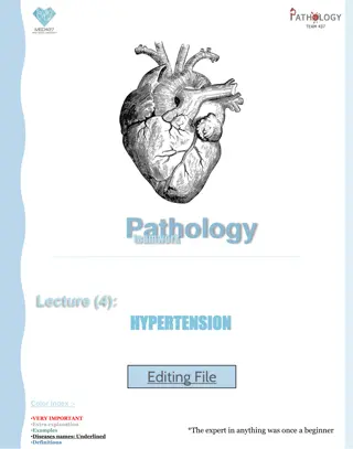
Normal red cell destruction Red cell destruction usually occurs after a mean lifespan of 120 days when the cells are removed extravascularly by the macrophages of the reticuloendothelial (RE) system, especially in the marrow but also in the liver and spleen. The breakdown of haem from haemoglobin liberates iron for recirculation via plasma transferrin mainly to marrow erythroblasts and protoporphyrin, which is broken down to bilirubin. Bilirubin circulates to the liver where it is conjugated to glucuronides, which are excreted into the gut via bile and converted to stercobilinogen and stercobilin (excreted in faeces)
Stercobilinogen and stercobilin are partly reabsorbed and excreted in urine as urobilinogen and urobilin. Globin chains are broken down to amino acids which are reutilized for general protein synthesis in the body. Haptoglobins are proteins in normal plasma which bind haemoglobin. The haemoglobin haptoglobin complex is removed by the RE system. Intravascular haemolysis (breakdown of red cells within blood vessels) plays little or no part in normal red cell destruction.
(a) Normal red blood cell (RBC) breakdown. This takes place extravascularly in the macrophages of the reticuloendothelial system. (b) Intravascular haemolysis occurs in some pathological disorders
Introduction to haemolytic anaemias Haemolytic anaemias are defined as anaemias that result from an increase in the rate of red cell destruction. The normal adult marrow, after full expansion, is able to produce red cells at 6 8 times the normal rate provided this is effective . It leads to a marked reticulocytosis. Therefore, anaemia due to haemolysis may not be seen until the red cell lifespan is less than 30 days.
Classification of haemolytic anaemias. 1-Hereditary Membrane Hereditary spherocytosis, hereditary elliptocytosis Metabolism G6PD deficiency, pyruvate kinase deficiency Haemoglobin Genetic abnormalities (Hb S, Hb C, unstable)
2-Acquired Immune Autoimmune Warm antibody type Cold antibody type Alloimmune Haemolytic transfusion reactions Haemolytic disease of the newborn Allografts, especially marrow transplantation Drug associated Red cell fragmentation syndromes See Table 6.6 March haemoglobinuria Infections Malaria, clostridia Chemical and physical agents Especially drugs, industrial/domestic substances, burns Secondary Liver and renal disease Paroxysmal nocturnal haemoglobinuria
Clinical features The patient may show pallor of the mucous membranes mild fluctuating jaundice and splenomegaly. There is no bilirubin in urine but this may turn dark on standing because of excess urobilinogen. Pigment (bilirubin) gallstones may complicate the condition and some patients (particularly with sickle cell disease) develop ulcers around the ankle Aplastic crises may occur, usually precipitated by infection with parvovirus which switches off erythropoiesis, and are characterized by a sudden increase in anaemia and drop in reticulocyte count Rarely, folate deficiency may cause an aplastic crisis in which the bone marrow is megaloblastic
Laboratory findings The laboratory findings are conveniently divided into three groups. 1 Features of increased red cell breakdown: (a) serum bilirubin raised, unconjugated and bound to albumin; (b) urine urinobilinogen increased; (c) serum haptoglobins absent because the haptoglobins become saturated with haemoglobin and the complex is removed by RE cells. 2 Features of increased red cell production: (a) reticulocytosis; (b) bone marrow erythroid hyperplasia; the normal marrow myeloid : erythoid ratio of 2 : 1 to 12 : 1 is reduced to 1 : 1 or reversed 3 Damaged red cells: (a) morphology (e.g. microspherocytes, elliptocytes, fragments); (b) osmotic fragility; (c) specific enzyme
Intravascular and extravascular haemolysis There are two mechanisms whereby red cells are destroyed in haemolytic anaemia. There may be excessive removal of red cells by cells of the RE system (extravascular haemolysis) or They may be broken down directly in the circulation (intravascular haemolysis)
Causes of intravascular haemolysis -Mismatched blood transfusion (usually ABO) -G6PD deficiency with oxidant stress -Red cell fragmentation syndromes -Some severe autoimmune haemolytic anaemias -Some drug and infection induced haemolytic anaemias -Paroxysmal nocturnal haemoglobinuria -March haemoglobinuria -Unstable haemoglobin -G6PD, glucose 6 phosphate dehydrogenase
The main laboratory features of intravascular haemolysis 1 Haemoglobinaemia and haemoglobinuria. 2 Haemosiderinuria. 3 Methaemalbuminaemia
Hereditary haemolytic anaemias Membrane defects Hereditary spherocytosis is the most common hereditary haemolytic anaemia in Northern Europeans. Pathogenesis HS is usually caused by defects in the proteins involved in the vertical interactions between the membrane skeleton and the lipid bilayer of the red cell The marrow produces red cells of normal biconcave shape but these lose membrane and become increasingly spherical (loss of surface area relative to volume) as they circulate through the spleen and the rest of the RE system. The loss of membrane may be caused by the release of parts of the lipid bilayer that are not supported by the skeleton. Ultimately, the spherocytes are unable to pass through the microcirculation, especially in the spleen, and die prematurely
The inheritance is autosomal dominant with variable expression; rarely it may be autosomal recessive. The anaemia can present at any age from infancy to old age. Jaundice is typically fluctuating and is particularly marked if the haemolytic anaemia is associated with Gilbert s disease (a defect of hepatic conjugation of bilirubin); splenomegaly occurs in most patients. Pigment gallstones are frequent Aplastic crises, usually precipitated by parvovirus infection, may cause a sudden increase in severity of anaemia
Hereditary elliptocytosis This has similar clinical and laboratory features to HS except for the appearance of the blood film usually a clinically milder disorder. It is predominantly discovered by chance on a blood film and there may be no evidence of haemolysis. Occasional patients require splenectomy. The basic defect is a failure of spectrin heterodimers to self associate into heterotetramers. A number of genetic mutations affecting horizontal interactions have been detected . Patients with homozygous or doubly heterozygous elliptocytosis present with a severe haemolytic anaemia termed hereditary pyropoikilocytosis.
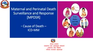
(a) Blood film in hereditary spherocytosis. The spherocytes are deeply staining and of small diameter. Larger polychromatic cells are reticulocytes (confirmed by supravital staining). (b) Blood film in hereditary elliptocytosis.
SouthEast Asian ovalocytosis This is common in Melanesia, Malaysia, Indonesia and the Philippines and is caused by a nine amino acid deletion at the junction of the cytoplasmic and transmembrane domains of the band 3 protein. The cells are rigid and resist invasion by malarial parasites. Most cases are not anaemic and are asymptomatic
Defective red cell metabolism Glucose 6 phosphate dehydrogenase deficiency Glucose 6 phosphate dehydrogenase (G6PD) functions to reduce nicotinamide adenine dinucleotide phosphate (NADP). It is the only source of NADPH which is needed for the production of reduced glutathione; a deficiency renders the red cell susceptible to oxidant stress
Epidemiology There is a wide variety of normal genetic variants of the enzyme G6PD. the most common being type B (Western) and type A in Africans. In addition, more than 400 variants caused by point mutations or deletions of the enzyme G6PD have been characterized which show less activity than normal. The inheritance is sex linked, affecting males, and carried by females who show approximately half the normal red cell G6PD values.
The female heterozygotes have an advantage of resistance to Falciparum malaria. The degree of deficiency varies with ethnic group, often being mild (10 60% of normal activity) in black African people, more severe in Middle Eastern and South East Asian people, and most severe in Mediterranean people (<10% of normal activity). Severe deficiency occurs occasionally in white people.
Clinical features G6PD deficiency is usually asymptomatic with a normal blood count between attacks of haemolysis. Although G6PD is present in all cells, the main clinical syndromes that occur are as follows. 1 Acute haemolytic anaemia in response to oxidant stress, e.g. drugs, fava beans or infections . Fava beans(Vica fava) contain an oxidant chemical, divicine. The acute haemolytic anaemia is caused by rapidly developing intravascular haemolysis with haemoglobinuria . The anaemia may be self limiting as new young red cells are made with near normal enzyme levels. 2 Neonatal jaundice. 3 Rarely, a congenital non spherocytic haemolytic anaemia. These syndromes may result from different types of severe enzyme deficiency
Agents that may cause haemolytic anaemia in glucose 6 phosphate dehydrogenase (G6PD) deficiency
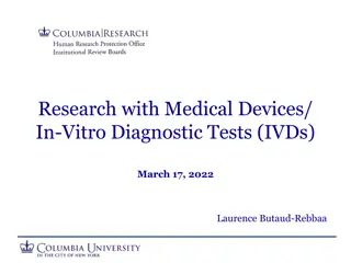
Blood film in G6PD deficiency with acute haemolysis after an oxidant stress. Some of the cells show loss of cytoplasm with separation of remaining haemoglobin from the cell membrane ( blister cells). There are also numerous contracted and deeply staining cells.
Diagnosis Between crises the blood count is normal. The enzyme deficiency is detected by one of a number of screening tests or by direct enzyme assay on red cells. During a crisis the blood film may show contracted and fragmented cells, bite cells and blister cells ,which have had Heinz bodies removed by the spleen. Heinz bodies (oxidized, denatured haemoglobin) may be seen in the reticulocyte preparation, particularly if the spleen is absent. There are also features of intravascular haemolysis. Because of the higher enzyme level in young red cells, red cell enzyme assay may give a false normal level in the phase of acute haemolysis with a reticulocyte response. Subsequent assay after the acute phase reveals the low G6PD level when the red cell population is of normal age distribution.
Treatment The offending drug is stopped, any underlying infection is treated, a high urine output is maintained and blood transfusion undertaken where necessary for severe anaemia. G6PDdeficient babies are prone to neonatal jaundice and in severe cases phototherapy and exchange transfusion may be needed. The jaundice is usually not caused by excess haemolysis but by deficiency of G6PD affecting neonatal liver function.
Glycolytic (EmbdenMeyerhof) pathway defects These are all uncommon and lead to a congenital non spherocytic haemolytic anaemia. The most frequently encountered is pyruvate kinase (PK) deficiency . This is inherited as an autosomal recessive, the affected patients being homozygous or doubly heterozygous. Over 100 different mutations have been described. The red cells become rigid as a result of reduced adenosine triphosphate (ATP) formation. The severity of the anaemia varies widely (haemoglobin (40 100 g/L) and causes relatively mild symptoms because of a shift to the right in the oxygen (O2) dissociation curve caused by a rise in intracellular 2,3 diphosphoglycerate (2,3 DPG). Clinically, jaundice is usual and gallstones are frequent. Frontal bossing may be present. The blood film shows poikilocytosis and distorted prickle cells, particularly post splenectomy.


 Normal red blood cell (RBC) breakdown. This takes place")


 Blood film in hereditary spherocytosis. The spherocytes")


")